1. Background
Herbs have long been used in treating various diseases in ancient medicine and play a key role in people’s health all around the world. Despite the advances in modern medicine, still about 25% of all new drugs are derived from medicinal plants (1). Rubia tinctorum L. (madder) is one of the useful plants, which belongs to the family of Rubiaceae that includes 450 genus and 6500 species (2). In traditional medicine, madder is still used for the treatment of cancers, tuberculosis, rheumatism, metrorrhagia, healing wounds, and inflammation (3), as well as also for the purposes of antilithiasic, diuretic, and aphrodisiac (4). The anthraquinones of Rubiaceae have biological activities such as antimicrobial, hypotensive, analgesic, antimalarial, antioxidant, antileukemic, and mutagenic effects (2). Studies in Rubia plants have led to the separation of biological components, including 148 anthraquinones and naphthoquinones, 65 terpenes, and 44 cyclopeptides, some of them have strong anticancer and antioxidant activities (3). On the other hand, some compounds of madder such as lucidin have shown mutagenic effects in bacterial and mammalian systems (5).
Anti-diarrhea activity of the crude aqueous extract of R. tinctorum L. roots has been reported in rat. It has protected rat against diarrhea caused by castor oil, and significantly inhibited the transmission in mice gastrointestinal tract (6). Moreover, madder has produced a dose-dependent reversible inhibition of jejunum contractions in rabbit as well as inhibition of carbachol- and KCl-induced contractions in rat jejunum. These effects strongly suggest an antispasmodic effect for R. tinctorum L. in gastrointestinal tract (6). However, the effect of madder on vascular tension is still unknown.
Various studies have shown the destructive effect of hyperglycemia on the endothelium of vessels. Prolonged exposure to high glucose concentration solution in vitro or in vivo has inhibited endothelial dependent relaxation in response to acetylcholine (7, 8). Oxidative stress induced by hyperglycemia has been contributed to the pathogenesis of vascular diseases by generating reactive oxygen species (ROS) (9, 10). There are some reports on the antioxidant properties of some Rubia plant species (2, 3) although the effect of madder on vascular complications induced by hyperglycemia is still unknown.
2. Objectives
Regarding the inhibitory effects of madder on the gastrointestinal tract smooth muscle contractions, the primary aim of this study was to investigate the effect of hydroalcoholic extract of R. tinctorum L. on rat isolated thoracic aorta smooth muscle ring tension. The secondary goal of this study was to examine the probable anti-oxidative protection of hydroalcoholic extract of R. tinctorum L. against the oxidative damage caused by high glucose concentration solution on rat isolated aorta endothelium.
3. Methods
3.1. Animals
Male Wistar rats (250 - 300 g) were obtained from the breeding colony of Semnan University of Medical Sciences, Semnan, Iran. Animals were housed in individual cages in a 12-hour light/dark cycle at 22 - 24°C, with food and water available ad libitum. The experimental protocol was approved by the ethical review board of Semnan University of Medical Sciences. All experimental trials were conducted in agreement with the national institutes of health guide for care and use of laboratory animals.
3.2. Preparation of Rat Thoracic Aortic Rings and Recording Isometric Contraction
Male rats were anesthetized with thiopental sodium (80 mg/kg, ip, Kwality Pharmaceutical Pvt. Ltd. India). The thorax was opened and a segment of the thoracic aorta was dissected and placed immediately in cold (4°C) Krebs-Henseleit solution (composition in mmol/L: NaCl 118, KCl 4.7, KH2PO4 1.2, MgSO4 1.2, CaCl2 2.5, Glucose 10, and NaHCO3 25, (all materials were ordered to Sigma-Aldrich), pH:7.4, and temperature: 36 ± 1°C). Aorta was cut into 5 mm sections in length. Endothelium was removed by gently rotating a metal wire in the lumen of the aorta. Then aorta ring was attached to an isometric force transducer (MLT0202, AD-instruments, Spain) coupled to the Power-Lab data acquisition system (ML866 Power-Lab 4/30, AD-Instruments, Australia) for aortic tension recording. Organ bath or chamber (double tissue bath-Harvard) containing Krebs solution was constantly gassed with 95% O2 and 5% CO2 and renewed every 15 minutes. Aortic ring was placed under 2.0 g resting tension for 60 minutes to reach the equilibrium. To verify the removal of the endothelium, acetylcholine (10-5 M, Sigma-Aldrich) was added to pre-contracted aorta ring with phenylephrine (10-6 M, Sigma-Aldrich). The lack of relaxation response to acetylcholine confirmed the absence of endothelium (11-13).
3.3. Experimental Groups
Sixty six male Wistar rats were randomly divided into 10 experimental groups:
In groups 1 - 4 (n = 7), the effect of additive madder (0.1, 0.5, 1, and 5 mg/mL) was measured on the aorta smooth muscle ring with intact or denuded endothelium pre-contracted by phenylephrine (10-6 M) or KCl (80 mM).
In groups 5 - 6 (n = 7), the effect of additive madder (0.1, 0.5, 1, and 5 mg/mL) was measured on the aorta ring with intact or denuded endothelium.
In groups 7 - 8 (n = 6), the effect of phenylephrine (10-6 M) plus madder (1 and 5 mg/mL) or madder alone (0.5, 1, and 5 mg/mL) was measured on the intact aorta ring in calcium-free Krebs solution plus EDTA (2 mM, Merck-Germany). EDTA was used to eliminate any trace of calcium in Krebs buffer.
In groups 9 - 10 (n = 6), response of intact aorta ring to phenylephrine (10-5 M) and acetylcholine (10-6 M) before and three hours after incubation with high glucose concentration solution (40 mM) with or without madder (0.5 mg/mL) was measured.
3.4. Plant and Extract Preparation
The plant madder (R. tinctorum L.) rhizome was obtained from the organization of promotion, education, and agricultural research center, Semnan, in summer 2014. The madder rhizome was dried in shadow, ground, and its hydroalcoholic extract was prepared by soxhlet apparatus via repeated distillation methods in 12 hours. The solution was then filtered and dried in oven at 40°C and finally stored at 4°C.
3.5. Statistical Analysis
The results were presented as mean ± standard error of mean (SEM); the criteria for statistical significant values was P < 0.05. The differences between groups were analyzed using paired or unpaired student’s t-test or repeated measures ANOVA for multiple comparisons (SigmaStat.3.0 - Germany).
4. Results
4.1. Effect of Madder on Aortic Ring Tension Pre-Contracted by Phenylephrine or KCl
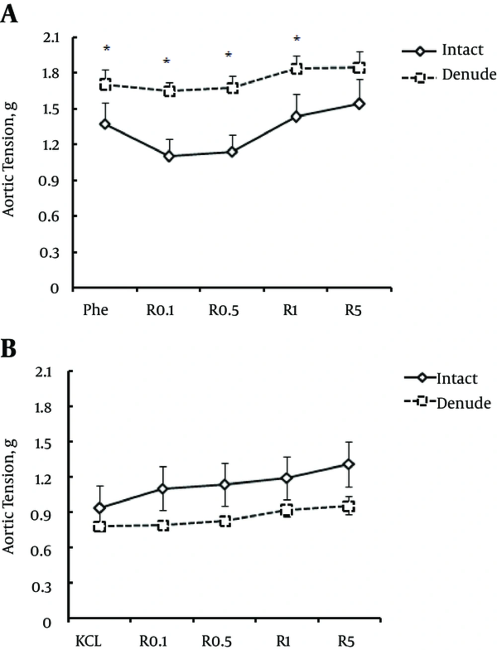
Isolated aorta ring with intact or denuded endothelium was contracted by phenylephrine (10-6 M) to a maximum of 1.37 and 1.7 g, respectively (P < 0.01). Madder was added to the chamber solution in stepwise increasing concentrations 0.1, 0.5, 1, and 5 mg/mL, with 1-minute intervals. Madder (5 mg/mL) increased the contraction produced by phenylephrine; however, this increase was not statistically significant (Figure 1A). Nevertheless, the contractile responses to phenylephrine plus madder were significantly different between aorta rings with intact and denuded endothelium (P < 0.01, Figure 1A).
The effect of different concentrations of madder on aortic smooth muscle tension (with intact and denuded endothelium) in response to phenylephrine (Phe) (a), KCl (b). (R - 0.1 to 5) represent madder concentrations from 0.1 to 5 mg/mL. (*) P < 0.01 indicates the significant value of aorta tension with intact versus denuded endothelium in response to phenylephrine followed by different concentrations of madder.
Aorta with intact or denuded endothelium was contracted by KCl (80 mM) to a maximum of 0.93 and 0.78 g, respectively. Madder was added to the chamber solution in stepwise increasing concentrations 0.1, 0.5, 1, and 5 mg/mL, with 1-minute intervals. Madder increased the contraction produced by KCl although the force of contraction was not different significantly compared to the initial contraction produced by KCl. In addition, comparing the contractile responses of aorta ring to KCl plus madder, there were significant differences between endothelium intact and endothelium denuded (Figure 1B).
4.2. Effect of Madder on Aorta Smooth Muscle Ring Tension
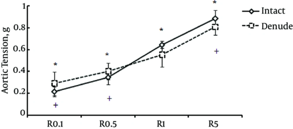
Madder (0.1, 0.5, 1, and 5 mg/mL) could induce contractions in aorta ring with intact or denuded endothelium in a concentration-dependent manner. The concentration of 5 mg/mL of madder caused significant contractions (P < 0.01) in the denuded endothelium aortic ring as compared to contractions induced by 0.1 and 0.5 mg/mL concentrations. No significant differences were observed in madder-induced contractile force between aorta rings with intact and denuded endothelium (Figure 2).
The effect of madder on aorta tension (with intact and denuded endothelium). (R - 0.1 to 5) represent madder concentrations from 0.1 to 5 mg/mL. (*) P < 0.01 comparison between contractile responses to madder in aorta within the intact endothelium group and (+) P < 0.01 the endothelium denuded group.
4.3. Effect of Madder on Aorta Smooth Muscle Tension in Free-Ca+2 Environment
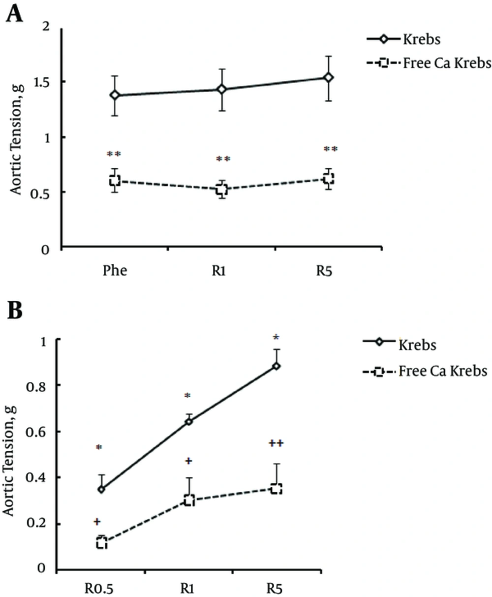
In order to investigate the dependence of madder-induced contraction on extracellular [Ca+2], the contractions produced by phenylephrine (10-6 M) plus madder (0.5, 1, and 5 mg/mL) or madder alone (0.5, 1, and 5 mg/mL) were measured in Ca+2-free Krebs. Phenylephrine-induced contraction in the aorta ring markedly declined in Ca+2-free environment and subsequently, the increased contractility induced by madder (1 and 5 mg/mL) significantly reduced (P < 0.01 Figure 3A). The contractile effect of madder alone (0.5, 1, and 5 mg/mL) was also abolished (Figure 3B). The reduction in madder-induced contraction in Ca+2-free Krebs was about 60%.
The effect of free Ca+2 -Krebs on contractile responses to phenylephrine (Phe) following madder (a) and madder alone (b). (R - 0.1 to 5) represent madder concentrations from 0.1 to 5 mg/mL. a: (**) P < 0.001 indicates the significant value of intact aorta tension in Krebs with Ca+2 versus Ca+2 free- Krebs. b: (*) P < 0.01 indicates the significant value of the effect of different concentrations of madder on intact aorta tension in Krebs with Ca+2. (++) P < 0.01 and (+) P < 0.05 indicate the significant value of intact aorta tension in Krebs with Ca+2 versus Krebs free of Ca+2 in response to madder.
4.4. Effect of Madder on Aortic Smooth Muscle Tension in High Glucose Concentration (Hyperglycemic) Environment
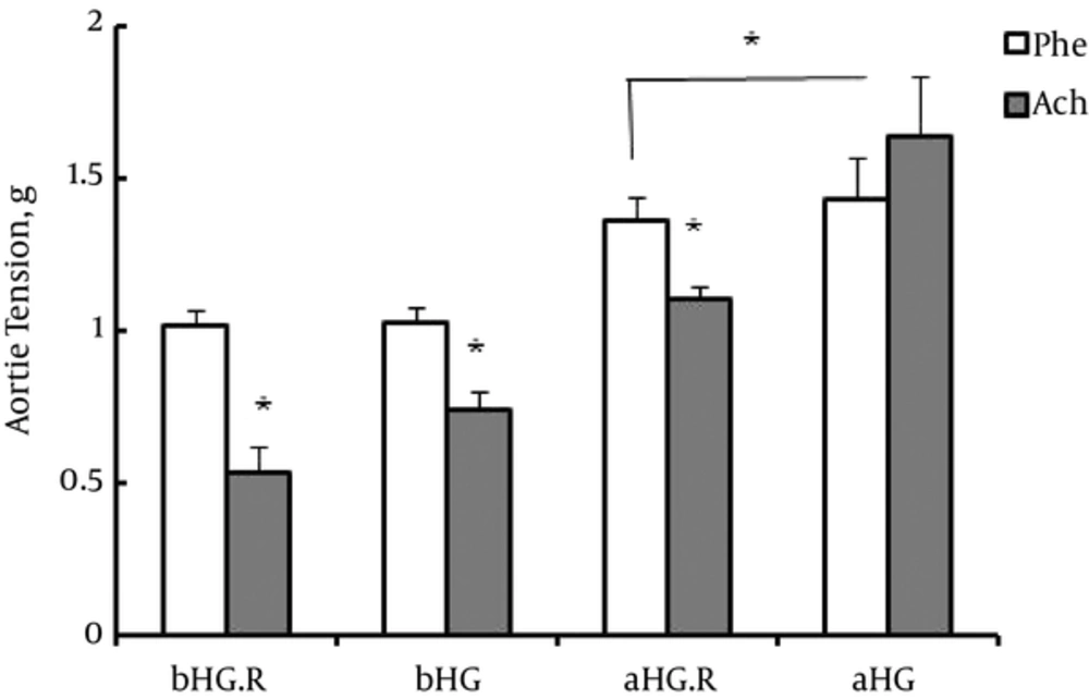
In order to investigate the effect of madder (0.5 mg/mL) on aorta smooth muscle tension in high glucose environment, the aorta ring was first incubated in high glucose concentration Krebs. The aorta ring response to acetylcholine (10-5 M) after phenylephrine (10-6 M) was observed as contraction (1.63 g) following 3 hours incubation in hyperglycemic environment. However, after 3 hours incubation of aorta ring with hyperglycemic solution plus madder (0.5 mg/mL), the response of aorta ring to acetylcholine was relaxation (1.1 g).
The effect of madder on aorta response to phenylephrine (Phe) and acetylcholine (Ach) following 3 hours incubation with high glucose or high glucose plus madder. bHG.R and aHG.R: before and after incubation with high glucose plus madder, respectively. (*) P < 0.05 indicates the significant value of aorta smooth muscle tension in response to phenylephrine versus acetylcholine and responses to phenylephrine in high glucose solution following adding madder versus incubation in solution without madder.
5. Discussion
This study showed that the hydroalcoholic extract of the plant madder mainly had constrictive effect on the isolated aorta smooth muscle. The madder additive concentrations following the phenylephrine did not have any significant effect on aortic contraction. Contractile response of aorta ring with denuded endothelium to madder was markedly stronger than the response of the ring with intact endothelium. The difference in madder contractile effects on endothelium denuded or intact aorta ring is in line with the demonstrated effect of phenylephrine on aorta smooth muscle in previous studies (13). Moreover, madder only slightly increased vascular contraction induced by KCl.
In fact, this study aimed to explore the effect of madder as a vaso-relaxant on the contractility of aorta smooth muscle. In previous studies, the inhibitory effects of madder on gastrointestinal motility were demonstrated and hence it was assumed as a smooth muscle relaxant. For instance, it was reported that aqueous extract of madder inhibited rabbit jejunum contraction in a concentration-dependent manner, as well as inhibited carbachol- and KCl-induced contractions in rat jejunum. These results suggest an antispasmodic effect for R. tinctorum, apparently by antagonizing the cholinergic effect on the visceral smooth muscle and calcium channel function. In addition, there are some reports indicating the protective effect of aqueous extract of madder against diarrhea caused by castor oil and inhibition of intestinal motility in rat (6). Therefore, based on previous studies on exploring the effect of madder on the gastrointestinal smooth muscle, we developed a hypothesis suggesting that madder may also induce a relaxant effect on the smooth muscle of great arteries, such as aorta. In order to test this hypothesis, we applied vasoconstrictors, such as phenylephrine or KCl, to induce contraction in the prepared isolated aorta smooth muscle ring and then added madder to evaluate its relaxant effect. However, the result was surprising since madder just tried to preserve the contraction effect of vasoconstrictors. In the next step, in order to explore whether or not the contraction induced by madder is dependent on vasoconstrictors, we applied different concentrations of madder “alone” to aorta ring. Madder could induce concentration-dependent contractions in intact and denuded endothelium aorta, with no significant difference mediated by endothelium. Our results showed the contractive effect of madder on vascular smooth muscle for the first time, which was independent of other vasoconstrictor effects.
The opposite effect observed in this study compared to what previously reported may be due to the type of the plant or extract, method of preparation of extract, method of study or using different tissues, or perhaps due to different intracellular pathways responsible for contractive or relaxant outcomes of madder relating to intracellular Ca2+ concentration (14). If the vasoconstrictor effect of madder is also displayed in vivo studies, the use of this plant or any of its product will be highly prohibited for hypertensive patients or those with certain types of vasculopathies.
The contractile response to KCl in the vascular smooth muscle is produced by membrane depolarization and therefore, activation of voltage gated calcium channels (VGCC), followed by increase in intracellular Ca2+concentration through release from sarcoplasmic reticulum and increase in Ca2+ sensitivity (15). In addition, α1-adrenoceptor agonist like phenylephrine increases intracellular Ca2+ by activating VGCCs or non-voltage dependent Ca2+ channels (16, 17). It has been shown that aorta smooth muscle contraction induced by α1-adrenoceptor activation is highly dependent on Ca2+ entry from extracellular environment (18, 19). In order to test the dependence of madder on the extracellular Ca2+ for producing its constrictive effect, we measured the madder-induced contractile force in Ca2+-free Krebs buffer. By adding madder to the Ca2+-free buffer, the constrictive response of aorta ring showed a marked decline compared to the respective contraction in the Krebs. Likewise, in Ca2+-free buffer, aortic contractile response to phenylephrine plus madder significantly reduced (60%). This experiment suggests that madder facilitates Ca2+ entry into the smooth muscle cell of aorta and causes contraction by increasing the sensitivity of cell to Ca2+. The other similarity of action of madder to the action of other vasoconstrictors in this study was the presence of endothelium and its effect on the level of contractile force. In endothelium intact aorta ring, the intensity of contraction induced by madder reduced.
As mentioned earlier, prolonged exposure of vascular endothelium to high glucose concentration would damage the endothelium by generating reactive oxygen species and oxidative stress (7-9). Since the antioxidant effect of some Rubia species has been previously reported (2, 3), we aimed to test the preventive effect of madder extract in aorta ring with endothelium damage caused by high glucose concentration. Three hours incubation with hyperglycemic solution caused the damage to aorta endothelium, so that its contractile response to phenylephrine increased and relaxing response to acetylcholine changed to contraction. However, incubation of aorta ring with hyperglycemic buffer plus madder (0.5 mg/mL) apparently preserved endothelium to some extent, since its responses to acetylcholine and phenylephrine were similar to the responses in Krebs buffer. Therefore, this study suggests that madder is capable of protecting aorta endothelium against probable oxidant damage due to high glucose concentration. Whether the displayed protective action of madder on endothelium is due to an antioxidant effect or due to other probable effects related to the plant, there is warrant for further studies, since clarifying the mechanism underlying this protective effect may become a useful pharmacologic tool in protecting diabetic patients from vasculopathies relating to high blood sugar in future.
There are many compounds derived from R. tinctorum root that may have anti-cancer or vascular protective effects; including alizarin, ruberythric acid, purpurin, lucidin, rubiadin, mulligan, scopolamine, and the glucosides (20). For instance, it is reported that mollugin has potential antioxidant and anti-inflammatory effects (21) that may involve in aortic endothelial protection against hyperglycemic environment. Similarly, it is reported that many agents such as, Propolise, Ramipril, and Hespiridin protect endothelium against damage induced by hyperglycemic environment through their antioxidant activities (22-24).
In summary, our results demonstrated madder vasoconstrictor effect on isolated aorta smooth muscle that might be mediated by calcium mobilization. In addition, it displayed madder protective effect on isolated aorta ring endothelium against damage caused by high glucose concentration environment, possibly through antioxidant activity.


