1. Background
The liver is one of the most important internal organs in living organisms and has an enormous regenerative capacity after injuries. Some of the most important liver functions include food processing and chemical detoxification. Oxidative stress on the liver is associated with liver function impairment. Free radicals, such as superoxide anion radical (O2-), nitrogen dioxide (NO2), and perhydroxyl radical (HO2·) are in vivo forms of reactive oxygen species (ROS). The production of ROS is a natural part of oxygen metabolism that is involved in cell signaling, homeostasis, and defense against microbes and toxins. Excessive amounts of ROS from exogenous sources are highly toxic (1). Fruits and vegetables containing antioxidant compounds effectively help the antioxidant defense system (2).
Tert-butyl hydroperoxide (t-BHP) is an experimental model that induces oxidative stress. Events that lead to the induction of oxidative stress by t-BHP are lipid peroxidation, GSH depletion, MDA formation, and leakage of ALT and lactate dehydrogenase (LDH) (3).
Pomegranate is an ancient plant of the Punicaceae family. Pomegranate is known to contain a variety of medicinal compounds and has proven pharmaceutical properties such as antioxidant, anti-atherosclerotic, anti-inflammatory, and anti-diabetic effects (4). The peel of pomegranate recently has attracted the attention of medical researchers, as it has been reported to contain polyphenols such as gallic acid, ellagic acid, anthocyanins, and flavonoids (5). An important factor in pomegranate’s profiles of total phenolic and antioxidant activity is its genotype. Black pomegranate is a rare cultivar of pomegranate that is used in traditional medicine for sore throat and lung diseases. Peel of this cultivar poses among the three pomegranates with the maximum amount of polyphenols content (6). In the present study, the in vitro protective effects of the BPPE against t-BHP induced oxidative stress were investigated in Wistar rats.
2. Objectives
The objective of this study was to investigate the protective effect of BPPE against oxidative hepatotoxicity induced by t-BHP in the liver tissues of Wistar rats.
3. Methods
3.1. Chemicals
In this study, t-BHP, 5,5’-dithiobis (2-nitrobenzoic acid) (DTNB), trichloroacetic acid (TCA), reduced glutathione (GSH), thiobarbituric acid (TBA), 1,1,3,3-tetramethoxypropane, bovine serum albumin (BSA), and Coomassie blue G dye were purchased from Sigma-Aldrich Company (St. Louis, MO), USA. All chemicals and reagents used were of analytical grades.
3.2. Preparation of BPPE
Black pomegranates (herbarium number: 248) were collected from Shiraz, Iran. The peel of the fruits was removed, shade-dried, and powdered. The powder was then soaked in 80% aqueous-methanol solution for three days. The extracted solution was filtered, and the solvent was removed by vacuum in a rotary evaporator.
3.3. DPPH• Free Radical Scavenging Assay
The DPPH• scavenging activity was estimated to evaluate the radical scavenging properties of the extracts (7). To this aim, 0.1 ml of the extract was added to 3.9 ml of DPPH solution (25 mg/mL), and the resulting mixture was vigorously stirred and kept in darkness for 30 minutes. The decrease in the solution absorbance was measured at a wavelength of 517 nm. The results were expressed in units of µg/mL.
3.4. Animals and Treatment
Adult male Wistar albino rats (200 - 250 g) were received from Ahvaz Jundishapur University of Medical Sciences animal house. Rats were maintained in polypropylene cages with free access to drinking water and standard rat chow at a controlled condition with a temperature of 20 ± 2°C and a 12:12 h light-dark cycle. The experiments were performed according to the guidelines of the Committee of Animal Experimentation of Ahvaz Jundishapur University of Medical Sciences (ethical code: IR.AJUMS.REC.1395.98).
Animals were classified into five groups (n = 6). Group I, the negative control, was given normal saline (5 mL/kg); Group II, the positive control, received normal saline orally for five days, and a single dose of t-BHP (0.2 mmol/kg, i.p.) (8) at the 6th day; groups III-V received BPPE orally at doses of respectively 100, 200, and 400 mg/kg, and a single dose of t-BHP at the 6th day 1 hour after administering the last dose of BPPE.
3.5. Sample Collection
Animals were anesthetized with a combination of ketamine/xylazine (60/6 mg/kg, i.p.) 24 hours after the last administration. Blood samples were collected, and serum was separated and stored at -20°C. Then, animals were sacrificed, and livers were isolated. A part of the tissue was preserved in 10% phosphate-buffered formalin for histological studies, and the remaining tissue was homogenized (1/10 w/v) in ice-cold Tris-HCl buffer for biochemical estimations. Protein content in homogenates was determined by Bradford assay (9).
3.6. Serum Biochemical Parameters
The colorimetric method was used for the determination of ALT and AST. As instructed by Belfield and Goldberg, the amount of pyruvate or oxaloacetate produced from the formation of 2, 4-dinitrophenylhydrazine was measured by the method of Reitman and Frankel (10). Moreover, ALP was assayed using the kits purchased from Randox Laboratories Co.
3.7. Reduced Glutathione Assay
The glutathione content (GSH) was measured by its reaction with Ellman’s reagent (DTNB) and the formation of a yellow color (11). In summary, 40 μl liver tissue suspension was added to 2 ml of Tris–EDTA buffer (pH = 8.6), then 40 μL of DTNB (10 mm in methanol) was added to the mixture. The mixture was incubated for 20 minutes at room temperature, and the yellow color was read at 412 nm with a spectrophotometer. The calibration curve was drawn using the glutathione standard.
3.8. Lipid Peroxidation
Lipid peroxidation of the tissues was investigated by measuring MDA. MDA reacts with a TBA solution and creates a pink color complex that has a maximum absorption of 532 nm (12). In brief, 0.5 mL of liver tissue suspension was mixed with 2.5 mL of TCA (10%, w/v) and then centrifuged at 3,000 rpm for 15 minutes. Then, 2 mL of supernatant was added to 1 mL of TBA (0.67%, w/v) in a test tube and placed in boiling water for 10 minutes. Finally, the absorbance was measured by a spectrophotometer at a wavelength of 532 nm.
3.9. Examination of CAT Activity
CAT activity in the tissue was assayed based on the rate of H2O2 decomposition following the method of Kalantar et al. (13). For this purpose, 50 μL of the tissue supernatant obtained after centrifugation of the tissue homogenate at 12,000 g for 20 min at 4°C and 200 µL of phosphate buffer were added to a cuvette with 250 µL of 0.066M H2O2 as a substrate. The enzymatic activity was measured in milliliters of H2O2 degraded per minute per mg of protein in the tissue supernatant. CAT activity measurement was performed using the molar extinction coefficient of 43.6 M-1.cm-1.
3.10. Histopathological Assessments
For the histological examination, 10% formalin was used to fix tissues. Tissues were then dehydrated by ethanol solutions and embedded in paraffin. Ultimately, 5-µm sections were prepared and stained with H & E dye and then examined under a light microscope. To score the samples, five microscopic fields were evaluated. Each sample was scored on a scale of 0 to 3 (none: 0, mild: 1, moderate: 2, and severe: 3) for each criterion.
3.11. Statistical Analysis
The results were expressed as mean ± SD. All statistical comparisons were made using a one-way ANOVA test followed by Tukey’s post hoc analysis. In all tests, P < 0.05 was considered statistically significant. Data were analyzed using Prism 5.0 statistical software package (San Diego, CA, USA).
4. Results
4.1. In vitro Antioxidant Activities of BPPE
The IC50 values for the radical-scavenging activity of BPPE are shown in Table 1. The DPPH• IC50 results of BPPE (74 µg/mL), which were used to measure its radical-scavenging ability, were lower than that of ascorbic acid (75.2 ± 5.4l µg/mL).
| Sample | DPPH• IC50, µg /mL |
|---|---|
| BPPE | 74 |
| Ascorbic acid | 75.2 ± 5.4 |
aValues are expressed as mean ± SD.
4.2. Effect of BPPE on Serum ALT, AST, ALP Levels
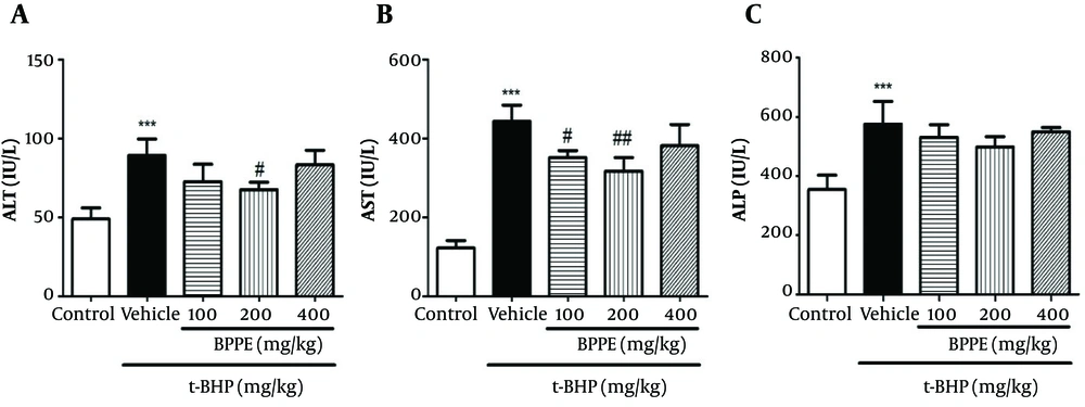
Twenty-four hours after injection of a single dose of t-BHP, the rats developed hepatotoxicity reflected in significantly elevation (P < 0.05) serum ALT, AST, ALP levels (Figure 1). The BPPE pretreatment reduced serum ALT, AST, and ALP levels at all doses, but the statistically significant reductions were seen in ALT at a dose of 200 mg/kg and in AST at doses of 100 and 200 mg/kg (P < 0.05).
4.3. Effect of BPPE on Liver Tissue Parameters
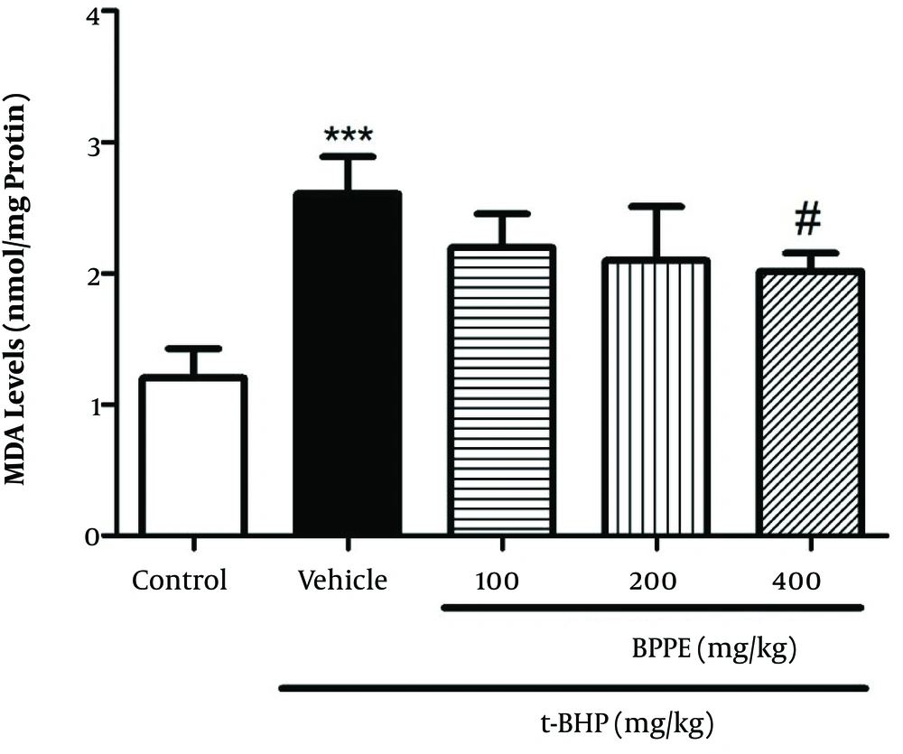
MDA levels in the group treated with 400 mg/kg of BPPE, and regarding the inhibition of lipid peroxidation, they were significantly lower than the group treated only with t-BHP (Figure 2).
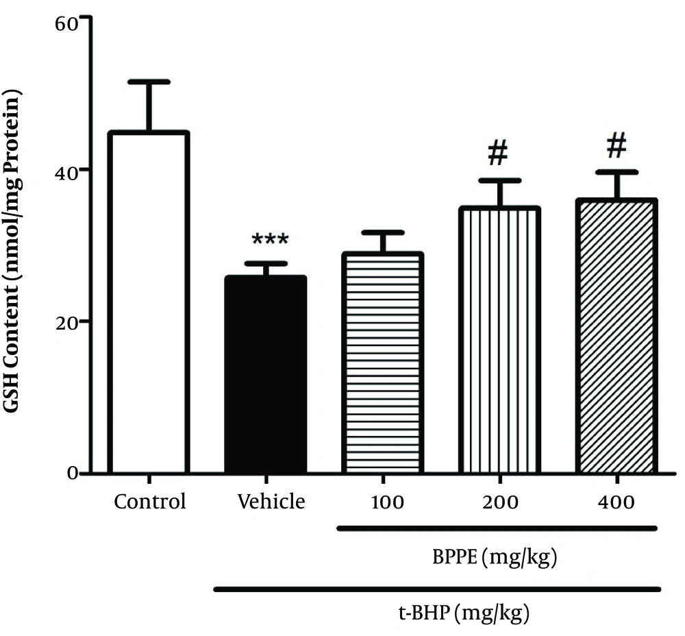
While administrating t-BHP led to significantly depleted GSH levels, BPPE pretreatment significantly recovered GSH level depletion (Figure 3). Also, the hepatic GSH levels were significantly increased by pretreatment with BPPE (200 and 400 mg/kg).
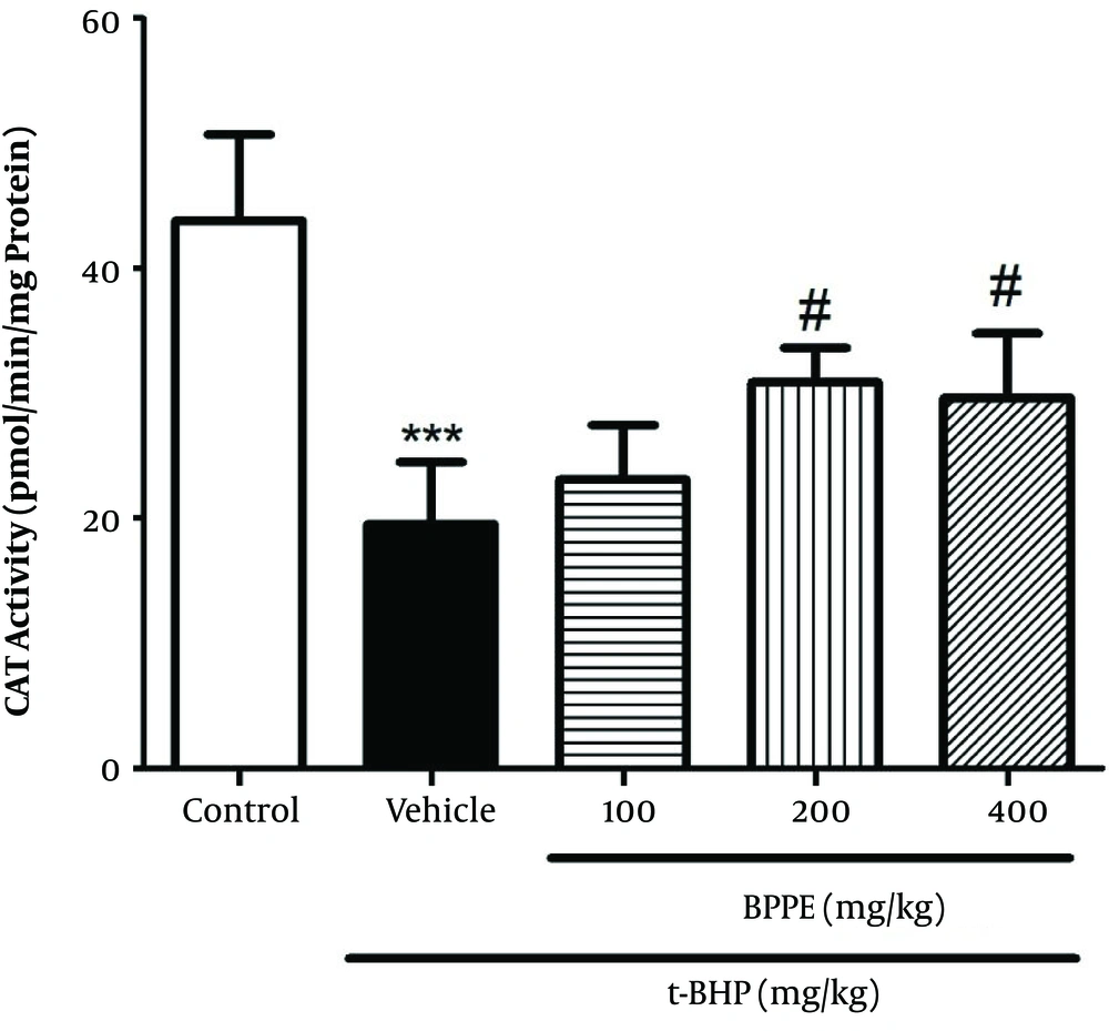
As shown in Figure 4, CAT activity was significantly lower in the t-BHP-only treated group than in the control group (P < 0.05). Pretreatment with BPPE (400 mg/kg) significantly inhibited the CAT activity reduction induced by t-BHP.
4.4. Light Microscopy Findings
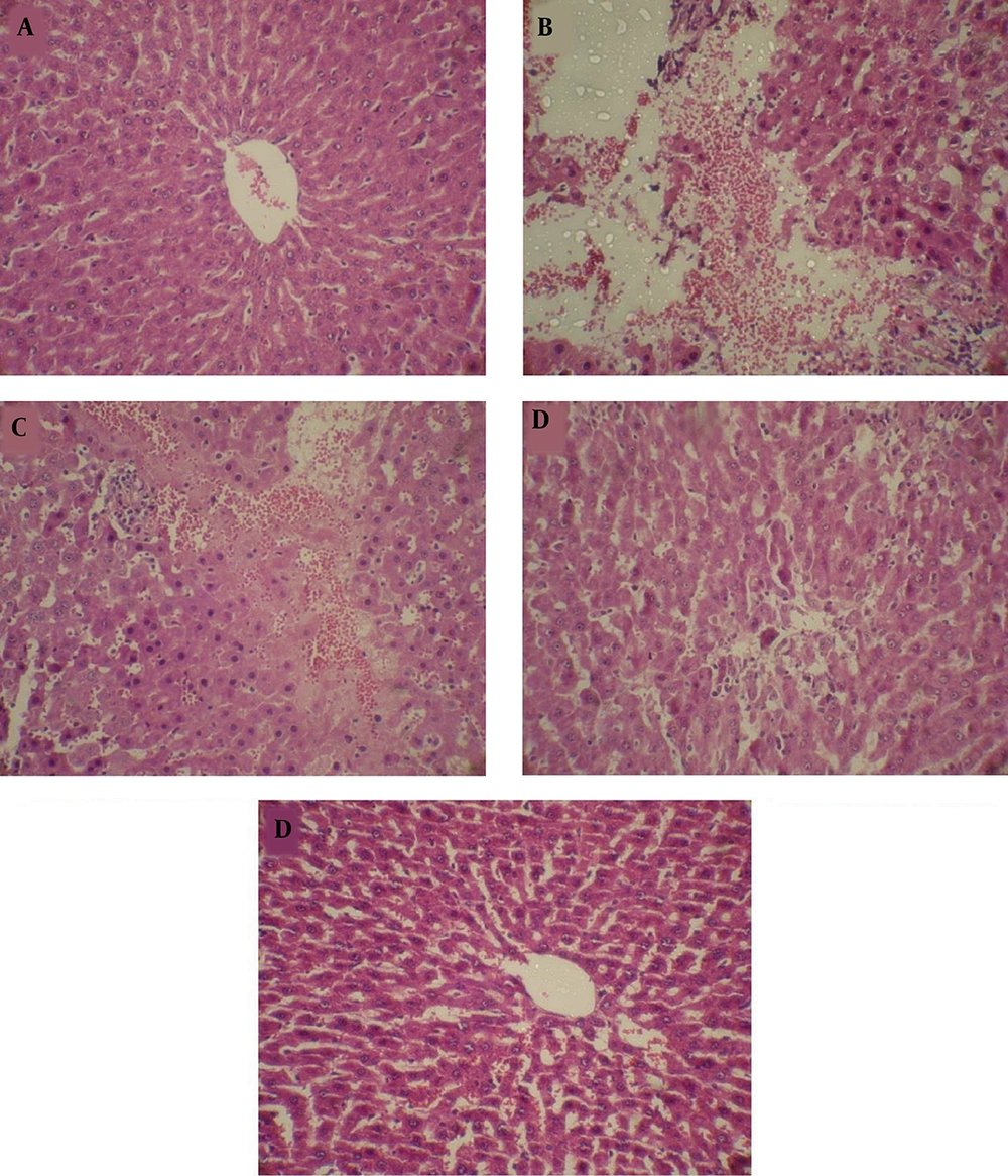
Examinations of histopathological changes in rat liver were performed by H&E stained sections (Figure 5). In the control group, the liver tissue is normal (Figure 5A), but in the t-BHP group, the radial hepatocyte arrangement was lost. The hepatocytes experienced apoptosis, and the sinusoids were dilated (Figure 5B). No recovery was observed in these images of treatment with 100 mg/kg of BPPE (Figure 5C). The tissue images of treatment with 200 mg/kg of BPPE exhibited to some extent improvement, and no apoptosis, tissue degradation, and sinusoidal dilatation were observed (Figure 5D). The tissue images of treatment with 400 mg/kg of BPPE showed that the liver tissue damages were significantly diminished, and no certain pathological changes were observed (Figure 5E). The microscopic scores of liver tissues of the t-BHP-injected group were significantly higher than the control group (P < 0.001). Microscopic scores reduced with BPPE treatment (P < 0.001) (Table 2).
Protective effect of BPPE on histopathological changes in t-BHP-induced hepatotoxicity in rats. A, control; B, rats treated with t-BHP; C, rats treated with BPPE (100 mg/kg) + t-BHP; D, rats treated with BPPE (200 mg/kg) + t-BHP; and E, rats treated with BPPE (400 mg/kg) + t-BHP. (H & E 400×).
aValues are expressed as mean ± SD.
bThe histological scores were compared by Kruskal-Wallis non-parametric test followed by Dunn post-test.
cSignificant difference compared to the control group (P< 0.01).
dSignificant difference compared to the t-BHP group (P < 0.05).
eSignificant difference compared to the control group (P < 0.05).
fSignificant difference compared to the t-BHP group (P < 0.01).
5. Discussion
In the current study, t-BHP induced liver injury has extensive use in the investigation of cell injury mechanisms that are initiated by oxidative stress in a variety of systems (14). Metabolism of t-BHP by cytochrome P450 leads to the production of free radical intermediates in the hepatocytes, which trigger ROS generation. Recently, researches have shown that oxidative stress is one of the causes of many diseases, such as cancer (15), cardiovascular (16), and neurodegenerative diseases (17).
The augmentation of cell oxidative defense capacity through the intake of antioxidants is of vital importance for preventing cellular injuries mediated by free radicals. Recently, natural antioxidants have received increasing research attention for their beneficial effects in biological systems. Most notably, phenolic phytochemicals of natural plant extracts are known to produce protective effects against oxidative damage (18). Various components, such as polyphenols and flavonoids play an important role in free radical scavenging due to their antioxidants capacity. Major phenolic acids are caffeic, neochlorogenic, and ferulic, and the main flavonoids are quercetin, luteolin, and kaempferol (19).
Black pomegranate (Punica granatum L.) exhibits high antioxidant capacity. This study explored the possible protective effect of BPPE against oxidative hepatotoxicity induced by t-BHP in rats. According to the results, t-BHP causes a significant increase in the levels of ALT, AST, and ALP in the serum. These results are in agreement with those presented in previous studies (20, 21). BPPE supplementation significantly alleviated the elevated levels of ALT, AST, and ALP in t-BHP-treated rats. An explanation of this finding is that the BPPE phyto-constituents are likely to improve the stability of the hepatocytes plasma membrane.
Histopathology examination of liver samples showed that t-BHP induced severe changes, including hepatocytes necrosis and congestion in central veins and sinusoids compared with the control group. Treatment with BPPE improved the t-BHP-induced hepatic damage, specifically severe degeneration and vacuolation of hepatocytes. BPPE administration can probably repair the hepatic tissue damages through the stimulation of protein synthesis and hepatocyte regeneration (22).
The oxidative stress that manifests as lipid oxidation is one of the mechanisms involved in cell damage. According to a proposed hypothesis, the reason for liver damage induced by t-BHP through lipid oxidation is due to free radicals, especially alkoxyl and peroxyl. In this study, it was observed that the MDA level was increased significantly, which could have occurred due to the accumulation of free radicals as a result of the inadequacy of antioxidant defense. The supplementation with BPPE reduces MDA levels. The phenolic compounds of BPPE may play a protective role by binding to lipid peroxidase or by chelating the metals involved in lipid oxidation. The protective effect of BPPE may be due to inhibition of the t-BHP metabolism via cytochrome P450, thus preventing the production of free radicals of lipid oxidation initiation. Also, the BPPE potential in preventing t-BHP induced lipid oxidation can be attributed to the content of its lipid antioxidants (tocopherols, tocotrienols, and carotenoids), and this may be related to their ability to share phenolic hydrogen (electron) with lipid peroxyl free radicals and these antioxidants are capable of penetrating the lipid layer of the cell membrane because of hydrophobicity and thus play a protective role against lipid oxidation (23).
Scientific evidence has shown that oxidative stress toxicities are associated with changes in levels of antioxidant enzymes (24). In this study, the CAT activity level was reduced in the rats exposed to t-BHP, and the BPPE supplementation increased the hepatic CAT activity to the normal level. The first enzyme in the ROS detoxification process is SOD, which converts superoxide free radicals to H2O2. The main role of CAT is to sweep the H2O2 produced by the SOD, and any increase in its activity may indicate an increase in H2O2 production or the expression of CAT-encoding genes. In the present study, it can be suggested that the modulation achieved in CAT activity is due to the function of the natural antioxidants in the BPPE.
Glutathione plays a role in the removal of free radicals and the stability of the thiol proteins of the cell membrane. In this study, the GSH level in the rats exposed to t-BHP was decreased, and the BPPE supplementation resulted in a significant increase in the GSH level, possibly due to the activity of BPPE phenolic antioxidants and increased detoxification capacity and the improved cellular redox/antioxidant status. As shown in previous studies, many of the phenolic compounds in plants can enhance the activity of the enzyme γ-glutamylcysteine synthetase (γ-GCS), a restriction enzyme in the GSH production (25, 26). and so another hypothesis is that the BPPE polyphenols may increase the γ-GCS expression and GSH content.
5.1. Conclusions
According to the results of the present study, we suggest that antioxidant-rich BPPE shows a protective effect against oxidative hepatotoxicity induced by t-BHP in Wistar rats. The results demonstrate the BPPE potency to reduce the level of liver marker enzymes, prevent lipid peroxidation, and adjust changes in antioxidant enzyme activity.

