


1. Background
2. Objectives
3. Methods
3.1. Preparation of Protoscolices and Hydatid Tissue
3.2. Herbal Extracts Preparation
3.3. Experiment Design
3.3.1. Effects of Herbal Extracts on Protoscolices In Vitro
3.3.2. Effect of Herbal Extracts on Hydatid Cyst Tissue In Vitro
3.3.3. Immunohistochemistry for Caspase-3 Detection
3.4. Statistical Analysis
4. Results
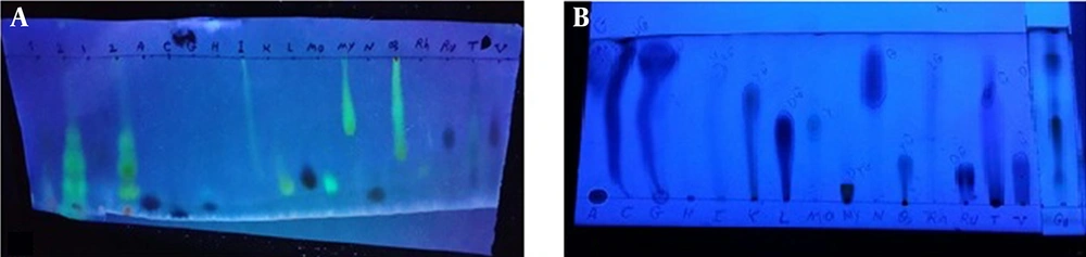
The result of chromatography. A) A. noeanum flavonoids extract chromatogram using descending paper chromatography in BAW solvent, B) A. noeanum flavonoids extract chromatogram using ascending thin layer chromatography in CAW solvent. Abbreviations: A (apigenin), C (chrysin), G (genistein), H (hesperidin), I (isorhamnetin), K (kaempferol), L (luteolin), Mo (morin), My (myricetin), N (naringenin), Q (quercetin), Rh (rhamnetin), Ru (rutin), T (tricine), and V (vitexin); G1 (1)= A. noeanum crude extract, G2 (2)= A. noeanum acid hydrolysis flavonoids extract.
| Mortality Over Time | Mortality (% ± SD) | |||||
|---|---|---|---|---|---|---|
| 30 Sec | 1 Min | 5 Min | 10 Min | 20 Min | 30 Min | |
| Extract concentrations (gr/mL) | ||||||
| 25% (0.122) | 4.7 ± 0.6 | 12 ± 2.6 | 31.7 ± 3 | 53.7± 5.9 | 78.7 ± 5.9 | 76.7 ± 3 |
| 50% (0.245) | 93.3 ± 1.5 | 95.7 ± 2.1 | 100 ± 0 | 100 ± 0 | 100 ± 0 | 100 ± 0 |
| 75% (0.367) | 100 ± 0 | 100 ± 0 | 100 ± 0 | 100 ± 0 | 100 ± 0 | 100 ± 0 |
| 100% (0.49) | 100 ± 0 | 100 ± 0 | 100 ± 0 | 100 ± 0 | 100 ± 0 | 100 ± 0 |
| Negative control (buffer) | 4 ± 1 | 5 ± 1.7 | 5 ± 0 | 5 ± 2 | 6 ± 1 | 6 ± 0 |
| Positive control (formalin) | 100 ± 0 | 100 ± 0 | 100 ± 0 | 100 ± 0 | 100 ± 0 | 100 ± 0 |
| Mortality Over Time | Mortality (% ± SD) | |||||
|---|---|---|---|---|---|---|
| 30 Sec | 1 Min | 5 Min | 10 Min | 20 Min | 30 Min | |
| Extract concentrations (gr/mL) | ||||||
| 25% (0.337) | 83.7 ± 4.7 | 92.3 ± 2.9 | 97 ± 1.7 | 100 ± 0 | 100 ± 0 | 100 ± 0 |
| 50% (0.675) | 91.7 ± 2.9 | 90.3 ± 2.5 | 100 ± 0 | 100 ± 0 | 100 ± 0 | 100 ± 0 |
| 75% (1.012) | 97.7 0.6± 0.6 | 100 ± 0 | 100 ± 0 | 100 ± 0 | 100 ± 0 | 100 ± 0 |
| 100% (1.35) | 100 ± 0 | 100 ± 0 | 100 ± 0 | 100 ± 0 | 100 ± 0 | 100 ± 0 |
| Negative control (buffer) | 4 ± 1 | 5 ± 1.7 | 5 ± 0 | 5 ± 2 | 6 ± 1 | 6 ± 0 |
| Positive control (formalin) | 100 ± 0 | 100 ± 0 | 100 ± 0 | 100 ± 0 | 100 ± 0 | 100 ± 0 |
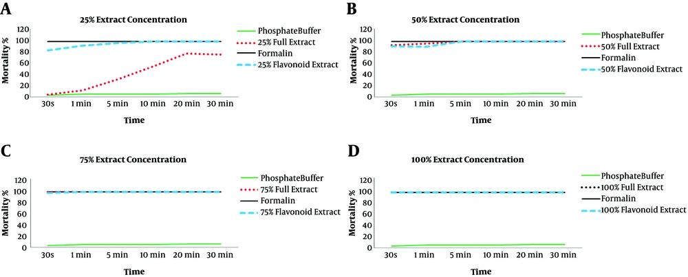
Comparison of the mortality rate of protoscolices in exposure to 25% (0.122 gr/mL), 50% (0.245 gr/mL), 75% (0.367 gr/mL) and 100% (0.49 gr/ml) concentrations of crude extract with mortality rate of the parasite in exposure to 25% (0.337 gr/mL), 50% (0.675 gr/mL), 75% (1.012 gr/mL) and 100% (1.35 gr/mL) concentrations of flavonoid extract
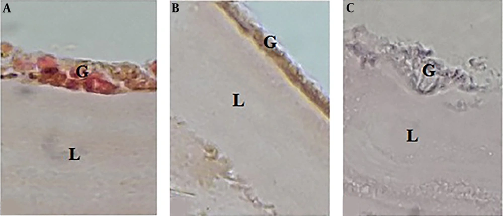
Histological changes in the hydatid cyst wall. A) After exposing the cyst to phosphate buffer solution, the germinal layer and its nuclei were clear, and the laminated layer was uniform and clear. B) After exposing the cyst to 100% concentration (0.49 gr/mL) of crude extract herbal extract, the germinal layer was found to be unclear, and its nuclei had disappeared. The laminated layer was also uneven and rough. C) By exposing the cyst to 100% concentration (1.35 gr/mL) of the flavonoid extract, the germinal layer was found to be completely destroyed, and the laminated layer was rough, curved, and uneven.
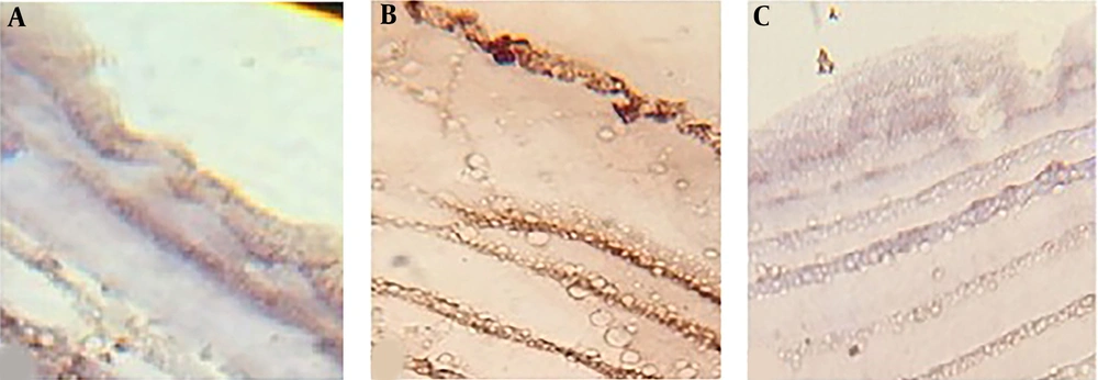
Caspase-3 expression in the hydatid cyst wall after 24 hours of exposure to A) phosphate buffer (control); B) 100% concentration (0.49 gr/mL) of crude extract; and C) 100% concentration (1.35 gr/mL) of flavonoid extract


