


1. Background
Papules and plaques affecting the plantar aspect of the foot are commonly encountered in dermatology practice, which can pose difficulties in accomplishing daily activities. The plantar wart, corn, and callus are the most common plantar lesions. The plantar wart is a benign tumor caused by the human papillomavirus (HPV), commonly by HPV-1 and other HPV-2, 4, 27, or 57 strains. A successive inoculation ensues the formation of a small shining ‘sago grain’ papules that soon assumes sharply defined rounded lesions having rough hyperkeratotic protrusions, which are painful (1).
Repetitive frictions and pressures are the main cause of the callus, also known as ‘tyloma’ (2), which is a diffuse hyperkeratotic lesion of relative thickness with undefined margins and a broad base (3). In secondary infections, callus often causes painful plantar plaque. A corn is a sharply demarcated hyperkeratotic lesion occurring over a bony prominence (2). Those who suffer from a corn present with painful thickened papule, which is deliberately progressive over time. It is consistently very well localized to the affected area. Callus and corn are a quotient of repeated mechanical trauma, which is associated with dynamic changes in the function of the foot. Plantar warts are more sensitive to lateral compression of the lesion whereas, corn, and callus are tender on direct pressure as it comes in contact with the body prominences (4). The most common site for corns and calluses is over the metatarsal heads and heel, while plantar warts can occur on any site. Although the clinical examination is sufficient to clinch an accurate diagnosis, atypical presentations indicate the need for invasive investigations like skin biopsy.
Dermoscopy is a simple, rapid, non-invasive, yet, precise diagnostic tool to visualize surface and subsurface morphological features that cannot be comprehended through naked eye exploration. Hence, it can be considered as a bridge between clinical examination and histopathological assessment (5). Dermoscopic descriptions of the above-mentioned conditions are limited in the literature (6, 7). Here, we attempted to characterize and discriminate dermoscopic patterns of painful plantar warts, corn, and callus. Also, an emphasis on the histopathological correlation of these dermoscopic patterns has been considered.
2. Methods
In this cross-sectional observational study, patients attending the dermatology outpatient department at a tertiary care center attached to S Nijalingappa Medical College in Southern India from June 2019 to January 2020 were recruited. Apart from institutional ethical clearance, written informed consent for performing dermoscopy and skin biopsy, as well as publishing the results with their photographs, was obtained from all participants. Patients with clinical features suggestive of plantar wart, corn, and callus were selected in this study. Demographic data (i.e., age, sex, occupation, duration, and site of the lesions) were registered for all cases. Those who were receiving topical or systemic therapy, either currently or during the past month, were excluded. Following a detailed clinical examination, a dermoscopic examination was performed using Illuco 1100 IDS, a handheld manual dermoscope with a magnification of 10 x attached to a smartphone was employed to capture the images.
Both polarized and non-polarized modes were pursued with ultrasonography gel as an interface medium. Histopathological examination was carried out to affirm the clinical diagnosis. Collected data were entered in a Microsoft Excel spreadsheet, and data analysis was administered by SPSS version 19 using percentages for qualitative data and Chi-square test for differences in the proportions. Statistical significance was considered when P-value < 0.05.
3. Results
Of 92 participants, plantar wart and corn were observed, respectively, in 56 (60.86%) and 22 (24.44%) patients. Fourteen (15.55%) patients had callus. The mean duration of lesions was 4 months. The mean age of participants was 20 years. Red dots and yellow halos were the most common features in warts in 50 (89.28%) and 46 (82.14%) patients, respectively, on dermoscopy (Figures 1A-F). None of the lesions of corn and callus revealed red dots or yellow halo. The translucent central core and whitish ring were distinguished only in patients who had corn, in 22 (100%) and 18 (81.81%) patients, respectively (Figures 2A-D). Yellowish areas were observed in 16 (72.72%) patients with corn. All callus lesions (100%) demonstrated opaque yellow areas (Figures 2E, 2F, 3A, 3B). Notably, dermatoglyphics were seen preserved in corn and callus. The characteristic dermoscopic features in wart, corn, and callus are depicted in Table 1. The statistical analysis was not significant in various dermoscopic patterns. Histopathology in wart showed hyperkeratosis, acanthosis, and dilated dermal capillaries (Figure 4A), whereas hyperkeratotic stratum, parakeratosis, and absence of granular layer were noted in corn (Figure 4B). Callus showed hyperkeratosis and acanthosis (Figure 4C). Localized and diffuse dermal fibrosis was noted in corn and callus, respectively.
| Dermoscopic Features | Plantar wart (n = 56) | Corn (n = 22) | Callus (n = 14) | P Value |
|---|---|---|---|---|
| Red dots | 50 (89.28) | - | - | - |
| Linear hemorrhages or streaks | 10 (10.71) | - | - | - |
| Yellow halo | 46 (82.14) | - | - | - |
| White halo | 10 (17.85) | - | ||
| Translucent central core | - | 22(100) | - | - |
| Yellowish areas | - | 16 (72.72) | 14 (100) | 0.08 |
| Preserved dermatoglyphics | - | 22 (100) | 14 (100) | |
| Absence of dermatoglyphics | 56 (100) | - | - | - |
| Whitish annular ring | - | 18 (81.81) | - | - |
| Focal white areas | - | - | 6 (42.85) | - |
aValues are expressed as No. (%) unless otherwise indicated.
![A, Clinical image of plantar wart showing hyperkeratotic papule on the sole. B, Dermoscopy shows dotted (yellow arrow) and linear (red arrow) vessels. Dermatoglyphics (black arrows) are absent abruptly in the involved skin [Illuco 1100 IDS, Polarized, 10×]. C, Clinical image of plantar wart showing grouped hyperkeratotic papules on the sole. D, Dermoscopy shows dotted vessels (yellow arrow) with the absence of dermatoglyphics, which are appreciated in adjacent normal skin (black arrows). E, Dermoscopic image of plantar wart shows dotted (yellow arrow) and globular (red arrow) vessels with absent dermatoglyphics [Illuco 1100 IDS, Polarized, 10×]. F, After paring bleeding, points are well appreciated in the plantar wart [Illuco 1100 IDS, Polarized, 10×].](https://services.brieflands.com/cdn/serve/3170b/097e78703758ef0cd8dd7186770a81732be08e46/jssc-116806-g001-F1-preview.webp "A, Clinical image of plantar wart showing hyperkeratotic papule on the sole. B, Dermoscopy shows dotted (yellow arrow) and linear (red arrow) vessels. Dermatoglyphics (black arrows) are absent abruptly in the involved skin [Illuco 1100 IDS, Polarized, 10×]. C, Clinical image of plantar wart showing grouped hyperkeratotic papules on the sole. D, Dermoscopy shows dotted vessels (yellow arrow) with the absence of dermatoglyphics, which are appreciated in adjacent normal skin (black arrows). E, Dermoscopic image of plantar wart shows dotted (yellow arrow) and globular (red arrow) vessels with absent dermatoglyphics [Illuco 1100 IDS, Polarized, 10×]. F, After paring bleeding, points are well appreciated in the plantar wart [Illuco 1100 IDS, Polarized, 10×].")
A, Clinical image of plantar wart showing hyperkeratotic papule on the sole. B, Dermoscopy shows dotted (yellow arrow) and linear (red arrow) vessels. Dermatoglyphics (black arrows) are absent abruptly in the involved skin [Illuco 1100 IDS, Polarized, 10×]. C, Clinical image of plantar wart showing grouped hyperkeratotic papules on the sole. D, Dermoscopy shows dotted vessels (yellow arrow) with the absence of dermatoglyphics, which are appreciated in adjacent normal skin (black arrows). E, Dermoscopic image of plantar wart shows dotted (yellow arrow) and globular (red arrow) vessels with absent dermatoglyphics [Illuco 1100 IDS, Polarized, 10×]. F, After paring bleeding, points are well appreciated in the plantar wart [Illuco 1100 IDS, Polarized, 10×].
![A, Clinical image of corn showing hyperkeratotic papule on the plantar aspect of the great toe. B, Dermoscopy shows a yellow area (black star) and preservation of dermatoglyphics (black arrow). Note the absence of vascular structures [Illuco 1100 IDS, Polarized, 10×]. Dermoscopic image of corn (C) and (D) shows the central translucent area (yellow star) with preserved dermatoglyphics (black arrow). Note the whitish ring (yellow arrow) [Illuco 1100 IDS, Polarized, 10×]. E, Clinical image of callus showing hyperkeratotic plaque on the sole. F, Dermoscopy shows the opaque yellow area (yellow star) and dermatoglyphics (black arrow) [Illuco 1100 IDS, Polarized, 10×].](https://services.brieflands.com/cdn/serve/3170b/8566819c05595b8aa9be758da572406565fad7a9/jssc-116806-g002-F2-preview.webp "A, Clinical image of corn showing hyperkeratotic papule on the plantar aspect of the great toe. B, Dermoscopy shows a yellow area (black star) and preservation of dermatoglyphics (black arrow). Note the absence of vascular structures [Illuco 1100 IDS, Polarized, 10×]. Dermoscopic image of corn (C) and (D) shows the central translucent area (yellow star) with preserved dermatoglyphics (black arrow). Note the whitish ring (yellow arrow) [Illuco 1100 IDS, Polarized, 10×]. E, Clinical image of callus showing hyperkeratotic plaque on the sole. F, Dermoscopy shows the opaque yellow area (yellow star) and dermatoglyphics (black arrow) [Illuco 1100 IDS, Polarized, 10×].")
A, Clinical image of corn showing hyperkeratotic papule on the plantar aspect of the great toe. B, Dermoscopy shows a yellow area (black star) and preservation of dermatoglyphics (black arrow). Note the absence of vascular structures [Illuco 1100 IDS, Polarized, 10×]. Dermoscopic image of corn (C) and (D) shows the central translucent area (yellow star) with preserved dermatoglyphics (black arrow). Note the whitish ring (yellow arrow) [Illuco 1100 IDS, Polarized, 10×]. E, Clinical image of callus showing hyperkeratotic plaque on the sole. F, Dermoscopy shows the opaque yellow area (yellow star) and dermatoglyphics (black arrow) [Illuco 1100 IDS, Polarized, 10×].
![Dermoscopic image of callus (A) and (B) shows opaque yellow area (yellow star) with focal white areas (red arrow) with preserved dermatoglyphics (black arrows) [Illuco 1100 IDS, Polarized, 10×]. C, Dermoscopic image of plantar wart shows globular vessels (yellow arrow) and adherent fabric fibres (red arrows). D, Dermoscopy of callus shows red, black, and brown dots, which are referred to as pseudo hemorrhagic structures [Illuco 1100 IDS, Polarized, 10×].](https://services.brieflands.com/cdn/serve/3170b/27dcb6a4b267b57e8a2aa1aa50466e41911656ff/jssc-116806-g003-F3-preview.webp "Dermoscopic image of callus (A) and (B) shows opaque yellow area (yellow star) with focal white areas (red arrow) with preserved dermatoglyphics (black arrows) [Illuco 1100 IDS, Polarized, 10×]. C, Dermoscopic image of plantar wart shows globular vessels (yellow arrow) and adherent fabric fibres (red arrows). D, Dermoscopy of callus shows red, black, and brown dots, which are referred to as pseudo hemorrhagic structures [Illuco 1100 IDS, Polarized, 10×].")
Dermoscopic image of callus (A) and (B) shows opaque yellow area (yellow star) with focal white areas (red arrow) with preserved dermatoglyphics (black arrows) [Illuco 1100 IDS, Polarized, 10×]. C, Dermoscopic image of plantar wart shows globular vessels (yellow arrow) and adherent fabric fibres (red arrows). D, Dermoscopy of callus shows red, black, and brown dots, which are referred to as pseudo hemorrhagic structures [Illuco 1100 IDS, Polarized, 10×].
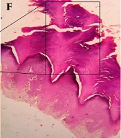
![A, Histopathology of plantar wart shows hyperkeratosis (black star), acanthosis, and few koilocytes (yellow circles). Note the elongated rete ridges and dilated capillaries [H & E, 10×]. B, Histopathology of corn showing hyperkeratosis (black star), acanthosis, and dermal fibrosis [H & E, 4×]. C, Histopathology of callus showing hyperkeratosis (black star), acanthosis, and fibrosis in the superficial dermis [H & E, 4×].](https://services.brieflands.com/cdn/serve/3170b/9206fb8264387e213ce845427c2750f87a808523/jssc-116806-g004-F4-preview.webp "A, Histopathology of plantar wart shows hyperkeratosis (black star), acanthosis, and few koilocytes (yellow circles). Note the elongated rete ridges and dilated capillaries [H & E, 10×]. B, Histopathology of corn showing hyperkeratosis (black star), acanthosis, and dermal fibrosis [H & E, 4×]. C, Histopathology of callus showing hyperkeratosis (black star), acanthosis, and fibrosis in the superficial dermis [H & E, 4×].")
A, Histopathology of plantar wart shows hyperkeratosis (black star), acanthosis, and few koilocytes (yellow circles). Note the elongated rete ridges and dilated capillaries [H & E, 10×]. B, Histopathology of corn showing hyperkeratosis (black star), acanthosis, and dermal fibrosis [H & E, 4×]. C, Histopathology of callus showing hyperkeratosis (black star), acanthosis, and fibrosis in the superficial dermis [H & E, 4×].
. Schematic diagram showing a dermoscopic and histopathological correlation in corn (D, E & F). Schematic diagram showing a dermoscopic and histopathological correlation in callus (G, H & I).")
Schematic diagram showing a dermoscopic and histopathological correlation in plantar wart (A, B & C). Schematic diagram showing a dermoscopic and histopathological correlation in corn (D, E & F). Schematic diagram showing a dermoscopic and histopathological correlation in callus (G, H & I).
4. Discussion
Plantar warts are benign tumors caused by HPV infection of the keratinocytes, which mostly occur in early adulthood. They are routinely procured from contact with water bodies, where moistened keratin is abraded by the uneven surface, which aids in inoculation of virus particles into the skin. The incubation period varies from few weeks to more than a year (1).
In this study, the dermoscopy of plantar warts illustrated red dots and short linear vessels or streaks, which were surrounded by a white or yellow halo. The absence of skin lines was striking. The aforementioned dermoscopic patterns are in line with previous reports (8-11). Interestingly, few linear structures of various colors were also noted. These were fabric fibers entangled to the eroded margins in plantar warts (Figure 3C), which is referred to as adherent fabric fiber sign (12). Fibres mimic linear vessels. However, the diameter of linear vessels varies inversely and are of red, black, or purplish color, whereas fibres have a uniform diameter and different colors.
Red dots represent thrombosed vessels in the projected papillae, and white halo was due to elongated and thickened rete ridges, hyperkeratotic keratin (Figure 5A-C). It should be noted that common warts and other variants of warts demonstrate a white halo around red dots (8). In contrast, a yellow halo was conspicuously noted in plantar warts and can be attributed to the significantly thickened stratum corneum and seepage of blood in the keratin layers. Linear streaks of vascular elements represented vertically arranged dermal capillaries, which were dragged along the growth of dermal papillae. Chronic and repeated high pressure on vascular elements attributes to linear vessels (13).
A callus is a broad-based diffuse area of hyperkeratosis, and a corn is a sharply demarcated lesion occurring over a bony prominence (2), which both relatively affect adults and older age groups with a slight female predilection. Repeated friction and pressure on the skin over the bony prominences result in the manifestation of corn and callus as a protective response. Hence, it causes the formation of hyperkeratotic skin, which is well localized to the affected area. Dermoscopy of corn revealed yellow area, whitish annular ring, and translucent central core. Preservation of dermatoglyphics was characteristic. Red dots and yellow or white halo were conspicuously absent. This observation is in line with previous reports (6). Histopathologically, corn demonstrates acanthosis, diminished granular layer, and parakeratosis with dense collagen (14). The thickening of the stratum corneum results in a yellow area under dermoscopy. The central translucent core representing the nucleus of the corn is due to localized fibrosis (Figure 5D-F). Prominent thickening of collagen correlated with the whitish ring.
Callus, under dermoscopy, demonstrated a prominent opaque yellow area with preservation of skin lines. Focal white areas were found. These findings are in line with previous descriptions. Vascular elements were absent, unlike plantar warts (15). Yellow areas were due to hyperkeratosis, orthokeratosis and acanthosis. Focal white areas were suggestive of white scales and were due to focal parakeratosis (Figure 5G-I). We also found brown and black globular structures in a few of the corn and callus lesions. These are correlated with dirt particles embedded within the skin lines and are called pseudo hemorrhagic structures (Figure 3D) (15). Nevertheless, these should not be confused with vessels. Thus, before scoping with a dermoscope, the plantar lesions should be wiped with an isopropyl alcohol solution.
In summary, plantar warts showed dotted vessels with a white halo. Dotted vessels were bright red or brownish-red in color, distributed irregularly; appearing as falooda seeds (16). Vascular elements were perceived in plantar warts but not in corn and callus. It is because of the upward projection of dermal papillae with dilated and thrombosed capillaries. Upward projection of dermal papillae is not a feature in both callus and corn; hence, vascular structures were absent. Even though paring of plantar papule may reveal thrombosed vessels, for the differentiation of these lesions and authentication of clinical diagnosis, dermoscope comes very handy in the recognition of vessels in a non-invasive fashion. These vascular patterns play a significant role in the therapeutic prediction of a plantar wart. The disappearance of dotted vessels is a sign of the resolution of the lesion (10).
In our study, with the added privilege of biopsy-proven diagnosis, the dermoscopic analysis and its correlation with histopathology have resulted in the uniqueness of the study. This study provides attestation of characteristic dermoscopic patterns in plantar papule or plaque is encountered, thus voids the need for biopsy. Further dermoscopy can be protracted in the therapeutic management of warts, wherein the presence of red dots with yellow halos indicates the need for continuation of treatment.
4.1. Conclusions
Dermoscopy can be useful in distinguishing clinically resembling painful plantar papules and plaques with characteristic patterns. Dermoscopic features were well correlated with histopathological changes. Hence, dermoscopy, as a rapid diagnostic tool, can eliminate the anxiety of patients scheduled for invasive procedures like skin biopsy. The authors recommend dermoscopic examination of confusing conditions to arrive at an accurate diagnosis for better management of a condition.
