1. Introduction
Granuloma faciale (GF), a chronic, benign, cutaneous vasculitis with characteristic clinical features, is seen mostly in middle age. Lesions are located predominantly in the regions exposed to light, manifested as plaques or papules (1). This condition has similar histopathological features to eosinophilic angiocentric fibrosis, mainly in the sinonasal cavity (2). Granuloma faciale describes a vasculitis characterized by infiltrated neutrophils, lymphocytes, and eosinophils in the grenz zone along with soft, well-defined plaques ranging in size from 0.5 to 1 cm, but extra facial lesions may also be observed (3). Diagnosis should be confirmed by excluding lesions like discoid lupus erythematous, sarcoidosis, Jessner’s lymphocytic infiltrate, mycosis fungoides, fixed drug eruption, and erythema elevatum diutinum (4).
In extra facial lesions without facial findings, lymphadenopathy, hepatosplenomegaly, or other systemic features are not associated. Thus, general investigations are necessary to exclude skin malignancies (5). Such a rare disease, GF, had only been reported in case reports or series. This case report describes a massive nasal case with a bizarre appearance, along with our management strategy.
2. Case Presentation
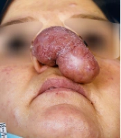
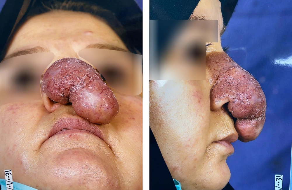
A 52-year-old woman was consulted for recurrent huge nasal lesions, resistant to previous treatments (Figure 1).
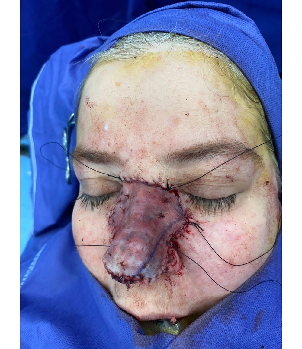
It first appeared ten years ago as a pustule and papule on the dorsum. At first, she received cryotherapy, pulsed dye laser (PDL), and fractional CO2 laser treatments. However, the disease progressed and became resistant to those treatments. Moreover, intra-lesion corticosteroid injections were repeated. Two cheek lesions were relieved by such treatments. However, the nasal lesion was growing, so three courses of rituximab were prescribed. Finally, she was referred by a dermatologist to cure and heal the patient’s self-confidence. All lesions were excised as far as the perichondrium layer was concerned until the perichondrium was spared for the next step. In the end, partial thickness skin grafts were used to complete the reconstruction (Figure 2). She had successful postoperative healing with good cosmetic results and satisfaction. Her instructions were sufficient to protect the grafted skin from the sun’s rays and dryness. A granuloma faciale was confirmed by histopathological examination.
3. Discussion
Numerous conservative and non-surgical treatments are available to slow the progression of the disease. However, none of them appear to be the best option. It is common to use intralesional corticosteroids, but long-term results are not satisfactory. Topical tacrolimus may modulate inflammatory infiltrate with fewer side effects. There is also a non-surgical treatment called Systemic Dapsone, which is limited due to its significant side effects.
Granuloma faciale generally responds to selective photothermolysis by PDL in the initial stages of illness, though short-term side effects of laser do not last long. Micallef and Boffa suggested that topical corticosteroids could improve the results of PDL. The patient had received dapsone 50 mg daily without significant response (6).
Marcoval et al. presented 11 cases with GF. The specimen showed a dermal, diffuse inflammatory infiltrate beneath the epidermis (7). These lesions were located in different layers of skin but mainly in the upper layers. In their assessment, neutrophils were abundant in all specimens. Eosinophils were also observable in all cases, but less than neutrophils (7).
Lindhaus and Elsner evaluated treatment protocols for GF in a systematic review and demonstrated that the first line was topical treatments like steroids and tacrolimus and intralesional steroid injection, followed by PDL laser and some systematic medications such as dapsone and cryotherapy and at last, there were cases treated by excision (8). They evaluated all treatments in divided groups and concluded that topical treatments might play a significant role because of their ease of use. However, they were unable to provide clear indications. The study also demonstrated that systemic medications such as dapsone, corticosteroid, and (clofazimine - an anti-leprosy drug) modulate inflammatory infiltrate to control disease; however, their side effects should always be considered (8). Prior studies generally focus on non-surgical treatments and have their reasons. However, in this case, the patient had a large, growing nasal GF resistant to standard treatment. In this case, surgical excision was logical, and we completed the procedure with a skin graft.
3.1. Conclusions
Granuloma faciale as a rare vasculitis in the face should be in mind to exclude other differential diagnosis cases which can be cured by excisional surgery and subsequent reconstructions.


