1. Background
Toxoplasma gondii is a protozoan of the Apicomplexa phylum with widespread prevalence in animal and human populations. It has been estimated that about one-third of the world's population is infected with this parasite. Toxoplasma gondii was first identified in 1908 by Nicole and Manceaux. The parasite is found in almost all continents, including Europe, North and South America, Asia, Africa, the Arctic, and Australia, except the South Pole (1, 2). Studies have shown that about 30% - 40% of Iranian people are infected with this parasite. The life cycle of T. gondii requires definite and intermediate hosts (3). The definite host of this parasite is cats, while the intermediate host is all warm-blooded animals, including domestic animals and humans (4). Symptoms have a range of asymptomatic, mild, severe, and even fatal. The disease is significant in humans and some animal species, such as goats and sheep, due to congenital infections and can harm the developing fetus or cause miscarriage. Humans become infected with the parasite by eating water or food contaminated with oocysts and eating uncooked meat (5, 6). Most people with a competent immune system are asymptomatic after infection, and the only sign of infection is a positive serological test, while in individuals with a defective immune system, the initial infection or reactivation of a previous infection can be severe and fatal (7).
A pregnant mother can transmit the infection to the fetus throughout pregnancy, and the older the gestational age, the greater the chance of transmission but the lower the chance of complications in the fetus (8, 9). Given that 90% of patients are asymptomatic and, determining the prevalence and serological measurement can be an effective step in diagnosing cases infected with T. gondii (10, 11). Mainly, to prevent the effects of disease before pregnancy, giving counseling before marriage about routine laboratory tests can be helpful for reproductive-age women. Whether they are susceptible to acute or chronic infection, therapeutic measures can be linked to the treatment so that non-immune individuals can benefit from special healthcare facilities before pregnancy. Several epidemiological studies have reported that the seroprevalence varies widely from 4.6% to 74.6% among childbearing age women in some provinces of Iran (3). However, there has been no study on the seroprevalence of infection in reproductive-age women in Birjand city. Therefore, this cross-sectional study aimed to determine the prevalence of anti-T. gondii IgG antibodies and its associated risk factors among reproductive age women referring to Birjand Comprehensive Health Center, East of Iran.
2. Objectives
This cross-sectional study aimed to determine the prevalence of IgG antibodies against T. gondii and its association with risk factors among reproductive-age women referring to Birjand Comprehensive Health Center, East of Iran.
3. Methods
3.1. Study Setting
This study was conducted in Birjand city, South Khorasan province in the East of Iran. The area of this province is 151.193 km2, and it is the third province of Iran in terms of area. According to the 2016 census, its population is 768,898 and in this regard is the 28th province in the country. The city is located in a mountainous area, with a semi-desert climate, cold winters, and hot and dry summers. The amount of rainfall in this city is low due to its climate, and the highest amount of rainfall occurs during December-May, and the average rainfall is 171 mm/year.
3.2. Sample Collection
The sample size was calculated based on a T. gondii seroprevalence rate of 30% (3), a confidence level of 95%, and P < 0.05. Using the following formula of sample size, this study was performed on 300 women of childbearing age who referred to Birjand Urban Health Center (Quds) in 2021 - 2022.
The inclusion criteria were: (1) women aged 15 - 50 years; (2) request for serological assessment for the immune status against this parasite; and (3) completion of the informed consent form. The exclusion criteria entailed (1) pregnant women under 18 years of age and over 50 years of age; (2) withdrawal from participating in the study. Blood samples collected from women (2 cc) were transported from the Central Parasitology Laboratory, Department of Microbiology, Birjand University of Medical Sciences, and were stored at -20°C until being tested with enzyme-linked immunosorbent assay (ELISA).
3.3. Questionnaire
Data were collected by a questionnaire to assess risk factors at the sampling time, as previously described (12). The questionnaire encompassed demographic data, including age, place of residence (urban or rural), and contact with cats. According to the opinion of scientific experts, the questionnaire was prepared, and its content validity was confirmed. The reliability of the questionnaire was also confirmed due to the clarity of the questions and its use in the previous studies.
3.4. Ethical Considerations
The present investigation was approved by the Ethics Committee of Birjand University of Medical Sciences, Iran (Ethic No. IR.BUMS.REC.1400.012). However, written informed consent was obtained from all participants before blood sampling.
3.5. Enzyme-Linked Immunosorbent Assay (ELISA)
All the serum samples were tested using the commercially available ELISA kit (Pishtaz Teb). Analyses were performed following the manufacturer’s instructions. Based on the ELISA kit, positive results for IgG were defined as values of C50 IU/mL. Moreover, negative results were defined as 50 IU/mL considered for IgG.
3.6. Assay Procedure
The desired number of coated wells were placed into the holder. We prepared 1: 40 dilution of test samples, negative control, positive control, and calibrators by adding 5 µL of the sample to 200 µL of sample diluent and mixing well. Next, 100 µL of diluted sera, calibrators, and controls were dispensed into the appropriate wells. For the reagent blank, 100 µL of sample diluent was dispensed in the well at the 1A position. The holder was tapped to remove air bubbles from the liquid, was mixed well, and was incubated at 37°C for 30 min. At the end of the incubation period, the liquid was removed from all wells. The microtiter wells were rinsed and flicked five times with diluted wash buffer (1x). Afterwards, 100 µL of enzyme conjugate was dispensed to each well, mixed gently for 10 sec, and incubated at 37°C for 30 min. The enzyme conjugate was removed from all wells, and the microtiter wells were rinsed and flicked five times with diluted wash buffer (1x). Next, 100 µL of TMB reagent was added to each well, mixed gently for 10 sec, and incubated at 37°C for 15 min. We added 100 µL of stop solution (1 N HCl) to stop the reaction and mixed gently for 30 sec. It is important to ensure that all the blue wells change to yellow completely. Note that there should not be air bubbles in each well before reading, and read OD at 450 nm within 15 min with a microwell reader.
3.7. Statistical Analysis
Analytical and descriptive statistics were carried out using the SPSS software version 20. Descriptive statistics were reported as a percentage and mean (SD). The chi-squared test was utilized to evaluate the univariate associations between independent variables and outcome. The significance level in the test was P < 0.05.
4. Results
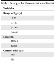
Out of 300 tested specimens, 25 were positive for T. gondii IgG, indicating that 8.3% of the test samples had anti-Toxoplasma antibodies. The chi-squared test indicated that living in rural regions was not significantly (P > 0.05) related to T. gondii seropositivity. Furthermore, the obtained results showed that contact with cats was significantly (P < 0.05) related to T. gondii seropositivity. Living in the urban or rural areas in none of the age ranges had a significant effect on positivity for T. gondii IgG (P > 0.05). The effect of living in a city × contact with cats on positivity for IgG was significant (P < 0.05), while living in a village × contact with cats had no significant effect on positivity for T. gondii IgG (P > 0.05). Moreover, living in rural regions was significantly (P < 0.05) related to T. gondii seropositivity (Table 1). The results showed that people in contact with cats had a higher rate of positivity for T. gondii IgG than people who did not have contact with cats (Table 2).
| Variables | P-Value | No. (%) | Toxoplasma gondii IgG+ |
|---|---|---|---|
| Range of age (y) | |||
| < 20 | 0.1 | 97 (32.3) | 8 (2.7) |
| 21 - 30 | 0.17 | 89 (29.6) | 8 (2.7) |
| 31 - 40 | 0.04 | 66 (22) | 7 (2.3) |
| 50 - 40 | 0.1 | 48 (16) | 2 (0.6) |
| Location | |||
| Urban | 0.01 | 34 (78) | 18 (6) |
| Rural | 0.17 | 66 (22) | 7 (2.3) |
| Contact with cats | |||
| Yes | 0.01 | 26 (8.6) | 9 (3) |
| No | 0.001 | 274 (91.4) | 16 (5.3) |
| Location and Contact with Cats | P-Value | No. (%) | Toxoplasma gondii IgG+ |
|---|---|---|---|
| Urban | |||
| Yes | 0.1 | 41 (13.6) | 6 (2) |
| No | 0.01 | 193 (64.3) | 12 (4) |
| Rural | |||
| Yes | 0.2 | 14 (4.6) | 2 (0.66) |
| No | 0.1 | 52 (17.3) | 5 (1.66) |
| Total | - | 300 (100) | 25 (8.3) |
a Values are expressed as No. (%).
5. Discussion
Toxoplasma gondii is a protozoan parasite with high prevalence in animal and human populations. The prevalence of this parasite varies in different parts of Iran. Exposure of women to this parasite can lead to miscarriage, premature delivery, and congenital malformations in children. It is necessary to obtain a level of awareness for the safety of women and girls against toxoplasmosis at the age of marriage. In our study, performed on women of reproductive age in Birjand, it was found that 8.3% of the people who participated in the study were positive for T. gondii IgG. Health education and familiarity with the ways of disease transmission are essential for all mothers at the reproductive age in South Khorasan. Ghadamgahi et al. examined toxoplasmosis and its relationship with risk factors in women who were referred to several laboratories in the North of Tehran, Iran. They showed that 28.3% of the subjects were positive for T. gondii IgG. In addition, they revealed that education and the consumption of raw vegetables and meat had no significant effect on the number of positive people. However, there was a significant relationship between age and disease frequency. In our study, age did not significantly affect the rate of positive results (13). A study conducted by Norouzi Larki et al. (14) on pregnant women in the South of the country showed that 8.9% of tested individuals were positive for T. gondii IgG, which was in line with the frequency of our target population. We can point out that the climate and, to some extent, the culture of the people of the South Khorasan region are the same as the South of the country. In research by Tavakoli Kareshk et al. on the women of reproductive age in Kerman province, it was shown that the frequency of positivity for T. gondii IgG increased with age (15-17).
Tavakoli kareshk et al. demonstrated that with an increase in age, the frequency of positivity for T. gondii IgG in blood donors in southern Iran rose and had a significant effect on the infection rate that does not correspond to our findings (18). As the report of Tavakoli Kareshk et al. (18) showed, similar to our study, women of childbearing age living in rural areas had a higher percentage of positivity for T. gondii IgG than those living in urban areas. The latter finding indicates that they were more likely to be exposed to T. gondii or to transmit disease in rural areas than in urban regions. Hygiene and familiarity with the ways of disease transmission are essential. In another research, Fallah and Shahbazi examined the frequency of T. gondii infection in girls from Ajabshir city, showing that people living in rural areas (25.9%) had a higher rate of positivity for T. gondii IgG than people living in urban areas (14.4%) (19). Sadooghian et al. in Khorasan Razavi province (Northeast of Iran) showed that women living in rural areas had a higher percentage of positivity for T. gondii IgG than women living in urban areas and place of residence had a significant effect on the level of pollution that is not in line with our results. The latter difference may be due to climatic differences (20).
In general, keeping cats in homes is not common, but due to the high movement of cats around the house and the repulsion of resistant oocysts, the possibility of infection with this parasite increases, which was confirmed by our results (20). Borna et al. studied the prevalence of T. gondii in women of childbearing age in a meta-analysis. These authors showed that contact with cats significantly augmented the incidence of T. gondii, which was consistent with our results (21, 22). Sharif et al. investigated the prevalence of T. gondii in butchers in Sari, Iran. They reported that 8% of people with a history of toxoplasmosis had contact with cats, which indicates that contact with cats is one of the most important ways of transmitting this infection (23, 24).
5.1. Limitations
The lack of funds to test more samples was one of the limitations of the current study. Therefore, it is suggested to perform serological tests on a larger sample size in future studies.
5.2. Conclusions
According to the results of the present study, education is needed to familiarize the woman at reproductive age with the ways of Toxoplasma transmission in South Khorasan.
