


1. Background
A congenital malformation is defined as any morphological anomaly in the formation of a tissue, organ, or body part that arises from abnormal development during embryonic or fetal life (1). Currently, congenital malformations are the leading cause of infant mortality in developed countries and contribute significantly to long-term morbidity, resulting in disabilities and handicaps (2).
The World Health Organization (WHO) attributes an additional 170 000 child deaths annually, from one month to five years of age, to congenital malformations. Furthermore, around 240,000 newborn deaths globally each year are due to congenital anomalies, accounting for approximately 7% of all neonatal fatalities (3). However, this proportion varies significantly across regions, from 5% in Southeast Asia to over 25% in Europe (4).
The causes of congenital malformations are multifaceted, involving a mix of genetic, environmental, social, economic, or sometimes unknown factors (5). In low- and middle-income countries, maternal infections such as syphilis and rubella, as well as conditions like diabetes mellitus or deficiencies in iodine or folic acid, are prominent causes of malformations (6). This diversity of risk factors leads to variations in the frequency and types of malformations worldwide (7).
Data from the Americas show a broad range in prevalence, with 3 to 5% of live births and 15% of stillbirths affected in Canada (8), 1.1 per 100 births in Latin America (9), and 63.8 per 10,000 births in Guatemala (10).
In Europe, the EUROCAT (European Surveillance of Congenital Anomalies) network reported a total prevalence of major congenital anomalies of 23.9 per 1 000 births, with 80% of these being live births (11). France, specifically, recorded a total prevalence of 3.2% of births (12).
For African countries, the prevalence is estimated at 7.85% of live births in Niger (13), 9.3 per 1 000 total births in Benghazi, Libya (14), and 30 per 10 000 live births in Algeria (15).
Morocco, recognizing the challenge posed by congenital malformations, has implemented several preventive and curative strategies. These include the national neonatal screening program for congenital diseases launched in 2012, systematic screening for congenital hypothyroidism, and the establishment of guidelines for the screening and management of childhood deafness (Ministry of Health and Social Protection Morocco, 2019) (16).
Despite these efforts, epidemiological data on congenital malformations are limited and not representative. The national prevalence of congenital malformations remains undefined, with most existing research being localized and focused on specific malformations. Moreover, the study of associations between congenital anomalies and various risk factors has been minimal in Morocco (17).
In the Marrakech-Safi region, the scarcity of epidemiological data on congenital malformations is particularly notable, even though this region ranks as the third most densely populated in Morocco, with an average density of 101.6 inhabitants per km2 (18). The rural areas of the region experience a relatively high poverty rate and elevated infant mortality (19) Reducing infant mortality here is crucial for meeting the 2030 Sustainable Development Goals and prioritizing the elimination of preventable deaths among newborns and children under five (20). This necessitates enhanced prevention and management of congenital malformations, alongside efforts to mitigate avoidable factors such as infections, nutritional deficiencies, vaccine-preventable diseases, and exposure to harmful substances like alcohol, tobacco, drugs, and chemicals (21).
2. Objectives
This study embodies the advanced regionalization approach Morocco has adopted, aiming to foster territorial development through the local management of projects and public policies that cater to the unique characteristics and potentials of each region (22). It represents the first endeavor in the Marrakech-Safi region to delineate the prevalence of various congenital malformations and their associated risk factors. The findings aim to bolster prevention efforts, inform prenatal diagnosis, and facilitate the planning of interventions tailored to the region's specific needs.
3. Methods
This descriptive, retrospective study aims to assess the prevalence and risk factors of congenital malformations diagnosed in the Marrakech-Safi region of central Morocco, utilizing obstetric records from January 1 to December 31, 2022.
The research was conducted in the maternity ward of the Mohammed VI University Hospital Center in Marrakech. This tertiary-level referral maternity hospital serves patients from the Marrakech-Safi region, encompassing the prefecture of Marrakech, the region's capital, and the seven provinces of Al Haouz, Chichaoua, El Kelâa des Sraghnas, Essaouira, Rhamna, Safi, and Youssoufia, in addition to the southern and central regions of Morocco. The facility is recognized as a specialized healthcare structure for mother and child health in the center and south of Morocco.
3.1. Participants
The study targeted newborns diagnosed with one or more congenital malformations, including both live births and stillbirths, provided the malformations were clearly defined in the records. A newborn was considered poly malformed if it had two or more congenital malformations (23).
After excluding files that were unavailable, lacked necessary study variables, or did not clearly specify the type of congenital malformation diagnosed, a total of 17 250 files were considered for the study.
A chart was completed for each malformed newborn, capturing sociodemographic, maternal, and neonatal data alongside details of the pregnancy and delivery. The independent variables included: (1) Origin; (2) maternal variables (maternal age, parity, medical and obstetric history); (3) pregnancy follow-up, and (4) ultrasound practice. Neonatal variables covered newborn status at birth, sex, Apgar score, and birth weight. Maternal age was categorized into three groups: Under 18 years, between 18 and 35, and over 35. Birth weight was classified into three categories: Hypotrophy (weight under 2500 g), normal weight (between 2500 g and 4200 g), and macrosomia (weight over 4200 g) (24). The Apgar score assessed the cardiorespiratory and neurological status of malformed newborns at birth, divided into three groups: A score of 7 to 10 at 5 minutes indicating normal status, 4 to 6 suggesting respiratory distress, and 0 to 3 indicating a critical state necessitating emergency care.
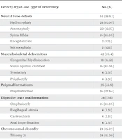
Malformations were coded according to the Royal College of paediatrics and child health (RCPCH) system, which is adapted from the 10th version of the International Classification of Diseases and Related Health Problems (ICD-10) (Table 1).
| Device/Organ and Type of Deformity | No. (%) | Prevalence/1000Births | ICD-10 CODE | 95% CI | |
| Lower | Upper | ||||
| Neural tube defects | 63 (39.62) | 3.65 | |||
| Hydrocephaly | 23 (15.09) | 1.33 | Q03 | 1.320 | 1.339 |
| Anencephaly | 20 (12.57) | 1.15 | Q00.0 | 1.143 | 1.156 |
| Spina Bifida | 16 (10.06) | 0.92 | Q05.9 | 0.915 | 0.924 |
| Encephalocele | 2 (1.25) | 0.11 | Q01.9 | 0.105 | 0.114 |
| Microcephaly | 2 (1.25) | 0.11 | Q02 | 0.105 | 0.114 |
| Musculoskeletal deformities | 42 (26.4) | 2.43 | |||
| Congenital hip dislocation | 18 (11.32) | 1.04 | Q65.2 | 1.036 | 1.043 |
| Varus equinus clubfoot | 16 (10.06) | 0.92 | Q66.0 | 0.915 | 0.924 |
| Syndactyly | 4 (2.51) | 0.23 | Q70 | 0.225 | 0.234 |
| Polydactyly | 4 (2.51) | 0.23 | Q69 | 0.225 | 0.234 |
| Polymalformations | 36 (22.6) | 2.08 | |||
| Polymalformed | 36 (22.64) | 2.08 | Q89.7 | 2.057 | 2.102 |
| Digestive tract malformation | 28 (17.6) | 1.62 | |||
| Omphalocele | 16 (10.06) | 0.92 | Q79.2 | 0.915 | 0.924 |
| Esophageal atresia | 4 (2.51) | 0.23 | Q39.0 | 0.225 | 0.234 |
| Gastroschisis | 4 (2.51) | 0.23 | Q79.3 | 0.225 | 0.234 |
| Anal imperforation | 4 (2.51) | 0.23 | Q42.1 | 0.225 | 0.234 |
| Chromosomal disorder | 24 (15.09) | 1.39 | |||
| Trisomy 21 | 24 (15.09) | 1.39 | Q90.9 | 1.386 | 1.393 |
| Facial malformation | 14 (8.8) | 0.81 | |||
| Cleft lip and palate | 8 (5.03) | 0.46 | Q37 | 0.452 | 0.467 |
| Cleft lip | 6 (3.77) | 0.34 | Q36 | 0.332 | 0.347 |
| Urogenital malformation | 12 (7.5) | 0.69 | |||
| Hypospadias | 8 (5.03) | 0.46 | Q54.9 | 0.452 | 0.467 |
| Sexual ambiguity | 4 (2.51) | 0.23 | Q56.4 | 0.225 | 0.234 |
Abbreviation: ICD-10, International Statistical Classification of Diseases and Related Health Problems, 10th revision.
3.2. Data Analysis
Statistical analysis was conducted using SPSS 21 software. Frequencies and percentages were calculated to describe the types of congenital malformations identified. pearson's chi-square tests analyzed the potential association between variables. A 5% level (P < 0.05) was considered statistically significant. The prevalence of each malformation was calculated with a 95% confidence interval to assess the accuracy of the prevalence identified.
3.3. Ethical Considerations
The study proceeded after securing authorization from the local and regional health services (Reference N°888/2021). Ethical principles such as anonymity and confidentiality were strictly observed. Hence, the evaluation forms utilized were coded and devoid of any patient names, the files were processed within the hospital to ensure confidentiality and anonymity, and the evaluation forms were disposed of after the study's completion.
4. Results
The study evaluated 17 250 cases at the maternity ward of Med VI University Hospital Center in Marrakech in 2022, identifying 159 newborns with congenital malformations. This equates to a prevalence of 0.92% (or 9.2/1 000 births).
Polymalformation syndrome was observed in 22.64% of the total population studied (Table 1). The highest frequency of congenital malformations occurred in mothers aged between 18 and 35 (62.26%) and in multiparous women (66%). Regarding the origin, the study revealed that 61% of cases originated from rural areas, in contrast to 39% from urban areas (Table 2), with a predominance of males, evidenced by a sex ratio of 1.38, i.e., 90 males for every 65 females (Table 3).
| Variables | Values a |
|---|---|
| Mother’s age (y) | |
| < 18 | 16 (10.06) |
| 18 to 35 | 99 (62.3) |
| > 35 | 44 (27.7) |
| Provenance | |
| Rural | 97 (61) |
| Urban | 62 (39) |
| Parity | |
| Primiparous | 54 (34) |
| Multiparous | 105 (66) |
| History of abortion | |
| Yes | 34 (21.4) |
| No | 125 (78.6) |
| Medical history | |
| Diabetes | 23 (14.5) |
| Anemia | 10 (6.3) |
| Hypothyroidism | 2 (1.25) |
| Heart disease | 2 (1.25) |
| Hypertension | 16 (10.06) |
| Nothing | 106 (66.7) |
| Pregnancy monitoring | |
| Yes | 50 (31.4) |
| No | 109 (68.6) |
| Progress of pregnancy | |
| Normal | 60 (37.7) |
| At risk | 99 (62.3) |
| Echography during pregnancy | |
| Yes | 66 (41.5) |
| No | 93 (58.5) |
| Method of delivery | |
| Vaginal delivery | 80 (50) |
| Cesarean section | 79 (50) |
| Gemellair pregnancy | |
| Yes | 0 (0) |
| No | 159 (100) |
a Values are expressed as No. (%).
| Variables | Values a |
|---|---|
| Sex | |
| Female | 65 (40.9) |
| Male | 90 (56.6) |
| Sexual ambiguity | 4 (2.51) |
| Weight | |
| Hypotrophy | 50 (31.4) |
| Normal | 99 (62.3) |
| Macrosomia | 10 (6.3) |
| Term of birth | |
| Premature | 71 (44.7) |
| Full term | 88 (55.3) |
| Status at birth | |
| Alive | 141 (88.7) |
| Stillborn | 18 (11.3) |
| Apgar score at birth | |
| 0 - 3 (state of apparent death) | 36 (22.6) |
| 4 - 6 (distressed) | 33 (20.7) |
| 7 - 10 (normal) | 90 (56.6) |
| Newborn evolution in the first 24 hours | |
| Alive | 64 (40.3) |
| Referred | 71 (44.6) |
| Deceased | 24 (15.1) |
a Values are expressed as No. (%).
The results (Table 1) indicate that neural tube defects were the most common, accounting for 39.62% of the malformations (hydrocephalus 15.09%, anencephaly 12.57%, spina bifida 10.06%, encephalocele, and microcephaly 2.5%), followed by musculoskeletal malformations at 26.4% (congenital hip dislocation 11.32%, clubfoot varus equines 10.06%, syndactyly and polydactyly each at 2.51%). Digestive tract malformations were present in 17.61% of cases (omphalocele 10.06%, esophageal atresia, gastroschisis, and anal imperforation each at 2.5%). Chromosomal malformations, specifically trisomy 21, constituted 15.09% of cases, facial malformations were observed in 8.8% of cases (cleft lip and palate 5.03%, cleft lip 3.77%), and urogenital malformations in 7.54% of cases (hypospadias 5.03% and sexual ambiguity 2.51%).
The maternal age group most commonly associated with congenital malformations was between 18 and 35, accounting for 62.3% of cases. Regarding origin, 61% of the women were from rural areas. Multiparous women represented 66% of the sample. A history of abortion was noted in 21.4% of the maternal population. Medical antecedents were observed in one-third of the mothers (33.3%), with 14.5% experiencing diabetes, 10.06% hypertension, and 6.3% anemia. The incidence of hypothyroidism and heart disease was identical in our study, each affecting 1.25% of cases. All malformed infants resulted from monofetal pregnancies. Regarding pregnancy monitoring, 68.6% of mothers did not receive prenatal care, although 62.3% of the pregnancies were considered high-risk. Obstetric ultrasound during pregnancy was conducted in only 41.5% of the cases. As for the delivery method, the proportions of vaginal deliveries and cesarean sections were equivalent.
Among the 159 malformed newborns, 56.6% were male, and 40.9% were female, resulting in a sex ratio of 1.38. The remaining 2.5% (4 cases) exhibited sexual ambiguity, associated with another congenital malformation as part of poly malformation syndrome. Nearly two-thirds of the newborns had a normal birth weight ranging between 2500 g and 4200 g. Hypotrophy, with a birth weight under 2500 g, was present in 31.4% of cases, and 6.3% were macrosomic, with a birth weight exceeding 4200 g.
In terms of the Apgar score at birth, 56.6% of newborns scored between 7 and 10, indicating a normal status. Meanwhile, 20.7% of cases were in distress (Apgar score between 4 and 6), and 22.6% were in a critical condition (score < 3), necessitating emergency resuscitation. Stillbirths among newborns with malformations accounted for eighteen cases (11.3%), but the overall mortality rate within the first 24 hours after birth was 15.1%.
4.1. Associated Factors
The statistical analysis of the data revealed a positive and significant correlation between the occurrence of one or more congenital malformations and several factors: maternal age (P = 0.001), parity (P = 0.002), medical history (P = 0.002), term of birth (P = 0.025), pregnancy monitoring (P = 0.008), progress of pregnancy (P = 0.017), ultrasound during pregnancy (P = 0.001), status at birth (P < 0.001), Apgar score (P < 0.001), sex of the newborn (P = 0.001), and birth weight (P < 0.001). Additionally, the analysis indicated that the p-values for the mother's provenance (P = 0.241) and history of abortion (P = 0.127) exceed 0.05. Consequently, these variables are not considered risk factors for congenital malformations in our study context (Table 4).
| Variables | Values a | Χ2 Value | P-Value |
|---|---|---|---|
| Mother’s age (y) | 13.486 a | 0.001 b | |
| < 18 | 16 (10.06) | ||
| 18 to 35 | 99 (62.3) | ||
| > 35 | 44 (27.7) | ||
| Provenance | 1.374 a | 0.241 | |
| Rural | 97 (61) | ||
| Urban | 62 (39) | ||
| Parity | 9.608 a | 0.002 b | |
| Primiparous | 54 (34) | ||
| Multiparous | 105 (66) | ||
| History of abortion | 2.329 a | 0.127 | |
| Yes | 34 (21.4) | ||
| No | 125 (78.6) | ||
| Medical history | 18.626 a | 0.002 b | |
| Diabetes | 23 (14.5) | ||
| Hypertension | 16 (10.06) | ||
| Anemia | 10 (6.3) | ||
| Heart disease | 2 (1.25) | ||
| Hypothyroidism | 2 (1.25) | ||
| Nothing | 106 (66.7) | ||
| Term of birth | 5.018 a | 0.025 b | |
| Premature | 71 (44.7) | ||
| Full term | 88 (55.3) | ||
| Apgar score at birth | 31.209 a | 0.000 b | |
| 0 - 3 (state of apparent death) | 36 (22.6) | ||
| 4 - 6 (distressed) | 33 (20.7) | ||
| 7 - 10 (normal) | 90 (56.6) | ||
| Pregnancy monitoring | 7.042 a | 0.008 b | |
| Yes | 50 (31.4) | ||
| No | 109 (68.6) | ||
| Progress of pregnancy | 5.716 a | 0.017 b | |
| Normal | 60 (37.7) | ||
| At risk | 99 (62.3) | ||
| Echography during pregnancy | 10.895 a | 0.001 b | |
| Yes | 66 (41.5) | ||
| No | 93 (58.5) | ||
| Sex of newborn | 14.905 a | 0.001b | |
| Female | 65 (40.9) | ||
| Male | 90 (56.6) | ||
| Sexual ambiguity | 4 (2.51) | ||
| Weight of newborn | 29.516 a | 0.000 b | |
| Hypotrophy | 50 (31.4) | ||
| Normal | 99 (62.3) | ||
| Macrosomia | 10 (6.3) | ||
| Status at birth | 20.594 a | 0.000 b | |
| Alive | 141 (88.7) | ||
| Stillborn | 18 (11.3) |
a Values are expressed as No. (%).
b Significant value (association between variable and congenital malformation).
5. Discussion
This study reported a prevalence of congenital malformations of 9.2 per 1000 births, a figure that aligns closely with those reported in other parts of Morocco: 1.02% of a percent at the Souissi maternity hospital in Rabat (25), 1.34% in the Oriental region (26), and 5.58 per 1000 at the “Les Orangers” hospital in Rabat (27). Comparatively, in other African countries, the prevalence stands at 9 per 1000 births in Cameroon (28), 3.2 per 1000 in Togo (29), and 3.6 per 1000 in Ivory Coast (30). In France, specifically the Paris region, the total prevalence of malformations and chromosomal anomalies among births and medical terminations of pregnancy was recorded at 32 per 1000 in 2000 (12). The variation in prevalence rates of congenital malformations can be attributed to multiple factors, such as geographical location, genetics, environmental exposures, and the efficacy of epidemiological surveillance, explaining the observed differences across countries and regions (31). These disparities might also stem from an underestimation of percentages, especially in regions with insufficient death registration systems, which may lead to the misclassification of deaths caused by congenital malformations (4). This emphasizes the necessity for a thorough understanding of the specific types and regional variations of congenital malformations to ensure effective prevention, management, and appropriate allocation of resources, taking into account these territorial differences (31).
In our series, poly malformation syndrome was observed in 22.64% of cases, a figure nearly identical to that reported by the “Les Orangers” hospital in Rabat, where the syndrome was present in 26.5% of cases (27). Regarding the prevalence distribution of the various malformations identified in our study, neural tube malformations emerged as the most prevalent, with a frequency of 39.62%, followed by musculoskeletal malformations at 26.4%. Malformations of the digestive tract were the fourth most common, behind poly malformation syndrome, affecting 17.61% of cases.
The predominance of neural tube defects has been highlighted in various studies and countries, including Pakistan, with a frequency of 20.35% of cases (32), and the Ivory Coast, with 51.16% of cases (30). The WHO has also acknowledged that neural tube defects, heart defects, and Down's syndrome are among the most serious and common congenital malformations (3). Our findings underscore the necessity to enhance the national program aimed at combating micronutrient deficiencies, particularly those of folic acid and iodine. Additionally, there's a need to increase women's awareness regarding the importance of pregnancy monitoring and the risks associated with the regular intake of Fenugreek (33).
Regarding the frequency of congenital malformations by gender, the literature suggests a higher predisposition among boys. At the Souissi maternity hospital in Rabat, Morocco, male newborns (57.9%) were more frequently affected by congenital malformations than female newborns (40.5%) (34). Shabbir et al. (32) also found that 57.52% of malformed newborns were boys and 42.47% were girls. These findings align with our study, where a significant association was observed between male sex and the occurrence of congenital malformations (P = 0.001).
In terms of birth weight, many studies report a higher frequency of malformations in newborns with normal birth weight (28, 35). Our findings agree with this observation, indicating a positive association between birth weight and the occurrence of congenital malformations (P < 0.001), with 62.3% of cases occurring in normal-weight newborns.
Kase and Visintainer noted that congenital malformations significantly impact the likelihood of preterm birth, contribute to the degree of prematurity (36), and increase neonatal morbidity risks for preterm infants (17). Newborns with congenital anomalies are more likely to be born preterm compared to those without, with the combined impact of prematurity and congenital anomalies being more than cumulative (37). Our study supports this, showing a significant relationship between congenital malformations and the term of birth (P = 0.025) despite a nearly equal distribution of malformations among preterm (44.7%) and full-term (55.3%) infants. This is akin to the findings of Kamla et al. (28), where 37% of malformed newborns were preterm.
In our study, a significant association was found between the Apgar score, the condition of the newborn at birth, and congenital malformations, each with a P-value of < 0.001. We observed an increase in the stillbirth rate among malformed newborns within the first twenty-four hours of life, from 11.3% to 15.1%. Additionally, 22.6% of newborns were in a critical state (Apgar score < 3), necessitating urgent intensive care. Indeed, low Apgar scores are associated with increased mortality and a higher risk of cerebral palsy (38). Enhanced access to health services, timely interventions, and the availability of professionals trained in neonatal resuscitation protocols can significantly decrease neonatal and infant morbidity and mortality (39).
Concerning the place of origin, Vrijheid et al. (40) demonstrate that the risk of non-chromosomal abnormalities escalates with socio-economic deprivation. In our study, the frequency of malformations among women from rural areas was 61%. However, our statistical analysis revealed no significant association (P = 0.241) between this variable and the occurrence of congenital malformations. This finding can be attributed to the demographic distribution in the Marrakech-Safi region, where 57% of the population resides in rural areas, compared to 43% in urban areas, underscoring the region's rural character (18). The socio-economically disadvantaged segments, especially those in isolated and mountainous areas, face challenges in accessing and availing health services for women and children and bear a higher burden of congenital malformations (31). Our results emphasize the need for the Ministry of Health to take further action to promote equity and reduce disparities both within and between regions. There's a call to enhance interventions at primary healthcare facilities and to improve practices within families and communities through a community health approach (41).
In the literature, multiparity and advanced maternal age are identified as factors that increase the risk of having a child with congenital anomalies (13, 29, 42). This aligns with our findings, which demonstrate a statistically significant association between multiparity and the occurrence of malformations in offspring (P = 0.002). According to Khoushnood et al. (43), an increase in maternal age significantly affects the risk of spontaneous abortion, maternal health, and pregnancy outcomes (including multiple births, prematurity, hypotrophy, and congenital anomalies). In our study, maternal age was also significantly associated with the occurrence of malformations (P = 0.001), with the highest incidence observed in the 18 - 35 age group (62.3%), indicating that mothers in this age group are most likely to give birth to malformed children. This result parallels findings from northern Togo, where the 26 - 30 age group was most affected by congenital malformations (29).
Previous research has highlighted that the frequency of congenital malformations significantly increases in women with chronic diseases, especially diabetes (44, 45). Our findings corroborate these results, showing a significant correlation between maternal medical history and the incidence of malformations (P = 0.002). This underscores the importance of primary prevention, aimed at reducing risks associated with low socio-economic status, poor diet, environmental contaminants, and chronic conditions such as hypertension and diabetes (46), particularly in the Marrakech-Safi region, which is among the most economically and socially challenged regions (41).
Our study revealed a significant link between the lack of pregnancy monitoring and the occurrence of congenital malformations (P = 0.008). Notably, 68.6% of mothers did not undergo pregnancy monitoring, although two-thirds (62.3%) had high-risk pregnancies. In Morocco, despite efforts to improve pregnancy and childbirth monitoring, only 53.5% of women receive the four prenatal consultations recommended by the Ministry of Health, with significant discrepancies based on the place of residence (65.6% in urban areas vs. 38.5% in rural areas) (47). This rate significantly correlates with a woman's education level and, inversely, with her socio-economic status and the stage of pregnancy (41). Indeed, a low socioeconomic status often relates to poor pregnancy follow-up and inadequate prevention of congenital malformations (29).
The findings of this study also show a significant link between undergoing ultrasound examinations during pregnancy and the incidence of malformations (P = 0.001), with 58.5% of the mothers of malformed newborns not having received an ultrasound examination during their pregnancies. Obstetrical ultrasound is recognized as the optimal method for studying and monitoring fetal malformations (30), facilitating the detection of Down syndrome and major structural anomalies in the first trimester, as well as severe fetal anomalies in the second trimester (3). The limited access to prenatal care and ultrasound examinations in our study can be attributed to the rural characteristics of the Marrakech-Safi region, the inaccessibility of healthcare facilities, and insufficient medical coverage, especially in isolated or mountainous areas (41). These factors complicate the provision of comprehensive prenatal care, genetic counseling, and specialized interventions for parents at risk of having children with congenital malformations (31).
In Morocco, the majority of deliveries occur in public establishments supervised by the Ministry of Health and Social Protection, with a minority of deliveries happening at home. Hence, data on these deliveries were not available for our study, which is a limitation. Additionally, data from private establishments was inaccessible.
Furthermore, during data collection, we encountered missing data for several variables, such as the mothers' consumption of harmful substances (tobacco, alcohol, drugs, psychotropics), parental consanguinity, diet, and exposure to environmental or occupational chemical contaminants, which could bias our results and potentially lead to an underestimation of prevalence.
5.1. Conclusions
This study has illuminated the prevalence of congenital malformations at 9.2 per 1000 births and identified associated risk factors in the Marrakech-Safi region, specifically maternal age, parity, maternal medical history, male sex, and newborn weight at birth. These insights will assist healthcare professionals in tailoring the activities of the national congenital disease screening program to the region's characteristics, emphasizing preventable congenital malformation factors. Enhancing genetic counseling and prenatal screening for high-risk parents and promoting the micronutrient deficiency combat program, particularly focusing on improving the skills of nursing staff, are vital strategies to address the incidence of birth defects. Additionally, reinforcing the neonatal death surveillance system and expanding it to all regions is crucial for tracking and reducing neonatal morbidity and mortality rates due to birth defects at both national and regional levels.
