Does Continuous Extremely Low-Frequency Electromagnetic Fields Exposure Harms Liver Tissue? A Microscopic Assay

authors:

avatar Vahid Semnani 1 , avatar Hadi Hasanzadeh 2 , 3 , * , avatar Amir Darbandi-Azar 4 , avatar Samaneh Seyyedi 5 , avatar Alireza Emadi 6 , avatar Mostafa Rezaei-Tavirani 7

Clinical Research Development Unit (CRDU), Kowsar hospital, Semnan University of Medical Sciences, Semnan, Iran
Cancer Research Center, Semnan University of Medical Sciences, Semnan, Iran
Department of Medical Physics, Semnan University of Medical Sciences, Semnan, Iran
Rajaie Cardiovascular Medical and Research Center, Iran University of Medical Sciences, Tehran, Iran
Aramesh molecular and genetic diagnostic laboratory, Department of Genetics, Tehran, Iran
Deputy of Research and Technology, Semnan University of Medical Sciences, Semnan, Iran
Proteomics Research Center, Shahid Beheshti University of Medical Sciences, Tehran, Iran

how to cite: Semnani V, Hasanzadeh H, Darbandi-Azar A, Seyyedi S, Emadi A, et al. Does Continuous Extremely Low-Frequency Electromagnetic Fields Exposure Harms Liver Tissue? A Microscopic Assay. Middle East J Rehabil Health Stud. 2018;5(1):e63105. https://doi.org/10.5812/mejrh.63105.

Abstract

Background:

Extremely low frequency electromagnetic fields (ELF-EMF) are commonly present in daily life all over the world. This study was designed to investigate the effects of the different exposure intensity of the low frequency electromagnetic fields on the liver tissue.

Methods:

Adult male Wistar rats (n = 30) were randomly allocated in one of the experimental groups including; group A, exposure to 0.5 millitesla (mT) for 2-week (n = 5); group B, exposure to 0.5 mT for 4-week (n = 5); group C, control without radiation (n = 5); group D, exposure to 1 mT for 2-week (n = 5); group E, exposure to 1 mT for 4-week (n = 5); and group F, sham group (n = 5). At the end of the experiment, their liver tissue was dissected and clinically assessed by a pathologist.

Results:

Our results show that for 1 month of exposure at 1 mT, focal hepatocitolysis and mild to moderate portal inflammation is visible and for 2 weeks exposure at this flux density, apoptosis and hepatocyte inflammation has occurred. However, for the groups exposed to 0.5 mT, 1 month exposure has caused hepatocyte focal inflammation, parenchymal hepatocitolysis, and focal apoptosis while, the exposure for 2 weeks did not affect the liver tissue.

Conclusion:

Exposure to ELF-EMF at the occupational limit cannot lead to irreversible changes and its effects on cells are enough below moderate changes. At the flux density of 1 mT, effects are moderate and irreversible and they are spread in larger areas than the lower flux density of 0.5 mT. Exposure time correlates with the level of stress on the cells; therefore, for a halftime exposure at occupational level, there exist no changes relative to the control group while for the fulltime exposure, some adverse effects were visible.

1. Background

During the past 3 decades, mankind has been exposed to many new physical and chemical agents. A major physical agent is electromagnetic field (EMF), which among other agents, is widespread and ubiquitous in modern daily life. Therefore, most people are exposed to ELF-EMF (extremely low frequency electric and magnetic fields) emitted by power lines, electrical panels, transformers, and domestic, electrical, and electronic devices (1-3). Associations between exposure to ELF-EMF and possible health hazards to mankind have been investigated in several epidemiological and experimental studies (4-8). Most of the epidemiological studies have shown that exposure to ELF-EMF has increased the incidence of certain types of cancer, especially acute childhood leukemia (9-11). Furthermore, several in vitro and in vivo studies have been published on the biological effects of ELF-EMF and some hypothetical mechanisms have been detected (12). A limited number of these studies have demonstrated that ELF-EMF promotes carcinogenic effects. Some available evidence appears that exposure to ELF-EMF can enhance or inhibit cell proliferation and apoptosis (13, 14). Several studies indicated that ELF-EMF exposure affects cell regulation and chromosomal structure (15-17). Moreover, other in vitro studies suggested that ELF-EMF can change the expression of some protein, which is involved in the control of cell proliferation (18, 19), while others demonstrate that exposure to ELF-EMF has no effects on cell proliferation, DNA replication, and cell regulation (20). As it seems, there exist some ambiguities in results. Some of these conflicting data might be due to the difference in frequency, intensity, duration, and also a certain type of cell lines (21).

A long-term study on the effects of ELF-EMF on the fertility and tumor promotion process has shown that exposure to 50 Hz magnetic fields is a significant risk factor for neoplastic development and infertility in mice; however, it has not increased the incidence of liver and lung tumor (22). It has shown that exposing liver cancer BEL-7402 cells to folic acid-modified magnetic nanoparticles (FA-MNPs) in the presence of 100 Hz ELF-EMF, significantly inhibited cell proliferation and induced higher apoptosis (23). Besides, it has shown that ELF-EMF exposure (50 Hz, 8 mT, 28 days) causes significant deterioration in learning and memory abilities (24).

The aim of this study was to determine the effects of ELF-EMF (50 Hz) on the rats’ liver tissue exposed to different intensities (0.5 and 1 mT) at different time durations (2 and 4 weeks) by analyzing microscopic changes in liver tissue.

2. Methods

2.1. Animals

Adult male Wistar rats, weighing 250 - 300 g were housed 3 to 4 per cage in a temperature-controlled colony room under light/dark cycle and had free access to water and food throughout the experiment. It is notable that the rats were exposed to these intensities 8 h/day and were housed in normal lighting conditions (12 hour light ON, 12 hour light OFF). This study was conducted in accordance with the policies stipulated in the guide for the care and use of laboratory animals (NIH) and was approved by the local ethics committee of Semnan University of Medical Sciences.

2.2. Exposure System

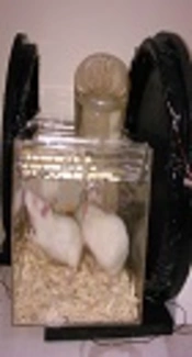
A calibrated ELF/EMF generator was used to produce an electromagnetic field. It was able to generate homogenous sinusoidal ELF-EMF, with the intensity of 0.5 and 1 mT as well as a frequency of 50 Hz with a 40 cm diameter Helmholtz coil placed around the exposure cage (Figure 1) 8 hrs/day for 2 and 4 weeks. Control rats were placed in the cage outside the coil. Sham exposed rats were maintained for an equal period of time inside the exposure cage with the generator off. The room was maintained at a constant temperature (25 ± 1.0°C) for all experiments.

Animals placed in the Plexiglas exposure Cage within the Helmholtz coils

2.3. Experimental Groups

The experimental groups consists of the following, in which 5 rats were considered in each group: Eexposure to 0.5 mT for 2 week (A), exposure to 0.5 mT for 4 week (B), control without radiation (which was not placed at the experimental condition) (C), exposure to 1 mT for 2 week (D), exposure to 1 mT for 4 week (E), and sham group (which was placed at the experimental setup cage with no exposure to consider the probable stress induced effects on the results) (F).

2.4. Slide Preparation

For the Histopathological assessment, the liver tissue was removed from anesthetized rat, fixed in 10% formaldehyde and embedded in paraffin wax. Then, 3-5 µm sections of tissue were cut and stained with Hematoxylin and Eosin (H and E). The prepared slides were used for light microscopy analysis in the 10 × and 40 × fields. In order to classify different experimental groups concerning cellular damage and tissue pathology, several parameters such as focal or distributed apoptosis, focal or distributed necrosis, portal, periportal or parenchymal inflammation (focal or distributed), and hepatocitolysis were assessed in different experimental groups by an anatomical pathologist who was blind to the groups.

3. Results

The results of light microscopy are brought in Figures 2 - 5. Figure 2 shows the images related to 1 mT exposure for 1 month; as it is observed, focal hepatocitolysis and mild to moderate portal inflammation is visible. In the case of 1 mT exposure for 2 weeks (Figure 3), apoptosis and hepatocyte inflammation has occurred. However, for the groups exposed to the lower field strength (0.5 mT), 1-month exposure (Figure 4) has caused hepatocyte focal inflammation, parenchymal hepatocitolysis and focal apoptosis. At this intensity, exposure for 2 weeks did not affect the liver tissue (Figure 5). Besides, in order to quantify our results, the chronic active hepatitis grading (25) was used, which results are brought in Table 1.

Table 1.

Modified Histological Activity Index-Grading Necro Inflammatory Scoresa

GroupABCDTotal ScoreReversible/Irreversible
1 mT-1 Month00325R
1 mT-2 Weeks00202R
0.5 mT-1 Month00101R
0.5 mT-2 Weeks00000R
Microscopic image (40 ×) of liver tissue in rats exposed to 1 mT magnetic field for 1 month, which shows mild to moderate portal inflammation (A) and focal hepatocitolysis (B)
Microscopic Image (40 ×) of Liver Tissue in Rats Exposed to 1 mT Magnetic Field for 2 Weeks, Which Shows Focal Hepatocyte Inflammation (A) and Apoptosis (B)
Microscopic image (40 ×) of liver tissue in rats exposed to 0.5 mT magnetic field for 1 month, which shows focal hepatocyte inflammation, parenchymal hepatocitolysis, and Focal Apoptosis (A and B)
Microscopic image (10 ×) of liver tissue in rats exposed to 0.5 mT magnetic field for 2 weeks, which shows no changes relative to the control Group

4. Discussion

Nowadays, innovations in technology have resulted in changing lifestyle. This change has been concomitant with some new factors, which have the potential to affect mankind health. One of these factors are electromagnetic waves, which are present everywhere around us with different sources, frequencies, and intensities that have motivated researchers to work on the biological effects of such agents at different levels from cell to the in vivo conditions (1-3). The ICNIRP (26) has considered an occupational reference level of the magnetic flux density of 0.5 mT and the public reference level of 0.1 mT. We considered a magnetic flux density at the occupational level and twice it as 1 mT. Besides, we selected a fulltime worker’s time (all working days within a month) and a halftime worker (2 working weeks within a month). Our results showed that for the 0.5 mT magnetic flux density, the group exposed for 2 weeks showed no change in liver tissue relative to the control group, while at this magnetic flux density, 1 month of exposure caused formation of focal inflammation, parenchymal hepatocitolysis, and focal apoptosis (Figure 4, Figure 5, Table 1). At the higher magnetic flux density of 1 mT, the 2-week exposure leads to focal apoptosis and focal hepatocyte inflammation, which for 1-month exposure results to focal hepatocitolysis and mild to moderate portal inflammation. As it is observed, exposure at the occupational limit cannot lead to irreversible changes and its effects on cells are enough below moderate changes. However, in the case of 1 mT exposure, the effects are moderate and irreversible and they are spread in larger areas than the lower flux density of 0.5 mT. Again, the exposure time affects the level of stress on the cells and therefore, 1 month of exposure induces more stress than the 2 weeks of exposure. The microscopic trauma is correlated with the molecular changes at the cell level such as changes in oxidative stress indicators, metabolism of free radicals and superoxide dismutase (SOD) (27-32). Our findings are in line with Emre et al., where by measuring oxidative stress indicators suggested a relation between exposure to the magnetic field and cell death (28), or Martinez et al., who used higher flux densities and studied molecular indicators (32). Canseven et al., (27) studied effects of 1 mT magnetic flux density with different exposure times on guinea pigs tissues and concluded that exposure time affects free radical formation as we found via microscopic study. Hashish et al., (29) by studying the effect of whole body exposure of mice, showed that a relation between exposure to ELF-EMF and oxidative stress exists, which in our study, microscopic outcomes of this exposure were recorded. Also, Liu et al., (31) reported similar changes. Besides, Cakir et al., studied the effect of exposure to ELF-EMF at a flux density of 0.97 mT and reported no change in liver weight after exposure, which is in accordance with our study as we showed no fibrosis and therefore, we do not expect any changes in liver weight. On the other hand, Zecca et al., (33) reported no pathological changes in animal growth rate and also morphology and histology of the liver tissue, which is due to the small magnetic flux density they implemented in their study.

4.1. Conclusion

A microscopic study on the effects of ELF-EMF exposure was performed on rat liver tissue at the occupational exposure level and twice that for the exposure time similar to a halftime and a fulltime worker. It was observed that for a halftime exposure at occupational level, there exist no changes in liver tissue while for the fulltime exposure, some adverse effects were visible. Besides, at the higher magnetic flux density of 1 mT, both the halftime and fulltime exposure resulted in adverse irreversible cellular effects. By the way, it seems it is better to conduct the experiment on a wider period of time (maybe up to 3 - 6 month) to observe more severe effects and simultaneously measure the level of some enzymes effective on the function of the liver.

Acknowledgements

References

  • 1.

    Grellier J, Ravazzani P, Cardis E. Potential health impacts of residential exposures to extremely low frequency magnetic fields in Europe. Environ Int. 2014;62:55-63. [PubMed ID: 24161447]. https://doi.org/10.1016/j.envint.2013.09.017.

  • 2.

    Ivancsits S, Diem E, Pilger A, Rudiger HW, Jahn O. Induction of DNA strand breaks by intermittent exposure to extremely-low-frequency electromagnetic fields in human diploid fibroblasts. Mutat Res. 2002;519(1-2):1-13. [PubMed ID: 12160887]. https://doi.org/10.1016/S1383-5718(02)00109-2.

  • 3.

    Karasek M, Woldanska-Okonska M. Electromagnetic fields and human endocrine system. ScientificWorldJournal. 2004;4 Suppl 2:23-8. [PubMed ID: 15517099]. https://doi.org/10.1100/tsw.2004.175.

  • 4.

    Hasanzadeh H, Rezaei-Tavirani M, Seyyedi SS, Zali H, Heydari Keshel S, Jadidi M, et al. Effect of ELF-EMF Exposure on Human Neuroblastoma Cell Line: a Proteomics Analysis. Iran J Cancer Prev. 2014;7(1):22-7. [PubMed ID: 25250144].

  • 5.

    Rezaei-Tavirani M, Hasanzadeh H, Seyyedi S, Zali H. Proteomic Analysis of the Effect of Extremely Low-Frequency Electromagnetic Fields (ELF-EMF) With Different Intensities in SH-SY5Y Neuroblastoma Cell Line. J Lasers Med Sci. 2017;8(2):79-83. [PubMed ID: 28652900]. https://doi.org/10.15171/jlms.2017.14.

  • 6.

    Rostami A, Shahani M, Zarrindast MR, Semnanian S, Rahmati Roudsari M, Rezaei Tavirani M, et al. Effects of 3 Hz and 60 Hz Extremely Low Frequency Electromagnetic Fields on Anxiety-Like Behaviors, Memory Retention of Passive Avoidance and Electrophysiological Properties of Male Rats. J Lasers Med Sci. 2016;7(2):120-5. [PubMed ID: 27330708]. https://doi.org/10.15171/jlms.2016.20.

  • 7.

    Safari M, Jadidi M, Baghian A, Hasanzadeh H. Proliferation and differentiation of rat bone marrow stem cells by 400muT electromagnetic field. Neurosci Lett. 2016;612:1-6. [PubMed ID: 26639423]. https://doi.org/10.1016/j.neulet.2015.11.044.

  • 8.

    Cuccurazzu B, Leone L, Podda MV, Piacentini R, Riccardi E, Ripoli C, et al. Exposure to extremely low-frequency (50 Hz) electromagnetic fields enhances adult hippocampal neurogenesis in C57BL/6 mice. Exp Neurol. 2010;226(1):173-82. [PubMed ID: 20816824]. https://doi.org/10.1016/j.expneurol.2010.08.022.

  • 9.

    Kheifets L, Ahlbom A, Crespi CM, Draper G, Hagihara J, Lowenthal RM, et al. Pooled analysis of recent studies on magnetic fields and childhood leukaemia. Br J Cancer. 2010;103(7):1128-35. [PubMed ID: 20877339]. https://doi.org/10.1038/sj.bjc.6605838.

  • 10.

    Schuz J. Exposure to extremely low-frequency magnetic fields and the risk of childhood cancer: update of the epidemiological evidence. Prog Biophys Mol Biol. 2011;107(3):339-42. [PubMed ID: 21946043]. https://doi.org/10.1016/j.pbiomolbio.2011.09.008.

  • 11.

    Zaryabova V, Shalamanova T, Israel M. Pilot study of extremely low frequency magnetic fields emitted by transformers in dwellings. Social aspects. Electromagn Biol Med. 2013;32(2):209-17. [PubMed ID: 23675624]. https://doi.org/10.3109/15368378.2013.776431.

  • 12.

    Wolf FI, Torsello A, Tedesco B, Fasanella S, Boninsegna A, D'Ascenzo M, et al. 50-Hz extremely low frequency electromagnetic fields enhance cell proliferation and DNA damage: possible involvement of a redox mechanism. Biochim Biophys Acta. 2005;1743(1-2):120-9. [PubMed ID: 15777847]. https://doi.org/10.1016/j.bbamcr.2004.09.005.

  • 13.

    Falone S, Grossi MR, Cinque B, D'Angelo B, Tettamanti E, Cimini A, et al. Fifty hertz extremely low-frequency electromagnetic field causes changes in redox and differentiative status in neuroblastoma cells. Int J Biochem Cell Biol. 2007;39(11):2093-106. [PubMed ID: 17662640]. https://doi.org/10.1016/j.biocel.2007.06.001.

  • 14.

    Ruiz Gomez MJ, De la Pena L, Pastor JM, Martinez Morillo M, Gil L. 25 Hz electromagnetic field exposure has no effect on cell cycle distribution and apoptosis in U-937 and HCA-2/1cch cells. Bioelectrochemistry. 2001;53(1):137-40. [PubMed ID: 11206921]. https://doi.org/10.1016/S0302-4598(00)00119-7.

  • 15.

    Piacentini R, Ripoli C, Mezzogori D, Azzena GB, Grassi C. Extremely low-frequency electromagnetic fields promote in vitro neurogenesis via upregulation of Ca(v)1-channel activity. J Cell Physiol. 2008;215(1):129-39. [PubMed ID: 17941084]. https://doi.org/10.1002/jcp.21293.

  • 16.

    Singh N, Lai H. 60 Hz magnetic field exposure induces DNA crosslinks in rat brain cells. Mutat Res. 1998;400(1-2):313-20. [PubMed ID: 9685689]. https://doi.org/10.1016/S0027-5107(98)00017-7.

  • 17.

    Winker R, Ivancsits S, Pilger A, Adlkofer F, Rudiger HW. Chromosomal damage in human diploid fibroblasts by intermittent exposure to extremely low-frequency electromagnetic fields. Mutat Res. 2005;585(1-2):43-9. [PubMed ID: 16009595]. https://doi.org/10.1016/j.mrgentox.2005.04.013.

  • 18.

    Reale M, Kamal MA, Patruno A, Costantini E, D'Angelo C, Pesce M, et al. Neuronal cellular responses to extremely low frequency electromagnetic field exposure: implications regarding oxidative stress and neurodegeneration. PLoS One. 2014;9(8). e104973. [PubMed ID: 25127118]. https://doi.org/10.1371/journal.pone.0104973.

  • 19.

    Seyyedi SS, Dadras MS, Tavirani MR, Mozdarani H, Toossi P, Zali AR. Proteomic analysis in human fibroblasts by continuous exposure to extremely low-frequency electromagnetic fields. Pak J Biol Sci. 2007;10(22):4108-12. [PubMed ID: 19090288].

  • 20.

    Griffin GD, Khalaf W, Hayden KE, Miller EJ, Dowray VR, Creekmore AL, et al. Power frequency magnetic field exposure and gap junctional communication in Clone 9 cells. Bioelectrochemistry. 2000;51(2):117-23. [PubMed ID: 10910159]. https://doi.org/10.1016/S0302-4598(99)00085-9.

  • 21.

    Vianale G, Reale M, Amerio P, Stefanachi M, Di Luzio S, Muraro R. Extremely low frequency electromagnetic field enhances human keratinocyte cell growth and decreases proinflammatory chemokine production. Br J Dermatol. 2008;158(6):1189-96. [PubMed ID: 18410412]. https://doi.org/10.1111/j.1365-2133.2008.08540.x.

  • 22.

    Qi G, Zuo X, Zhou L, Aoki E, Okamula A, Watanebe M, et al. Effects of extremely low-frequency electromagnetic fields (ELF-EMF) exposure on B6C3F1 mice. Environ Health Prev Med. 2015;20(4):287-93. [PubMed ID: 25939981]. https://doi.org/10.1007/s12199-015-0463-5.

  • 23.

    Wen J, Jiang S, Chen Z, Zhao W, Yi Y, Yang R, et al. Apoptosis selectively induced in BEL-7402 cells by folic acid-modified magnetic nanoparticles combined with 100 Hz magnetic field. Int J Nanomedicine. 2014;9:2043-50. [PubMed ID: 24790442]. https://doi.org/10.2147/IJN.S60457.

  • 24.

    Duan Y, Wang Z, Zhang H, He Y, Lu R, Zhang R, et al. The preventive effect of lotus seedpod procyanidins on cognitive impairment and oxidative damage induced by extremely low frequency electromagnetic field exposure. Food Funct. 2013;4(8):1252-62. [PubMed ID: 23764910]. https://doi.org/10.1039/c3fo60116a.

  • 25.

    Rosai J. Rosai and Ackerman's Surgical Pathology E-Book. Elsevier Health Sciences; 2011.

  • 26.

    International Commission on Non-Ionizing Radiation P. Guidelines for limiting exposure to time-varying electric and magnetic fields (1 Hz to 100 kHz). Health Phys. 2010;99(6):818-36. [PubMed ID: 21068601]. https://doi.org/10.1097/HP.0b013e3181f06c86.

  • 27.

    Canseven AG, Coskun S, Seyhan N. Effects of various extremely low frequency magnetic fields on the free radical processes, natural antioxidant system and respiratory burst system activities in the heart and liver tissues. Indian J Biochem Biophys. 2008;45(5):326-31. [PubMed ID: 19069844].

  • 28.

    Emre M, Cetiner S, Zencir S, Unlukurt I, Kahraman I, Topcu Z. Oxidative stress and apoptosis in relation to exposure to magnetic field. Cell Biochem Biophys. 2011;59(2):71-7. [PubMed ID: 20824388]. https://doi.org/10.1007/s12013-010-9113-0.

  • 29.

    Hashish AH, El-Missiry MA, Abdelkader HI, Abou-Saleh RH. Assessment of biological changes of continuous whole body exposure to static magnetic field and extremely low frequency electromagnetic fields in mice. Ecotoxicol Environ Saf. 2008;71(3):895-902. [PubMed ID: 17996303]. https://doi.org/10.1016/j.ecoenv.2007.10.002.

  • 30.

    Jian W, Wei Z, Zhiqiang C, Zheng F. X-ray-induced apoptosis of BEL-7402 cell line enhanced by extremely low frequency electromagnetic field in vitro. Bioelectromagnetics. 2009;30(2):163-5. [PubMed ID: 19051321]. https://doi.org/10.1002/bem.20461.

  • 31.

    Liu Y, Weng E, Zhang Y, Hong R. [Effects of extremely low frequency electromagnetic field and its combination with lead on the antioxidant system in mouse]. Zhonghua Lao Dong Wei Sheng Zhi Ye Bing Za Zhi. 2002;20(4):263-5. [PubMed ID: 14694649].

  • 32.

    Martinez-Samano J, Torres-Duran PV, Juarez-Oropeza MA, Elias-Vinas D, Verdugo-Diaz L. Effects of acute electromagnetic field exposure and movement restraint on antioxidant system in liver, heart, kidney and plasma of Wistar rats: a preliminary report. Int J Radiat Biol. 2010;86(12):1088-94. [PubMed ID: 20701462]. https://doi.org/10.3109/09553002.2010.501841.

  • 33.

    Zecca L, Mantegazza C, Margonato V, Cerretelli P, Caniatti M, Piva F, et al. Biological effects of prolonged exposure to ELF electromagnetic fields in rats: III. 50 Hz electromagnetic fields. Bioelectromagnetics. 1998;19(1):57-66. [PubMed ID: 9453708]. https://doi.org/10.1002/(SICI)1521-186X(1998)19:1<57::AID-BEM7>3.0.CO;2-3.