


1. Background
Cancer is the second leading cause of death behind cardiovascular diseases (CVDs) worldwide. In less developed countries, it’s the third leading cause of death after CVDs and accidents (1). Breast cancer is the most recurrent malignancy and the leading cause of cancer death in women worldwide (2).
In Iran, breast cancer accounts for 32% of all female cancers (3). It is the major cause of death in women aged 35 to 45 years worldwide, yet in Iran, this age is 10 years lower than in other countries (4). Breast cancer is defined as the out of control growth of cells in breast tissue in the mammary glands (lobules) or in the ducts that connect the lobules to the nipple (5). Risk factors for breast cancer include family history, age at first pregnancy, early and late-onset of menstruation, obesity, alcohol consumption, and physical inactivity (6). There are four systemic treatments for breast cancer: surgery, radiation therapy, chemotherapy, and hormone therapy (7, 8). As a key component of cancer treatment, especially to reduce tumor recurrence, nearly 52% of patients receive radiation therapy at least once during their cancer treatment (9, 10). Radiation therapy uses gamma radiation or X-ray or particle accelerators to damage the genes of cancer cells (11). The most common techniques of radiation therapy include external and internal radiotherapy (brachytherapy) (12). The basis of radiation therapy is the exposure of malignant cells to ionizing radiation, which destroys the cells it passes through. The radiotherapy should be focused on tumor cells, so the damage to normal cells will be minimized (11). Most patients experience side effects, including acute skin complications. The highest damage occurs in highly proliferative tissues. The skin, gastrointestinal mucosa, and bone marrow are among the most vulnerable tissues, and the majority of side effects occur in the skin (13). Radiodermatitis, a reaction caused by secondary skin, is a frequently occurring side effect of radiotherapy that nearly 95% of patients undergoing radiotherapy experience this complication. Initial skin reactions include erythema, dry scaling, and itching. However, in later stages, more severe reactions such as wet scaling, wounds, necrosis, and bleeding appear and often are accompanied by pain and discomfort (14). The more severe the complications, the lower will be the quality of life (QoL). Which, in turn, negatively affects the daily activities. Even in the case of severe complications, treating doctors may have to change the treatment plan and reduce the anti-tumor effects of radiation (15). Hence, as most of the complications occur in the skin, skin care is an important point during radiotherapy. Gosselini et al. believed that skincare should be intended to relieve symptoms (primarily pain), helping the patient to achieve a sense of well-being, and to promote QoL (16). Nevertheless, evidence is not sufficient to determine the right method to prevent or treat radiotherapy-induced dermatitis. Therefore, most medical centers use a combination of different interventions such as promoting public hygiene, washing the site with mild soap and water, and using herbs such as aloe vera, corticosteroids, and honey ointments (17-19). Although topical agents are used to preventing or treating radiation-induced dermatitis, preventive care remains an integral part of radiotherapy (20). However, since it’s an outpatient healthcare service, patients have the main role, which indicates the importance of patients education. Guidelines that are aimed at minimizing the effects of radiation therapy on the skin should be presented in a consistent and understandable manner which is acceptable to patients and in accordance with their learning style and abilities (21). Patients education is an important part of the health care team’s jobs that empowers them to change their behaviors and improve QoL (13). Several studies have acknowledged the positive impacts of patients education on the QoL of cancer patients. For example, D'haese et al. (2010) reported that implementation of skin care protocols raised standards of care (22). Dodd et al. (2010) reported that patients who were educated on how to deal with the side effects of chemotherapy and radiotherapy showed better and earlier self-care activities than untrained patients (23). In a study titled “Exploring the impact of educational protocols during radiotherapy and their relationship with skin toxicity and self-esteem in patients with breast cancer”, Mohammad et al. concluded that health education and implementation of educational protocols to care for the site under radiation therapy had positive effects on the rate of skin complications. Regarding the management of skin reactions during radiotherapy, Kumar et al. proposed bathing as an effective measure to maintain skin hydration (18). Depending on the patients’ status, different recommendations can be provided to patients who receive radiation therapy. Most of the studies on the prevention of radiodermatitis have been focused only on one aspect of the problem; in addition, there are insufficient clinical studies that refute or confirm the obtained results. On the other hand, it is crucial to provide comprehensive training about radiation therapy and prevention of radiodermatitis to patients before the onset of complications.
2. Objectives
The current study aimed to determine the effect of preventive-care education on radiotherapy-induced dermatitis in patients with breast cancer who had been admitted to two teaching hospitals in Zahedan in 2019.
3. Methods:
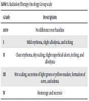
After obtaining the approval of the Ethics Committee of Zahedan University of Medical Sciences (IR.ZAUMS.REC.1398.186), the current quasi-experimental study was conducted on patients with breast cancer undergoing radiotherapy in Khatam al-Anbia and Ali ibn-Abi Taleb hospitals in Zahedan in 2019. The inclusion criteria included being a candidate for radiotherapy, no history of allergies, no previous radiotherapy, no unhealed scar at the site of radiation therapy, no active infection at the site of radiotherapy, not having diabetes, no severe chest deformity or history of hyperpigmentation, being aged 30-65 years, and willingness to participate in the study. The exclusion criteria were discontinuation of radiotherapy for any reason, absence from a training session, prescribed use of dermatological drugs to treat radiodermatitis, and recurrence of cancer. A total of 80 patients were selected based on the sample size formula and using convenience sampling (24). A random number table was prepared according to the sample size. Then, a moving point to the right was randomly selected. Odd and even numbers were considered to represent the intervention and control groups, respectively. Data were collected using a demographic form, including age, level of education, BIM, disease stage, marital status, smoking, and albumin level, and the Radiation Therapy Oncology Group (RTOG) scale (Table 1).
| Grade | Description |
|---|---|
| zero | No difference over baseline |
| I | Mild erythema, slight allodynia, and itching |
| II | Clear erythema, dry scaling, slight superficial ulcers, itching, and allodynia |
| III | Wet scaling, secretion of light green or yellow exudate, formation of sores, and edema |
| IV | Hemorage and necrosis |
The RTOG contains five grades, as follows: grade zero indicates no difference between the skin of the treated area and that of other areas; grade 1 indicates mild erythema, slight allodynia, and itching emerge; Grade 2 is accompanied by clear erythema, dry scaling, slight superficial ulcers, itching, and allodynia; Grade 3 is associated with wet scaling along with the secretion of light green or yellow exudate and the formation of sores and edema; Finally, bleeding and necrosis occur at grade 4. This instrument was published by the Oncology Radiotherapy Group Association in 2014 and has been used by several studies in Iran (14, 24-27).
Before providing the intervention, written informed consent was obtained from all patients. The intervention group received three 30-minute sessions of individual face-to-face training with educational content approved by the oncologist (Table 2). The training sessions were organized before starting radiotherapy. Thus, in the last three weeks of chemotherapy, with an interval of one week, we referred to the chemotherapy wards of Khatam al-Anbia and Ali ibn Abi Talib hospitals, to select the eligible patients. Then, after allocating them to groups of control and intervention, the intervention was provided. To evaluate radiodermatitis, the scale was filled three times (At the end of the first, third, and sixth weeks of radiotherapy) by interviewing with participants in the intervention group. The interviews were performed under the supervision of an oncologist. The control group received routine care. In the control group, the RTOG was completed by one of the researchers under the supervision of an oncologist. To observe ethical considerations, after finishing the study, participants in the control group were provided with the educational booklet.
| Session | Description |
|---|---|
| 1 | Introduction, cancer definition, treatment methods (chemotherapy, surgery, radiotherapy) |
| 2 | Providing explanations on chemotherapy and radiotherapy care, radiodermatitis, prevention, and radiodermatitis care |
| 3 | Further explanations about radiodermatitis, and questions and answers related to radiodermatitis |
Data were analyzed in SPSS version 22. Descriptive statistics were used to determine percentage, mean, standard deviation, minimum, and maximum [values in the two groups]. The Shapiro–Wilk test was used to evaluate the normality of data. The independent t-test was used to compare the means in the two groups. Eventually, to compare the frequency of qualitative variables of the two groups, the Mann-Whitney test, chi-squared test, Fisher's exact test, and the generalized estimation equation (GEE) model were used. A P value of < 0.05 was considered statistically significant.
4. Results:
There was no statistically significant difference between the two groups concerning the age, level of education, albumin level, BMI, disease stage, marital status, and smoking (Table 3).
Based on the findings, there was no difference concerning the frequency of radiodermatitis at the end of the first week of treatment in the two groups, but at the end of the third and sixth weeks, its frequency was higher in the control group than the intervention group (Tables 4 and 5).
The GEE test showed that over time the probability of developing higher grades of radiodermatitis increases by 1.074. This probability was 1.355 higher in the control group than the intervention group (P value = 0.03). Meanwhile, this test indicated no significant interaction between the two groups and over time (P value = 0.09) (Table 6).
| Group, Variable | Intervention | Control | P Value |
|---|---|---|---|
| Age | 7.57 ± 46.1a | 7.89 ± 45.5 | 0.81b |
| Education | 5.007 ± 6.45 | 4.49 ± 7.27 | 0.45b |
| Albumin level | 0.36 ± 3.89 | 0.37 ± 3.90 | 0.85c |
| BMI | 2.25 ± 24.22 | 2.3 ± 24.85 | 0.21c |
| Disease stage, No (%) | 0.79d | ||
| Stage 2 | 10 (25) | 9 (22.5) | |
| Stage 3 | 30 (75) | 31 (77.5) | |
| Marital status | 0.74d | ||
| Married | 35 (87.5) | 34 (85) | |
| Single | 5 (12.5) | 6 (15) | |
| Smoking | 1e | ||
| Yes | 1 (2.5) | 2 (5) | |
| No | 39 (97.5) | 38 (95) |
avalues are expressed as (Mean ± SD).
bMann-Whitney test.
cIndependent t-test.
dChi-squared test.
eFisher's exact test.
| Group, Radiodermatitis Severity | Intervention, No (%) | Control, No (%) |
|---|---|---|
| No change | 37 (92.5) | 29 (72.5) |
| Mild erythema | 3 (7.5) | 11 (27.5) |
| Total | 40 (100) | 40 (100) |
| Group, Radiodermatitis Severity | Intervention, No (%) | Control, No (%) |
|---|---|---|
| Clear erythema and dry scaling | 37 (92.5) | 26 (65) |
| Wet scaling | 3 (7.5) | 12 (30) |
| Ulcer and bleeding | 0 (0) | 2 (10) |
| Total | 40 (100) | 40 (100) |
| Variable | Odds Ratio | Standard Error | 95% Confidence Interval | P Value |
|---|---|---|---|---|
| Time | 1.074 | 0.158 | (1.38, 0.76) | 0.0001 |
| Group | 1.355 | 0.643 | (2.61, 0.09) | 0.03 |
| Time and group interaction | 0.39 | 23 | (0.06, -0.84) | 0.09 |
5. Discussion:
This study indicated that the designed educational intervention had a positive effect on the severity of radiodermatitis in patients with breast cancer undergoing radiation therapy. The frequency of radiodermatitis severity at the end of the first week of treatment was similar (i.e., zero) between both groups. In line with our study, Cui et al. (2015) investigated the effect of topical use of olive oil on the prevention of radiodermatitis in patients with nasopharynx cancer. General skin care recommendations were provided to the control group, and olive oil was administered for patients in the intervention group according to the prescribed protocol. Radiodermatitis severity was assessed weekly for nine weeks. The authors reported that until the third week, no case of radiodermatitis was observed in both groups (28). Schneider et al. (2014) evaluated the effect of Calendula officinalis in the prevention and treatment of radiodermatitis in patients with head and neck cancer. They studied this effect at the end of the first session and, then, once every five sessions until the end of treatment and 30 days after the last session. The results revealed no radiodermatitis in the two groups from the first to the fifth session (i.e., the end of the first week of radiation therapy) (29). The findings of the aforementioned studies are in good agreement with the present study concerning the severity of radiodermatitis at the end of the first week (i.e., its absence in this stage). Since radiation directly affects the DNA structure, it causes damage to all skin cell lines, which results in disrupted natural processes of cell proliferation and differentiation. Consequently, it impairs the cell replacement rate and causes clinical symptoms. The most common skin reactions appear approximately 10-14 days after the start of treatment (30).
In the present study, at the end of the third and sixth weeks of radiotherapy, the frequency of radiodermatitis in the intervention group was lower than the control group. Also, the intervention group had a lower risk of developing high grades of radiodermatitis, which confirms the positive impact of the educational intervention. Bauer et al. (2016) performed a study on using the 4MAT teaching model to promote skin care during radiotherapy. The authors adopted a structured theoretical framework to educate patients and reported that this approach empowered patients to better implement the educational program, which reduced the severity of radiodermatitis (31). In the above study, the educational content was instructed to the nurses of the oncology ward, and they taught the materials to the patients in one session. However, we adopted a gradual approach to educate the patients, so that the educational content was divided, and in each session, only some parts were taught. Besides, the patients could ask their questions. It could be assumed that the gradual presentation of materials, devoting part of the program to answering questions, and the temporal proximity of training sessions with the start of radiotherapy have positively affected the outcomes insofar as the prevention of radiodermatitis was concerned.
In a systematic review study by Bolderston et al. (2006) that aimed to develop practical guidelines for the prevention of acute skin reactions during radiotherapy, the results suggested that skin cleansing training is the only factor that could significantly prevent skin reactions (32). In this research, the results of various interventions (washing with soap and water, use of steroid compounds, calendula ointment, dressing, etc.) were discussed, and the authors reported that washing the skin with lukewarm soap and water could prevent skin reactions. Therefore, in the present study, training participants to wash their skin with mild soap and water can be an important contributor to the reduced incidence of radiodermatitis. Consistent with our study, Bernier et al. (2008) proposed that one can maintain skin hydration and mitigate the severity of radiodermatitis by educating and encouraging patients to wash their skin with mild soap and water (33). Likewise, Torabi Parizi et al. (2015) emphasized that educational programs should empower patients to enhance their abilities and skills to accept and adapt to chemotherapy and radiation therapy while considering the potential side effects (34).
Because radiotherapy and prolonged exposure to radiation cause damage to the skin, the body tries to reduce the damage by replacing the damaged cells, which leads to clinical symptoms such as dryness and desquamation. Such complications are unavoidable; however, their severity can be reduced through empowering the patients to participate in their treatment by self-care. Teaching preventive strategies such as public hygiene, washing the burn site with mild soap and water, avoiding tight clothing in the treatment area, and avoiding contact with physical substances such as metals and rhinestones or chemicals such as powder, perfume, and lotions can be effective in controlling radiodermatitis. This study showed that the educational program could reduce the frequency of radiodermatitis, which reinforces the impact of teaching preventive strategies in this regard.
In conclusion, the results emphasize the positive impact of preventive-care education on the severity of radiodermatitis in patients with breast cancer. Since during breast cancer radiotherapy, most patients experience radiodermatitis, such complications can be addressed by educating patients on how to prevent [or reduce] radiodermatitis through self-care activities.
