

1. Introduction
Addison’s disease diagnosis is sometimes very difficult due to the non-simultaneous occurrence of typical symptoms. The renal symptoms reported in hospitalized patients include hyponatremia (70% - 90%), hyperkalemia (40% - 60%), hypercalcemia (6%), and Acute Kidney Injury (AKI) (6%) (1).
Since adrenal failure is a rare disease with non-specific manifestations, clinical suspicion is a key factor in the early diagnosis of the disease. This case report describes the patient reporting adrenal failure manifested as renal failure and hyperkalemia without other laboratory manifestations and hypotension. The patient was affected by idiopathic CKD and was referred to Imam Hossein Hospital of Tehran. It is a rare case of Addison’s disease reported in a CKD patient.
2. Case Presentation
The patient was a 34-year-old man who had been referred to Imam Hossein Hospital Clinic in 2019 with the complaint of occasional nausea, weakness, and lethargy. The patient did not report the symptoms of weight loss, diarrhea, vomiting, and fever. However, he reported similar symptoms over the past three to four months. The patient did not report any underlying diseases, trauma, and using medications except for vitamin supplements. His mother had diabetes and renal failure. He was married and had a daughter (born in 2011) born to his first wife. He did not report smoking and drug abuse.
He had been referred to the emergency ward with complaints of similar symptoms about two weeks ago. In the first time of referring to the emergency, his vital signs were recorded as follows: Blood pressure (BP): 135/85 mmHg, pulse rate (PR): 80 beats/min, oral temperature (T): 37.1°C, and Respiratory Rate (RR): 13 cycles/min. His body mass index (BMI) was 27 kg/m2. There was no obvious observation in clinical examinations, and he did not report the symptoms of dehydration and edema. The results of the examination performed in the first time of referring were recorded as follows: urea: 92 mg/dL, creatinine (Cr): 3.2 mg/dL, sodium (Na): 134 meq/L, potassium (K): 8.5 meq/L, calcium: 10.4 mg/dL, venous blood gas (VBG) pH: 7.29, HCO3: 17.2, and PCO2: 40. Other laboratory tests such as fasting blood sugar (FBS), HbA1C, complete blood count (CBC), lactate, and urine analysis were normal. Due to the high potassium level, the patient underwent hemodialysis twice. Then, the potassium level became normal, while the creatinine level was still high (2.3 - 2.5 mg/dL). The results of the kidney ultrasound were reported as normal. During the first period of hospitalization, the patient showed the following status: K: 5 - 8.5 meq/L, Na: 132 - 137 meq/L, systolic blood pressure: 135 - 120 mmHg, and diastolic blood pressure: 95 - 80 mmHg. Due to the normal kidney size and no decrease in the creatinine level over the 10 days of hospitalization, a kidney biopsy was done for the patient, and the patient was discharged with a report of creatinine of 2.3 mg/dL. In the visit of the subsequent week, the patient’s status was reported as follows: normal blood pressure, Cr: 2.4 mg/dL, Na: 135 meq/L, and K: 5 meq/L. The results of kidney biopsy performed by optical microscopy, periodic acid Schiff, hematoxylin and eosin, jones and Congo red staining, and immunofluorescence were reported as follows:
Corticomedullary renal tissue with the presence of 39 glomeruli that 4 of them were obsolete. The other glomeruli were almost preserved. There was no evidence of GBM thickening or crescent formation, but 3 - 4 glomeruli showed prominent juxtaglomerular apparatus. Nearly all the tubules were preserved. There were no significant pathologic changes in the interstitium.
No amyloid deposition in the Congo-Red stained slides. In the third visit at Imam Hossein Hospital Clinic, the patient complained of weakness, lethargy, anorexia, and nausea. The patient’s vital signs were reported as follows: BP: 110/70, PR: 80, RR: 12, and T: 37°C. The patient was alert during the clinical examination and answered questions. The patient’s mucus seemed to be slightly dry. Cardiac and pulmonary auscultation was normal. No lymphadenopathy was observed in the head and neck examination, and the thyroid size and consistency were normal without any nodularity. No organomegaly was observed in the abdominal examination. The upper limbs and the lower limbs had a normal force and reflex. No scrotum was found in the genital examination of the left testicle. The other clinical examinations reported normal results. Regarding the testicular reexamination, some questions were asked about the patient’s sexual status. Despite denial in previous examinations, the patient reported being affected by infertility in the past two years. Due to the record of hyperkalemia, the patient was hospitalized, and the results of the tests were reported as follows: K: 7.2 meq/L, Na: 134 meq/L Cr: 3.6 mg/dL, urea: 144, Ca: 10.4 mg/dL, VBG pH: 7.12, HCO3: 13, PCO2: 37.5, SO2: 55, lactate: 1.2 mmol/L, FBS: 121 mg/dL, HbA1C: 6.8%, anion gap: 12, and chloride: 109 meq/L. The results of urine analysis and CBC were normal.
Regarding the report of AKI, the patient was treated with normal saline serum and calcium gluconate and insulin, with glucose starting for the treatment of hyperkalemia. The results of rhabdomyolysis and urate nephropathy were negative. A high potassium level (6.8 meq/L) and acidosis were again observed in his tests. Thus, hemodialysis was done for the patient. The patient was reexamined and asked about any changes in his skin color. He reported the generalized darkness of the skin over the recent period. Regarding the probability of adrenal failure, serum ACTH and cortisol were assessed and hydrocortisone treatment started. The results were reported as follows: ACTH: 1250 pg/mL and Cortisol: 4.3 mic/dL.
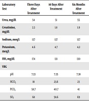
Due to no significant result in the examination of the left testicle, ultrasound was done and reported the following results: Left testicular size: 17 × 28 mm, right testicular size: 10 × 27 mm observed in the inguinal canal. No vascularity was observed in the Hyperhetroechoic region region with the size of 10 × 6.5 × 6 mm and unclear limits of right testicle parenchyma that was an indicator of a tumoral lesion. An endocrinologist checked the patient’s condition, and the following tests were requested for detecting the cause of adrenal failure considering the existence of testicular cancer. Tumor markers such as lactate dehydrogenase, Beta HCG, Alpha-fetoprotein were normal. Besides, 21-hydroxylase autoantibodies were checked, and the result was negative. Regarding the relationship between Addison’s disease and autoimmune polyglandular syndrome, thyroid function was tested, and results were reported as euthyroid. Concerning the high level of creatinine, ANA, anti-dsDNA, and ANCA tests were done, and the results were reported negative. The urologist offered an abdominal CT scan to check the testicular tumor, and the result indicated no metastasis and lymphadenopathy. The results of the tests performed for the patient over the three to four-day treatment period by hydrocortisone and fludrocortisone, as well as serum therapy, are reported in Table 1. The results of the tests performed 10 days after the treatment are indicated in Table 1. The patient was discharged with prednisolone and fludrocortisone.
| Laboratory Test | Three Days After Treatment | 10 Days After Treatment | Six Months After Treatment |
|---|---|---|---|
| Urea, mg/dL | 54 | 51 | 55 |
| Creatinine, mg/dL | 2.3 | 1.8 | 1.9 |
| Sodium, meq/L | 137 | 137 | 137 |
| Potassium, meq/L | 4.6 | 4.7 | 4.2 |
| FBS, mg/dL | 174 | 131 | 130 |
| VBG | |||
| pH | 7.33 | 7.35 | 7.38 |
| HCO3 | 18 | 23.6 | 23 |
| PCO2 | 58.7 | 40.7 | 41 |
| SO2 | 68 | 58.6 | 69 |
Laboratory Tests After Three and 10 Days and Six Months of Treatment
To detect the cause of Addison’s disease, the chest CT scan and patient’s symptoms were reviewed again, and tuberculosis was ruled out. Corticosteroid treatment increased the patient’s blood sugar, and diabetes treatment started under the supervision of an endocrinologist. Because of the history of infertility, a karyotype test was also done as suggested by the endocrinologist, and it was reported as normal. The results of the tests performed six months after starting the treatment are included in Table 1.
3. Discussion
The prevalence of AKI in adrenal failure is not known. However, it accounts for 4% of the causes of mortality in these patients (2). The connection between AKI and hyperkalemia in patients makes the diagnosis delayed (3). The lack of typical symptoms such as hyponatremia, hypoglycemia, and hypotension in the reported case made it difficult to diagnose the adrenal failure. However, unproportioned hyperkalemia to the kidney failure and acidosis raised the probability of adrenal failure in the second hospitalization.
Several studies have investigated the manifestation of adrenal failure as kidney failure and hyperkalemia. In a similar study, a patient referred with K: 7.3 mmol/L, Cr: 1.9 mg/dL, Na: 131 mmol/L, and BP: 95/69 mmHg. The unproportioned hyperkalemia to the uremia degree was a guiding indicator (4). In three cases in which the patients had been referred to the doctor with a high level of creatinine, all the three patients showed normal saline-resistant hypotension (1, 5). In the studied case, hypotension was not observed as a manifestation of the disease, which can be justified by CKD.
Nagler et al. (6) reported a 66-year-old man hospitalized because of symptomatic bradycardia and progressive muscle weakness. The patient reported unusual metastatic lung cancer (adenocarcinoma). The laboratory analyses indicated pH = 7.2, increased creatinine level (127 µmol/L), and serum urea (21.4 mmol/L) observed for the first time. There was no obvious reason for hyperkalemia. A low-dose ACTH stimulation test indicated the inadequate cortisol response (184 nmol/L). The low level of aldosterone (0.1 nmol/L) and the high activity of plasma rennin (33.7 mU/L) approved the primary assumption of hypoaldosteronism. Therefore, adrenal metastasis indicated by tomography scan revealed the adrenal failure (6).
Concerning the clinical importance of adrenal failure and the existence of effective treatments, this case report was proposed to emphasize the necessity of paying more attention to the clinical and laboratory symptoms indicating adrenal failure accompanied by kidney failure and hyperkalemia. Disproportionate hyperkalemia to uremia that was treatment-resistant was a useful indicator for diagnosis in this patient with CKD. Therefore, the careful investigation of clinical and laboratory symptoms can improve the attitudes towards diagnosis.
