1. Introduction
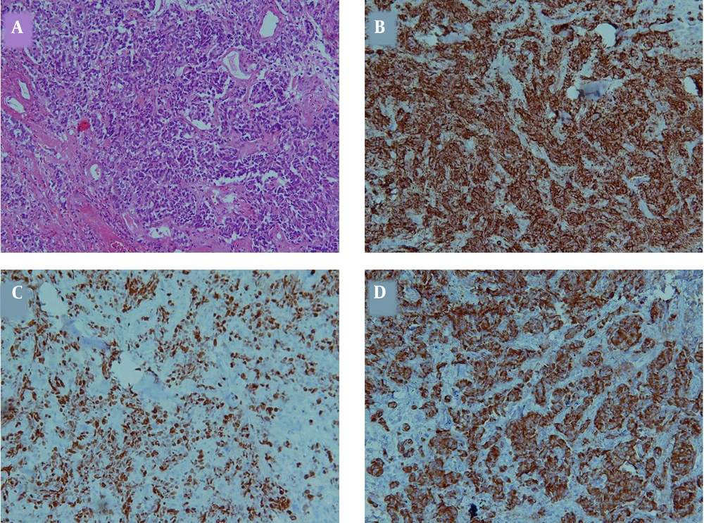
Most primary small cells carcinoma develop from the lung, and extra-pulmonary small cell carcinoma accounts for less than 5% of all small cell carcinoma cases (1). The most common site for extra-pulmonary small cell carcinoma is gastrointestinal, followed by genitourinary and head and neck regions. The larynx and salivary glands are the most common sites of involvement in the head and neck; however primary involvement of the palate is extremely rare (2). The extra-pulmonary small cell carcinoma is similar to pulmonary involvement regarding pathology features, which drives from pluripotential stem cells with neuroendocrine characteristics including round small cells containing dense and inconspicuous nuclei, sparse cytoplasm, high mitotic index, formation of sheets and trabecular patterns, and possible necrotic areas (1).
There is growing evidence indicating the ability of extra-pulmonary small cells carcinoma to originate from pluripotent basilar cells with neuroendocrine phenotype known as trans-differentiation phenomenon (1). The differentiation between primary and metastatic extra-pulmonary small cell carcinoma is challenging. There is also no standard guideline for its treatment as it is a rare occasion, and there is no consensus between radiation oncologists and cancer surgeons about the best treatment strategy.
In the present study, a highly rare case of hard palate small cell carcinoma with neuroendocrine pathology features is presented. To our best of knowledge, this is the third case report of extra-pulmonary small cell carcinoma involving the palate in the literature review. Knowing the clinical presentation and pathology characteristics of such rare tumor in addition to follow-up outcome can be highly useful to establish a reliable guideline for hard palate small cell carcinoma management.
2. Case Presentation
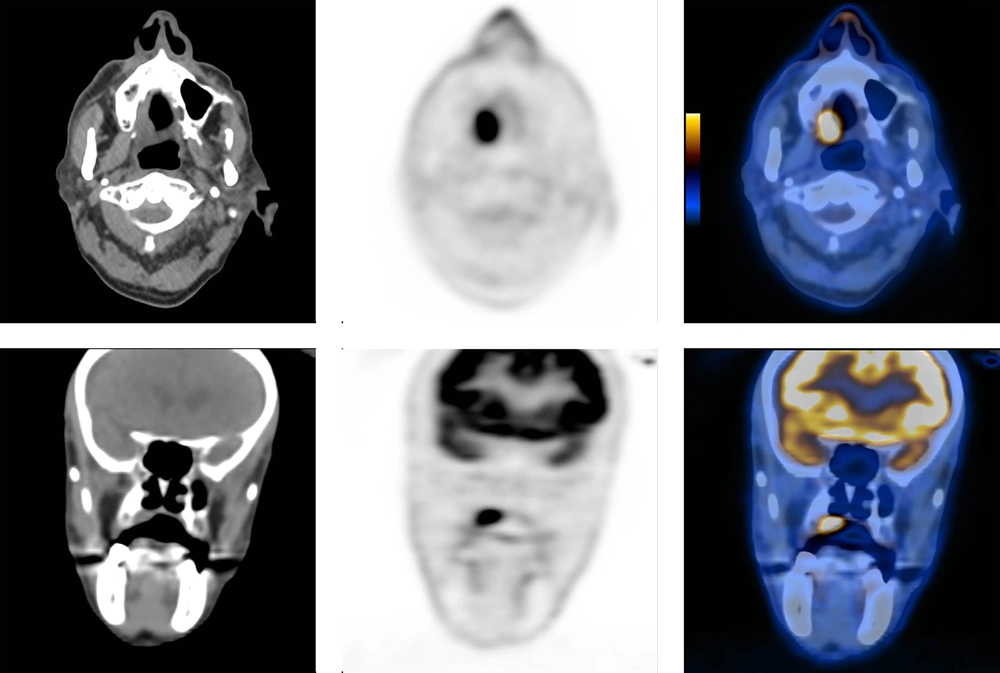
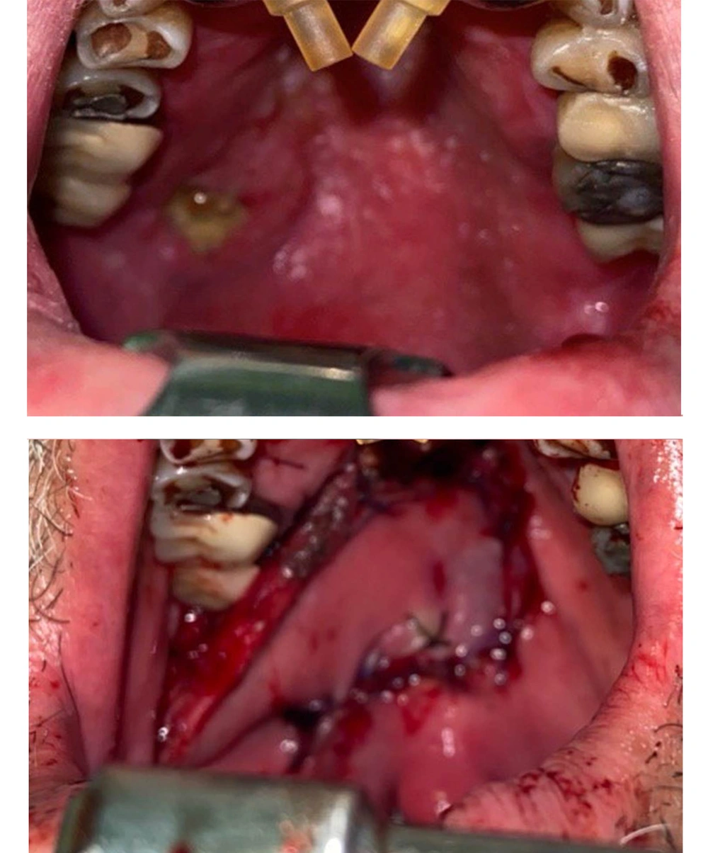
We present a rare case of extra-pulmonary small cell carcinoma of the hard palate in a 75-year-old non-smoker, non-alcoholic male patient, presented with an ulcerative lesion on the right aspect of the hard palate for at least 6 months before his first otolaryngology visit. The punch biopsy demonstrated neoplastic cells positive for AE1/AE3 and chromogranin and negative for S100, P63, LCA, TTF1, Cyclin D1, and GATA-3. The Ki67 was 95%, compatible with a small round cell tumor (small cell carcinoma) (Figure 1). Following consultation with the hospital tumor board, the patient was sent for 18 F-FDG PET/CT scan to exclude distant metastasis. The PET/CT images demonstrated intensely hypermetabolic hard palate lesion with no evidence of FDG-avid regional or distant metastasis (Figure 2). Additional prior imaging investigations, including chest CT scan and head and neck MRI, also failed to demonstrate any regional or pulmonary metastasis.
Finally, the lesion was excised with a safe margin and reconstructed with a local buccinator-based myomucosal flap (Figure 3). The permanent pathology report demonstrated squamous mucous membrane involved by nests, cords, and small sheets of small cells with hyperchromatic nuclei and scant cytoplasm, representing small round cell tumor consistent with high-grade neuroendocrine carcinoma. Additional evaluation for HPV infection was reported negative.
3. Discussion
Extra-pulmonary small cell carcinoma usually occurs in the sixth and seventh decades of life. Smoking and alcohol are considered risk factors; however, genetic and developmental factors are more contributory than environmental factors (1). Biopsy from an ulcerative oral lesion is easy to obtain. Fine-needle aspiration cytology evaluation can also be performed with the presence of an experienced cytopathologist. It is better to obtain the biopsy specimen from the edges of the ulcerative tumor to avoid necrotic central parts with possible false-negative results.
Extra-pulmonary small cell carcinoma usually has aggressive nature. Advanced locoregional disease, regional lymphadenopathy, and distant metastasis are also common (1). Evaluation for Immunohistochemical markers including cytokeratin, neuron-specific enolase (NSE), epithelial membrane antigen (EMA), CEA, chromogranin A, and synaptophysin is highly useful to consolidate the diagnosis. Thyroid transcription factor-1 (TTF-1) can be used to differentiate small cell carcinoma from other neuroendocrine tumor types because small cell tumor cells are usually positive for this marker despite other neuroendocrine tumors (3). Our case was positive for chromogranin but negative for TTF-1.
For small cell tumors, it is of crucial importance to exclude regional or distant metastases before the final treatment planning. With the widespread availability of PET/CT scans, many oncology patients undergo this imaging procedure for metastatic evaluation according to the approved guidelines. There are several different PET radiotracers available, some of which are more specific, like 68-Gallium DOTATATE, which is recommended for evaluation of neuroendocrine tumors. However, our case demonstrated a very high rate of Ki67 (95%), which is compatible with high-grade and atypical neuroendocrine tumors. For evaluation of high-grade neuroendocrine tumors with high Ki67 and mitotic index, 68-Ga DOTATATE may demonstrate false-negative results because of the less differentiated nature of the tumor. Therefore, we used Fludeoxyglucose (18F) as the radiotracer to evaluate our patient for metastasis (4). Fortunately, regional or distant metastases were excluded in our patients with preoperative imaging, as discussed earlier.
The differential diagnosis for our case included metastatic squamous cell carcinoma, Merkel cell tumors, other types of neuroendocrine tumors, lymphoma, metastatic melanoma, and poorly differentiated carcinoma. The extra-pulmonary small cell carcinoma staging system is not well established, and patients are divided into limited disease (LD) and extensive disease (ED) (1). Interestingly, this rare disease is more common in the head and neck region, similar to our case. There is no standard guideline for the treatment of extra-pulmonary small cell carcinoma. Its prognosis is poor. Complete excision is recommended when complete resection is possible with minimal morbidity. Similar chemotherapeutic regimens are used for pulmonary small cell carcinoma (combination of cisplatin + etoposide or cisplatin + irinotecan). Radiotherapy also has an effective role (1).
In a study by Ochsenreither et al., long-term survival with combination therapy, including surgery, in addition to chemo- or chemoradiotherapy has been reported (5). It is showed that extra-pulmonary small cell carcinoma with head and neck origin has a better outcome rather than other sites. This may be partly attributed to earlier detection and more intensive treatment (1). Palate small cell carcinoma is a highly rare occasion. Complete excision and chemoradiotherapy are recommended. The PET/CT is useful for staging the disease. Standard guidelines should also be established for its treatment.
.jpg)


