Abstract
Background:
Both low-level laser therapy (LLLT) and platelet-rich plasma (PRP) are demonstrated to promote the repair of mandibular defects.Objectives:
This study investigated the mechanical properties and calcium content at the tooth extraction site in a rat model exposed to LLLT (λ = 808 nm) with or without PRP.Methods:
In this experimental rat model study, the left first molar maxillary teeth were extracted in twenty male rats. Then, the animals were randomly divided into four groups. Group one: after extraction, the extraction sockets were treated with 0.9 W gallium-aluminum-arsenide (GaAlAs) diode laser irradiation for five minutes every 72 hours for the next 12 days (4 times overall); group two: PRP was placed in the extraction sockets; group three: a combination of both treatments (LLLT+PRP) was applied; group four: the extraction sockets remained untreated (the control group). All rats were sacrificed 30 days post-operative. All bone blocks of the extracted socket were prepared for mechanical strength and calcium content analyses. Mann-Whitney test, one-way ANOVA test, and post hoc Fisher’s least significant difference (LSD) were used to analyze the data. A P-value less than 0.05 was considered significant. All analyses were performed by SPSS 16.0. The graph is illustrated in the graph pad 5.Results:
The compressive strength in the laser group was significantly higher than in the control and PRP-treated groups (P = 0.0001 and 0.00044, respectively). Compared to the control and PRP groups, the effects of a combination of PRP and LLLT mechanical strength were statistically similar. Calcium content was influenced by none of the treatments.Conclusions:
The mechanical strength of the bone blocks was significantly stronger in the LLLT group than in the other groups. Platelet-rich plasma alone or combined with LLLT demonstrated a synergistic impact on neither mechanical strength nor calcium content.Keywords
Low-level Laser Therapy Platelet-rich Plasma Alveolar Bone Tooth Extraction
1. Background
Bone healing is a complex biological process that can be clinically challenging (1-3). Bone defects in the jaws may originate from congenital or developmental malformation, tumor resection, cyst enucleations, trauma, infections, and tooth removal. Tooth extraction is one of the most common procedures performed in a dental office. Healing is uneventful in most cases, but some extraction wounds require longer to heal and may be painful or lead to bone defects.
Previous studies have introduced several methods to accelerate bone repair, including treating the tooth sockets with various bioactive materials (4) and using specific growth factors such as bone morphogenetic proteins (BMPs) (5), vitamin D (6), calcium phosphate (7), hormones, plant extracts (8), low-intensity ultrasound mechanical stimulation (9, 10), electromagnetic fields (11) and LLLT (12, 13). Low-level laser therapy is also known as ''photo-bio-modulation therapy" and has extensive applications in many clinical and experimental studies. Several studies have concluded that LLLT can be beneficial for several specific applications in dental practice (14). Low-level laser therapy accelerates repair with or without the administration of various osteoinductive and osteoconductive biomaterials (15). The laser affects tissues in numerous ways, such as hemostasis, microbial decontamination, tissue ablation, and vaporization, and influences biological processes, including bio-stimulation (photo-bio-modulation), which have many therapeutic effects (16). One of the promising uses of LLLT is the acceleration of bone metabolism (17). The laser applies a directional non-ionizing electromagnetic radiation, which is monochromatic and coherent (18), and its wound healing stimulation effects have been demonstrated with various power densities. Laser energy can enhance osteoblastic processes, epithelial cells and fibroblast proliferation, collagen synthesis by fibroblasts, lymphatic system activation, angiogenesis, and bone formation (19). Low-level laser therapy has also been demonstrated to boost the osteogenic properties of some biomaterials.
Bone healing takes a long time, and spontaneous bone repair can be complicated if the bone defects are more extensive than a specific size. Therefore, researchers have extensively studied the osteoinductive and osteoconductive effects of different biological and chemical agents to accelerate this process. One such biological agent is platelet-rich plasma (PRP) (20-22). There is a growing interest in applying PRP for bone repair as it reduces injured tissue's recovery time. Similarly, PRP promotes bone healing through its many bone-forming ingredients, such as specific proteins, cytokines, chemokines, and several growth factors of the plasma and platelets. Platelets have various functions, such as wound repair, re-epithelialization, and homeostasis. They also release several growth factors, stimulate angiogenesis, promote fibroblast proliferation, and accelerate collagen synthesis (23).
Platelet-rich plasma contains fibrin and various growth factors that can promote bone growth, such as vascular endothelial growth factor (VEGF), transforming growth factor-beta (TGF- β), epithelial growth factor (EGF), platelet-derived growth factor (PDGF), and fibroblast growth factor (FGF) (24). They can all be gelified as a scaffold for filling injured tissues (25). Moreover, some PRP growth factors (such as PDGF) can mobilize adjacent tissues’ mesenchymal cells to proliferate, migrate, and differentiate into osteoblasts. Previous studies have demonstrated that PRP can facilitate the migration of osteoblasts and periodontal ligament cells (26). Moreover, the synergistic effect of PRP and other biomaterials and therapeutic interventions has been confirmed in several studies (27, 28).
The combination of low-level laser and PRP has been used for stimulating regeneration in orthopedics (29-31), dermatology (using fractional CO2 laser (32, 33) and low-level laser (34)), muscle repair (35), dental implant (36), healing of periodontal defects (37), periodontal regeneration in fenestration defects (38), bone regeneration (39), and Medication-Related Osteo-Necrosis of the Jaw (MRONJ) management (40). Their combination can presumably enhance different cell regeneration processes.
2. Objectives
Considering the above gaps in the literature, the present study aimed to illuminate any potential synergic impact of biogenic materials, such as platelet-rich plasma and LLLT, on bone calcium content and tooth extraction site mechanical properties in maxillary bone defects in a rat model.
3. Methods
3.1. Ethical Statement
The University ethical committee approved the study protocol, and all procedures complied with ethical principles of animal handling and treatment (IR.SUMS.DENTAL.REC.1400.020). The ARRIVE guidelines were followed in reporting the present study.
3.2. Study Design, Sample Size, and Experimental Procedures
The current work is an experimental study. In the present study, four groups were compared: the control group, laser group, PRP group, and laser-PRP combination group. The sample size was determined according to a previous study (41) and was modified based on our limitations and constraints (ethics and time). Considering an α = 0.05 and a power of 80%, five rats were required for each group (n = 5).
The rats were procured from the Animal Laboratory of the Shiraz University of Medical Sciences. All rats were kept in large, well-controlled environments with a temperature of 22 ± 2°C and 12 h of light/dark cycles, adequate ventilation, and unlimited access to food and water.
Twenty healthy Sprague–Dawley male rats, approximately 80 - 90 days old and 180 - 200 grams, were procured from the Animal Laboratory of the University of Medical Sciences. Anesthetic methods and medications were similar in all groups.
3.3. Inclusion and Exclusion Criteria
Healthy rats were included in the study, and animals with abnormal health conditions were excluded.
3.4. Randomization and Blinding
Laboratory personnel labeled and allocated rats randomly; therefore, authors were blinded to sample groups during and at the end of the experiment.
3.5. Outcome Measures
Our primary outcome measures were the calcium content and mechanical properties of the extraction socket of rats’ first left maxillary molar.
3.6. Experimental Procedures
All rats were anesthetized by intramuscular administration of a 4:1 solution of ketamine (Vetased, Romania)/xylazine (Sedaxyl, Belgium) at a dose of 0.15ml per 100g of body weight. After the onset of anesthesia, the maxillary left first molar teeth of all animals were extracted. All animals were divided randomly into one control group (n = 5) and three experimental (case) groups: laser- (n = 5), PRP- and laser-PRP-treated (n = 5) group. In the PRP- and laser-PRP-treated groups, approximately 100 µL of PRP (after preparation) was transferred into the socket immediately after extraction, and then, 100 λ CaCl2 was added to the jellified PRP. A figure of eight sutures using vicryl 4-0 was placed over the socket to prevent the gelatinized PRP from extrusion.
The tooth sockets of both the laser group and the laser-PRP group were then exposed to gallium aluminum arsenide diode laser every 72 h after extraction for the next 12 days (4 times overall).
All rats were euthanized by CO2 70% 30 days after the extraction (42). The maxilla was harvested, and the overlying soft tissue was removed using a scalpel. Tooth sockets with a rim of peripheral bone were separated and immersed in 10% buffered neutral formalin for 48 hrs. Using a digital caliper (Digit-Cal SM, TESA S.A., Switzerland), the length and width of all bone blocks were measured as 4 ± 0.1 mm and 3 mm ± 0.1 mm, respectively. The thickness of the blocks was measured using the same caliper. All blocks were stored at -20ºC.
3.7. Preparation of PRP
Similar to our previous study (43), three mL of the rat blood was transferred into a tube containing citric acid anti-coagulant and centrifuged at 5000 rpm for 15 minutes. A micropipette was used to remove the plasma and buffy coat, which were stored in sterile tubes. Plasma was centrifuged (IntraSpin® system, BioHorizons, Birmingham, Alabama, USA) for the second time at 2000 rpm for 10 min, and the bottom one-third was considered PRP. To gelatinize PRP and activate platelets, it was fortified with 100 λ of 2.5% CaCl2 immediately after transferring into the tooth sockets and socket sutured.
3.8. Laser Device
The laser device was gallium-aluminum-arsenide (GaAlAs) (OSRAM LD 808, Germany), and its parameters were set as follows: 808 nm wavelength, 0.9 W output, 1459 J/cm2 dose, and continuous irradiation mode for five minutes (44, 45). The laser device had a beam area of 1 cm2 and a continuous wavelength of 808nm and was applied for five minutes in the vertical direction in close contact with the sockets.
3.9. Mechanical Test Assessment
All samples were simulated in terms of mass and volume. A scale with an accuracy of 0.1 mg was used to measure the mass. Archimedes' principle of solids' volume was used to evaluate the samples' volume.
The removed bone samples were longitudinally fixed for mechanical testing and exposed to axial compression mechanical tests using a Zwick/Roell machine (Germany) at a speed of 0.5mm/min.
3.10. Calcium Content Assessment
After the mechanical test, we collected all bone block pieces (generated by breaking blocks caused by the mechanical test) and prepared them for calcium content measurement. This preparation procedure started by heating in a furnace to leave the fragments of bone blocks at 520ºC for 20 h in the oven (Gallenkamp Muffle Furnace, England). After that, the ashed bones were powdered, and 0.03 grams of bone powder was dissolved in 250 µL HCl and diluted with 31 mL distilled water. The calcium contents were measured with atomic absorption (Thermo scientific iCE 3500, USA) at a wavelength of 422.7 nm.
3.11. Statistical Methods
The mechanical test data were statistically analyzed by the Mann-Whitney test. Bone calcium contents were compared by the one-way ANOVA test. Intergroup statistical differences were conducted by Fisher’s least significant difference (LSD) as post hoc tests. A P-value less than 0.05 was considered significant for calcium content and mechanical strength. All analyses were performed by SPSS 16.0 for Windows. The graph is depicted in graph pad 5.
4. Results
All rats tolerated the extraction procedure without any noticeable inflammation and were able to eat a regular diet after the procedure.
4.1. Mechanical Test
The thickness of the newly-formed bone in laser and laser/PRP groups (Range = 1.2 - 1.4 mm) was higher than the control group (Range = 0.7 - 0.8 mm).
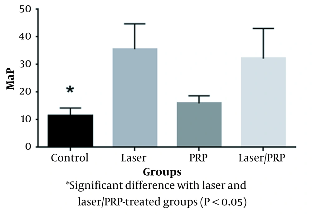
The compressive strength in the laser group was significantly higher than in the control group (P = 0.0001, Figure 1). Also, a significant difference was observed between the laser/PRP and control groups (P = 0.00044). Moreover, the results showed that the mechanical strengths of PRP-treated and control groups were statistically similar (Figure 1).
The comparison of the compressive strength of the reparative bone in tooth sockets treated with different methods. Control versus laser P = 0.0001, Control versus laser/PRP P = 0.000448, PRP versus laser P = 0.001, PRP versus laser/PRP P = 0.003
4.2. Calcium Content Finding
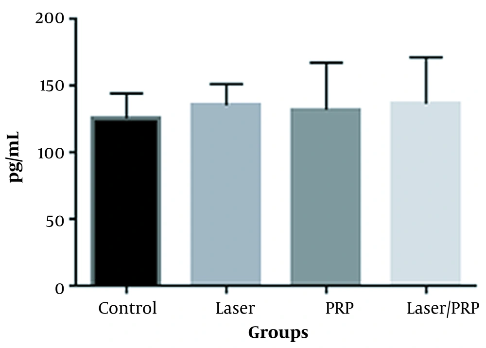
Figure 2 displays the calcium content quantity of the reparative bone in tooth sockets of different groups. The data demonstrated that neither laser nor PRP could significantly impact Ca contents. The combined laser and PRP administration also had no significant influence on Ca content (Figure 2).
The comparison of the calcium content between the different groups
5. Discussion
In the postoperative phase, bone regeneration is visible, gradually filling the defect. The bone healing process usually requires four to six months, and a longer time is needed for bone remodeling (46).
Recent studies comparing the bone-healing processes in rats and humans have observed numerous similarities, but this process is generally faster in rats (47). This was the rationale for choosing rats as the experimental model for the present study (principles of the 3Rs: replacement, reduction, and refinement) (42). A previous study demonstrated differences between human and small rodents’ bones which have the basic bone structures without Haversian canals (48).
Degranulation of the PRP promotes the discharge of several growth factors and substances such as VEGF, TGF beta-1, PDGF, FGF, connective tissue growth factor, transforming growth factors such as insulin or stimulatory (IGF-1), epidermal growth factor, platelet thromboplastin, calcium, serotonin, and fibrinogen hydrolytic enzymes (49, 50). Previous studies demonstrated that PRP could promote bone regeneration by releasing growth factors such as TGF-β1 and PDGF (22) after platelet degranulation (2).
Low-level laser therapy procedure has several limitations, such as the lack of an established optimization and a standard protocol for exposure time, power intensity, and wavelength. Moreover, different studies have used other experimental models with various exposure durations, which complicates comparing the outcomes. Low-level laser therapy effect is demonstrated to be dose-dependent, and a single exposure has no significant effect on bone repair (1, 51). In the current study, although we have applied four consecutive doses of LLLT, we observed that LLLT did not increase calcium contents.
Previous in vivo and in vitro studies demonstrated that LLLT could induce osteoblast proliferation and differentiation. Low-level laser-like GaAlAs at 830 nm wavelength stimulates osteoblastic cell growth and increases alkaline phosphatase (ALP) and osteocalcin in osteogenic cell line culture (52). ALP activity and osteoclastic expression are associated with the mineralization of newly-formed bones. The viability of osteocytes around dental implants is demonstrated to be higher after low-level laser therapy (53). Even though literature indicates a higher number of osteoblasts, higher ALP activity, and osteocalcin expression level associated with LLLT administration, our data did not show any related increase in calcium content. Therefore, the observed increase in mechanical strength seems unrelated to calcium content, increased osteoblast mineralization capacity, or ALP activity. A previous study found that LLLT can accelerate bone healing in irradiated tooth sockets of albino rats (54). It has also been demonstrated to stimulate bone formation in large calvaria defects of ovariectomized rats (3). In a recent immunohistochemical study, LLLT administration led to an increase in bone formation, collagen synthesis (55), bone metabolism, tooth movement (56), angiogenesis, and osteoid formation (57). Therefore, the observed increase in mechanical strength may be attributed to an increase in collagen content. In contrast to our findings, Nicola et al. found that applying LLLT increased mineral apposition rate and bone volume in the femur (58). The femur is a load-bearing bone, while alveolar sockets are exposed to low mechanical pressure after tooth extraction. Therefore, the different results between our findings and those of Nicola et al. can be attributed to physiological differences in mechanical stimulation.
The study of photo-bio-modulation therapy is expected to define a standardized protocol for specific wavelengths and radiation doses. The wavelength of 808 nm (used in the present study) penetrates the tissue surface (mucosa) into the underlying bone (maxilla) and may be more effective for bone-generation purposes (59). Earlier studies have demonstrated that several regenerative strategies, biomaterials, and additional therapies (such as LLLT and PRP) can accelerate bone repair and growth (1). Although we did not observe any calcium content alternation, our study’s findings indicated that LLLT administration could increase the mechanical strength of a repaired bone.
In the present study, we showed that LLLT enhances bone biomechanical properties. In addition, compared to the control group, we observed no improvement in bone biomechanical properties in the animals treated with PRP. Technically, one would expect that the combination of PRP and LLLT could reinforce bone regeneration, which would be notably superior to the application of each individual treatment (3, 22). However, we observed no significant improvement in bone biomechanical properties using a combination of LLLT/PRP treatment compared to the laser alone. In the present study, the rationale behind the increased compressive strength of newly-formed bone might be attributed to the local effects of laser stimulation of the differentiation of mesenchymal stem cells and the proliferation of fibroblasts and osteoblasts (59).
To prevent PRP removal during chewing, the edges of the tooth sockets were sutured. Suturing has been used to avoid bone exposure and the consequent necrosis that may impair bone healing (60). Our data revealed that neither PRP nor suturing (used together) demonstrated any beneficial effect on the calcium content and mechanical strength. As we did not find any PRP and LLLT synergistic effect, and noted that the PRP preparation method could influence its effects (27), the use of PRP prepared with different procedures is recommended.
5.1. Conclusions
Mechanical and biochemical analyses of the bone blocks demonstrated that PRP treatment does not improve bone biomechanical properties. However, LLLT may influence the reinforcement of bone biomechanical properties. Calcium content analysis demonstrated that PRP and LLLT, alone or in combination, were not effective.
5.2. Limitations
As PRP preparation is vital in its effects on bone repair, using just one PRP preparation method can be one of the study’s limitations. Although this method has proved effective in a previous study (43), a standard preparation protocol is lacking. Another limitation that has not been evaluated in the present study could be the evaluation of bone-specific markers, such as those involved in mineralization, which could indicate the degree of mineralization more precisely. We used the axial compression test in the present study, which was also used in previous studies to examine the biomechanical properties of bone (61). The present study examined repaired bone’s calcium content and mechanical properties, and we did not acquire histological specimens.
References
-
1.
Obradovic RR, Kesic LG, Pesevska S. Influence of low-level laser therapy on biomaterial osseointegration: a mini-review. Lasers Med Sci. 2009;24(3):447-51. [PubMed ID: 18566853]. https://doi.org/10.1007/s10103-008-0573-z.
-
2.
Mariano R, Messora M, de Morais A, Nagata M, Furlaneto F, Avelino C, et al. Bone healing in critical-size defects treated with platelet-rich plasma: a histologic and histometric study in the calvaria of diabetic rat. Oral Surg Oral Med Oral Pathol Oral Radiol Endod. 2010;109(1):72-8. [PubMed ID: 19926499]. https://doi.org/10.1016/j.tripleo.2009.08.003.
-
3.
Garcia VG, da Conceicao JM, Fernandes LA, de Almeida JM, Nagata MJ, Bosco AF, et al. Effects of LLLT in combination with bisphosphonate on bone healing in critical size defects: a histological and histometric study in rat calvaria. Lasers Med Sci. 2013;28(2):407-14. [PubMed ID: 22370617]. https://doi.org/10.1007/s10103-012-1068-5.
-
4.
El-Ghannam A. Bone reconstruction: from bioceramics to tissue engineering. Expert Rev Med Devices. 2005;2(1):87-101. [PubMed ID: 16293032]. https://doi.org/10.1586/17434440.2.1.87.
-
5.
McCarthy TL, Ji C, Centrella M. Links among growth factors, hormones, and nuclear factors with essential roles in bone formation. Crit Rev Oral Biol Med. 2000;11(4):409-22. [PubMed ID: 11132763]. https://doi.org/10.1177/10454411000110040201.
-
6.
Doetsch AM, Faber J, Lynnerup N, Watjen I, Bliddal H, Danneskiold-Samsoe B. The effect of calcium and vitamin D3 supplementation on the healing of the proximal humerus fracture: a randomized placebo-controlled study. Calcif Tissue Int. 2004;75(3):183-8. [PubMed ID: 15386160]. https://doi.org/10.1007/s00223-004-0167-0.
-
7.
Higgins TF, Dodds SD, Wolfe SW. A biomechanical analysis of fixation of intra-articular distal radial fractures with calcium-phosphate bone cement. J Bone Joint Surg Am. 2002;84(9):1579-86. [PubMed ID: 12208914]. https://doi.org/10.2106/00004623-200209000-00010.
-
8.
Isler SC, Demircan S, Cakarer S, Cebi Z, Keskin C, Soluk M, et al. Effects of folk medicinal plant extract Ankaferd Blood Stopper on early bone healing. J Appl Oral Sci. 2010;18(4):409-14. [PubMed ID: 20835578]. [PubMed Central ID: PMC5349063]. https://doi.org/10.1590/s1678-77572010000400015.
-
9.
Hasuike A, Sato S, Udagawa A, Ando K, Arai Y, Ito K. In vivo bone regenerative effect of low-intensity pulsed ultrasound in rat calvarial defects. Oral Surg Oral Med Oral Pathol Oral Radiol Endod. 2011;111(1):e12-20. [PubMed ID: 21176808]. https://doi.org/10.1016/j.tripleo.2010.09.061.
-
10.
Findik Y, Baykul T. Effects of low-intensity pulsed ultrasound on autogenous bone graft healing. Oral Surg Oral Med Oral Pathol Oral Radiol. 2014;117(3):e255-60. [PubMed ID: 23026154]. https://doi.org/10.1016/j.oooo.2012.05.023.
-
11.
Kooistra BW, Jain A, Hanson BP. Electrical stimulation: Nonunions. Indian J Orthop. 2009;43(2):149-55. [PubMed ID: 19838363]. [PubMed Central ID: PMC2762246]. https://doi.org/10.4103/0019-5413.50849.
-
12.
Kazancioglu HO, Ezirganli S, Aydin MS. Effects of laser and ozone therapies on bone healing in the calvarial defects. J Craniofac Surg. 2013;24(6):2141-6. [PubMed ID: 24220424]. https://doi.org/10.1097/SCS.0b013e3182a244ae.
-
13.
Nagata MJ, Santinoni CS, Pola NM, de Campos N, Messora MR, Bomfim SR, et al. Bone marrow aspirate combined with low-level laser therapy: a new therapeutic approach to enhance bone healing. J Photochem Photobiol B. 2013;121:6-14. [PubMed ID: 23474527]. https://doi.org/10.1016/j.jphotobiol.2013.01.013.
-
14.
Walsh LJ. The current status of low level laser therapy in dentistry. Part 1. Soft tissue applications. Aust Dent J. 1997;42(4):247-54. [PubMed ID: 9316312]. https://doi.org/10.1111/j.1834-7819.1997.tb00129.x.
-
15.
Jonasson TH, Zancan R, de Oliveira Azevedo L, Fonseca AC, Silva MCD, Giovanini AF, et al. Effects of low-level laser therapy and platelet concentrate on bone repair: Histological, histomorphometric, immunohistochemical, and radiographic study. J Craniomaxillofac Surg. 2017;45(11):1846-53. [PubMed ID: 28935484]. https://doi.org/10.1016/j.jcms.2017.08.008.
-
16.
Aoki A, Sasaki KM, Watanabe H, Ishikawa I. Lasers in nonsurgical periodontal therapy. Periodontol 2000. 2004;36:59-97. [PubMed ID: 15330944]. https://doi.org/10.1111/j.1600-0757.2004.03679.x.
-
17.
Hsu LF, Tsai MH, Shih AH, Chen YC, Chang BE, Chen YJ, et al. 970 nm low-level laser affects bone metabolism in orthodontic tooth movement. J Photochem Photobiol B. 2018;186:41-50. [PubMed ID: 30005205]. https://doi.org/10.1016/j.jphotobiol.2018.05.011.
-
18.
Schindl A, Schindl M, Pernerstorfer-Schon H, Schindl L. Low-intensity laser therapy: a review. J Investig Med. 2000;48(5):312-26. [PubMed ID: 10979236].
-
19.
Kawasaki K, Shimizu N. Effects of low-energy laser irradiation on bone remodeling during experimental tooth movement in rats. Lasers Surg Med. 2000;26(3):282-91. [PubMed ID: 10738291]. https://doi.org/10.1002/(sici)1096-9101(2000)26:3<282::aid-lsm6>3.0.co;2-x.
-
20.
Nikolidakis D, Meijer GJ, Oortgiesen DA, Walboomers XF, Jansen JA. The effect of a low dose of transforming growth factor beta1 (TGF-beta1) on the early bone-healing around oral implants inserted in trabecular bone. Biomaterials. 2009;30(1):94-9. [PubMed ID: 18838161]. https://doi.org/10.1016/j.biomaterials.2008.09.022.
-
21.
Wiltfang J, Kloss FR, Kessler P, Nkenke E, Schultze-Mosgau S, Zimmermann R, et al. Effects of platelet-rich plasma on bone healing in combination with autogenous bone and bone substitutes in critical-size defects. An animal experiment. Clin Oral Implants Res. 2004;15(2):187-93. [PubMed ID: 15008930]. https://doi.org/10.1111/j.1600-0501.2004.00980.x.
-
22.
Marx RE, Carlson ER, Eichstaedt RM, Schimmele SR, Strauss JE, Georgeff KR. Platelet-rich plasma: Growth factor enhancement for bone grafts. Oral Surg Oral Med Oral Pathol Oral Radiol Endod. 1998;85(6):638-46. [PubMed ID: 9638695]. https://doi.org/10.1016/s1079-2104(98)90029-4.
-
23.
Monto RR. Platelet rich plasma treatment for chronic Achilles tendinosis. Foot Ankle Int. 2012;33(5):379-85. [PubMed ID: 22735279]. https://doi.org/10.3113/FAI.2012.0379.
-
24.
Oryan A, Alidadi S, Moshiri A. Platelet-rich plasma for bone healing and regeneration. Expert Opin Biol Ther. 2016;16(2):213-32. [PubMed ID: 26561282]. https://doi.org/10.1517/14712598.2016.1118458.
-
25.
Sadeghi-Ataabadi M, Mostafavi-Pour Z, Vojdani Z, Sani M, Latifi M, Talaei-Khozani T. Fabrication and characterization of platelet-rich plasma scaffolds for tissue engineering applications. Mater Sci Eng C Mater Biol Appl. 2017;71:372-80. [PubMed ID: 27987720]. https://doi.org/10.1016/j.msec.2016.10.001.
-
26.
Aoki A, Mizutani K, Schwarz F, Sculean A, Yukna RA, Takasaki AA, et al. Periodontal and peri-implant wound healing following laser therapy. Periodontol 2000. 2015;68(1):217-69. [PubMed ID: 25867988]. https://doi.org/10.1111/prd.12080.
-
27.
Shahsavari-Pour S, Aliabadi E, Latifi M, Zareifard N, Namavar MR, Talaei-Khozani T. Evaluation of the Possible Synergic Regenerative Effects of Platelet-Rich Plasma and Hydroxyapatite/Zirconia in the Rabbit Mandible Defect Model. Iran J Med Sci. 2018;43(6):633-44. [PubMed ID: 30510340]. [PubMed Central ID: PMC6230930].
-
28.
Tschon M, Veronesi F, Contartese D, Sartori M, Martini L, Vincenzi F, et al. Effects of pulsed electromagnetic fields and platelet rich plasma in preventing osteoclastogenesis in an in vitro model of osteolysis. J Cell Physiol. 2018;233(3):2645-56. [PubMed ID: 28786478]. https://doi.org/10.1002/jcp.26143.
-
29.
Prodromos CC, Finkle S, Dawes A, Dizon A. Intra-Articular Laser Treatment Plus Platelet Rich Plasma (PRP) Significantly Reduces Pain in Many Patients Who Had Failed Prior PRP Treatment. Medicines (Basel). 2019;6(3). [PubMed ID: 31315168]. [PubMed Central ID: PMC6789467]. https://doi.org/10.3390/medicines6030075.
-
30.
Barbosa D, de Souza RA, de Carvalho WR, Xavier M, de Carvalho PK, Cunha TC, et al. Low-level laser therapy combined with platelet-rich plasma on the healing calcaneal tendon: a histological study in a rat model. Lasers Med Sci. 2013;28(6):1489-94. [PubMed ID: 23307438]. https://doi.org/10.1007/s10103-012-1241-x.
-
31.
Allahverdi A, Sharifi D, Takhtfooladi MA, Hesaraki S, Khansari M, Dorbeh SS. Evaluation of low-level laser therapy, platelet-rich plasma, and their combination on the healing of Achilles tendon in rabbits. Lasers Med Sci. 2015;30(4):1305-13. [PubMed ID: 25759233]. https://doi.org/10.1007/s10103-015-1733-6.
-
32.
Chang HC, Sung CW, Lin MH. Efficacy of Autologous Platelet-Rich Plasma Combined With Ablative Fractional Carbon Dioxide Laser for Acne Scars: A Systematic Review and Meta-Analysis. Aesthet Surg J. 2019;39(7):NP279-87. [PubMed ID: 30809666]. https://doi.org/10.1093/asj/sjz048.
-
33.
Schoenberg E, Wang JV, Zachary CB, Saedi N. Treatment of acne scars with PRP and laser therapy: an up-to-date appraisal. Arch Dermatol Res. 2019;311(8):643-6. [PubMed ID: 31144021]. https://doi.org/10.1007/s00403-019-01936-7.
-
34.
Gentile P, Dionisi L, Pizzicannella J, de Angelis B, de Fazio D, Garcovich S. A randomized blinded retrospective study: the combined use of micro-needling technique, low-level laser therapy and autologous non-activated platelet-rich plasma improves hair re-growth in patients with androgenic alopecia. Expert Opin Biol Ther. 2020;20(9):1099-109. [PubMed ID: 32678725]. https://doi.org/10.1080/14712598.2020.1797676.
-
35.
Ozaki GA, Camargo RC, Koike TE, Garcia TA, Castoldi RC, Pereira JD, et al. Analysis of photobiomodulation associated or not with platelet-rich plasma on repair of muscle tissue by Raman spectroscopy. Lasers Med Sci. 2016;31(9):1891-8. [PubMed ID: 27649962]. https://doi.org/10.1007/s10103-016-2067-8.
-
36.
Sleem S, Zayet MK, El-Ghareeb TI, Saleh HAK. Evaluation of The Bio-Stimulatory Effect of Platelet Rich Fibrin Augmented by Diode LASER Compared to Platelet Rich Fibrin Alone on Dental Implant Replacing Posterior Mandibular Teeth. Randomised Clinical Trial: Split Mouth Study. Open Access Maced J Med Sci. 2019;7(5):869-75. [PubMed ID: 30962853]. [PubMed Central ID: PMC6447333]. https://doi.org/10.3889/oamjms.2019.183.
-
37.
Hemaid S, Saafan A, Hosny M, Wimmer G. Enhancement of Healing of Periodontal Intrabony Defects Using 810 nm Diode Laser and Different Advanced Treatment Modalities: A Blind Experimental Study. Open Access Maced J Med Sci. 2019;7(11):1847-53. [PubMed ID: 31316672]. [PubMed Central ID: PMC6614254]. https://doi.org/10.3889/oamjms.2019.484.
-
38.
Nagata MJ, de Campos N, Messora MR, Pola NM, Santinoni CS, Bomfim SR, et al. Platelet-rich plasma, low-level laser therapy, or their combination promotes periodontal regeneration in fenestration defects: a preliminary in vivo study. J Periodontol. 2014;85(6):770-8. [PubMed ID: 24001046]. https://doi.org/10.1902/jop.2013.130318.
-
39.
Surmeli Baran S, Temmerman A, Salimov F, Ucak Turer O, Sapmaz T, Haytac MC, et al. The Effects of Photobiomodulation on Leukocyte and Platelet-Rich Fibrin as Barrier Membrane on Bone Regeneration: An Experimental Animal Study. Photobiomodul Photomed Laser Surg. 2021;39(4):245-53. [PubMed ID: 33434103]. https://doi.org/10.1089/photob.2020.4943.
-
40.
Tenore G, Zimbalatti A, Rocchetti F, Graniero F, Gaglioti D, Mohsen A, et al. Management of Medication-Related Osteonecrosis of the Jaw (MRONJ) Using Leukocyte- and Platelet-Rich Fibrin (L-PRF) and Photobiomodulation: A Retrospective Study. J Clin Med. 2020;9(11). [PubMed ID: 33138266]. [PubMed Central ID: PMC7693575]. https://doi.org/10.3390/jcm9113505.
-
41.
Buchignani VC, Germano EJ, Dos Santos LM, Gulinelli JL, Ishikiriama BLC, Orcini WA, et al. Effect of low-level laser therapy and zoledronic acid on bone repair process. Lasers Med Sci. 2019;34(6):1081-8. [PubMed ID: 31154596]. https://doi.org/10.1007/s10103-019-02810-8.
-
42.
Cirak E, Ozyurt A, Peker T, Omeroglu S, Gungor MN. Comparative evaluation of various low-level laser therapies on bone healing following tooth extraction: An experimental animal study. J Craniomaxillofac Surg. 2018;46(7):1147-52. [PubMed ID: 29805068]. https://doi.org/10.1016/j.jcms.2018.05.012.
-
43.
Nabavizadeh SS, Talaei-Khozani T, Zarei M, Zare S, Hosseinabadi OK, Tanideh N, et al. Attenuation of osteoarthritis progression through intra-articular injection of a combination of synovial membrane-derived MSCs (SMMSCs), platelet-rich plasma (PRP) and conditioned medium (secretome). J Orthop Surg Res. 2022;17(1):102. [PubMed ID: 35177103]. [PubMed Central ID: PMC8851803]. https://doi.org/10.1186/s13018-021-02851-2.
-
44.
Peimani A, Eslammanesh T, Sadeghi M, Rahati R. Effect of Diode Low-level Laser Irradiation Time on Socket Healing. J Contemp Dent Pract. 2020;21(6):640-4. [PubMed ID: 33025933].
-
45.
Hamad SA, Naif JS, Abdullah MA. Effect of Diode Laser on Healing of Tooth Extraction Socket: An Experimental Study in Rabbits. J Maxillofac Oral Surg. 2016;15(3):308-14. [PubMed ID: 27752199]. [PubMed Central ID: PMC5048318]. https://doi.org/10.1007/s12663-015-0842-x.
-
46.
Del Fabbro M, Bortolin M, Taschieri S. Is autologous platelet concentrate beneficial for post-extraction socket healing? A systematic review. Int J Oral Maxillofac Surg. 2011;40(9):891-900. [PubMed ID: 21602028]. https://doi.org/10.1016/j.ijom.2011.04.009.
-
47.
Elsubeihi ES, Heersche JN. Quantitative assessment of post-extraction healing and alveolar ridge remodelling of the mandible in female rats. Arch Oral Biol. 2004;49(5):401-12. [PubMed ID: 15041488]. https://doi.org/10.1016/j.archoralbio.2003.12.003.
-
48.
Nunamaker DM. Experimental models of fracture repair. Clin Orthop Relat Res. 1998;(355 Suppl):S56-65. [PubMed ID: 9917626]. https://doi.org/10.1097/00003086-199810001-00007.
-
49.
Debus ES, Schmidt K, Ziegler UE, Thiede A. [The role of growth factors in wound healing]. Zentralbl Chir. 2000;125 Suppl 1:49-55. German. [PubMed ID: 10929647].
-
50.
Shanaman R, Filstein MR, Danesh-Meyer MJ. Localized ridge augmentation using GBR and platelet-rich plasma: case reports. Int J Periodontics Restorative Dent. 2001;21(4):345-55. [PubMed ID: 11519703].
-
51.
Garcia VG, Sahyon AS, Longo M, Fernandes LA, Gualberto Junior EC, Novaes VC, et al. Effect of LLLT on autogenous bone grafts in the repair of critical size defects in the calvaria of immunosuppressed rats. J Craniomaxillofac Surg. 2014;42(7):1196-202. [PubMed ID: 24657115]. https://doi.org/10.1016/j.jcms.2014.02.008.
-
52.
Ozawa Y, Shimizu N, Kariya G, Abiko Y. Low-energy laser irradiation stimulates bone nodule formation at early stages of cell culture in rat calvarial cells. Bone. 1998;22(4):347-54. [PubMed ID: 9556134]. https://doi.org/10.1016/s8756-3282(97)00294-9.
-
53.
Dortbudak O, Haas R, Mailath-Pokorny G. Effect of low-power laser irradiation on bony implant sites. Clin Oral Implants Res. 2002;13(3):288-92. [PubMed ID: 12010159]. https://doi.org/10.1034/j.1600-0501.2002.130308.x.
-
54.
Korany NS, Mehanni SS, Hakam HM, El-Maghraby EM. Evaluation of socket healing in irradiated rats after diode laser exposure (histological and morphometric studies). Arch Oral Biol. 2012;57(7):884-91. [PubMed ID: 22365421]. https://doi.org/10.1016/j.archoralbio.2012.01.009.
-
55.
Ribeiro LNS, de Figueiredo FAT, da Silva Mira PC, Arnez MFM, Matsumoto MAN, de Menezes LM, et al. Low-level laser therapy (LLLT) improves alveolar bone healing in rats. Lasers Med Sci. 2022;37(2):961-9. [PubMed ID: 34002343]. https://doi.org/10.1007/s10103-021-03340-y.
-
56.
Zheng J, Yang K. Clinical research: low-level laser therapy in accelerating orthodontic tooth movement. BMC Oral Health. 2021;21(1):324. [PubMed ID: 34182967]. [PubMed Central ID: PMC8237464]. https://doi.org/10.1186/s12903-021-01684-z.
-
57.
Dalirsani Z, Ghazi N, Delavarian Z, Pakfetrat A, Esmaily H, Davaji M, et al. Effects of diode low-level laser therapy on healing of tooth extraction sockets: a histopathological study in diabetic rats. Lasers Med Sci. 2021;36(7):1527-34. [PubMed ID: 33644838]. https://doi.org/10.1007/s10103-021-03270-9.
-
58.
Nicola RA, Jorgetti V, Rigau J, Pacheco MT, dos Reis LM, Zangaro RA. Effect of low-power GaAlAs laser (660 nm) on bone structure and cell activity: an experimental animal study. Lasers Med Sci. 2003;18(2):89-94. [PubMed ID: 12928818]. https://doi.org/10.1007/s10103-003-0260-z.
-
59.
Batista JD, Sargenti-Neto S, Dechichi P, Rocha FS, Pagnoncelli RM. Low-level laser therapy on bone repair: is there any effect outside the irradiated field? Lasers Med Sci. 2015;30(5):1569-74. [PubMed ID: 25975746]. https://doi.org/10.1007/s10103-015-1752-3.
-
60.
H Koshak H. Dental Suturing Materials and Techniques. Glob J Otolaryngol. 2017;12(2):27-37. https://doi.org/10.19080/gjo.2017.12.555833.
-
61.
Palacio EP, Muller SS, Sardenberg T, Mizobuchi RR, Galbiatti JA, Durigan AJ, et al. Detecting early biomechanical effects of zoledronic Acid on femurs of osteoporotic female rats. J Osteoporos. 2012;2012:162806. [PubMed ID: 23304634]. [PubMed Central ID: PMC3529494]. https://doi.org/10.1155/2012/162806.