1. Introduction
Oncocytic cysts of the larynx are benign, uncommon, and slow-growing lesions that mostly or exclusively consist of oncocytes (1-4). The most common locations of these lesions are false vocal cords and laryngeal ventricles (5). These cysts most frequently occur in middle-aged and elderly people (6-8) and cause various clinical symptoms depending on their location and size (9, 10). We present two male patients referring with the chief complaint of prolonged voice hoarseness, who were finally diagnosed with oncocytic laryngeal cysts.
2. Case Presentation
2.1. Case 1
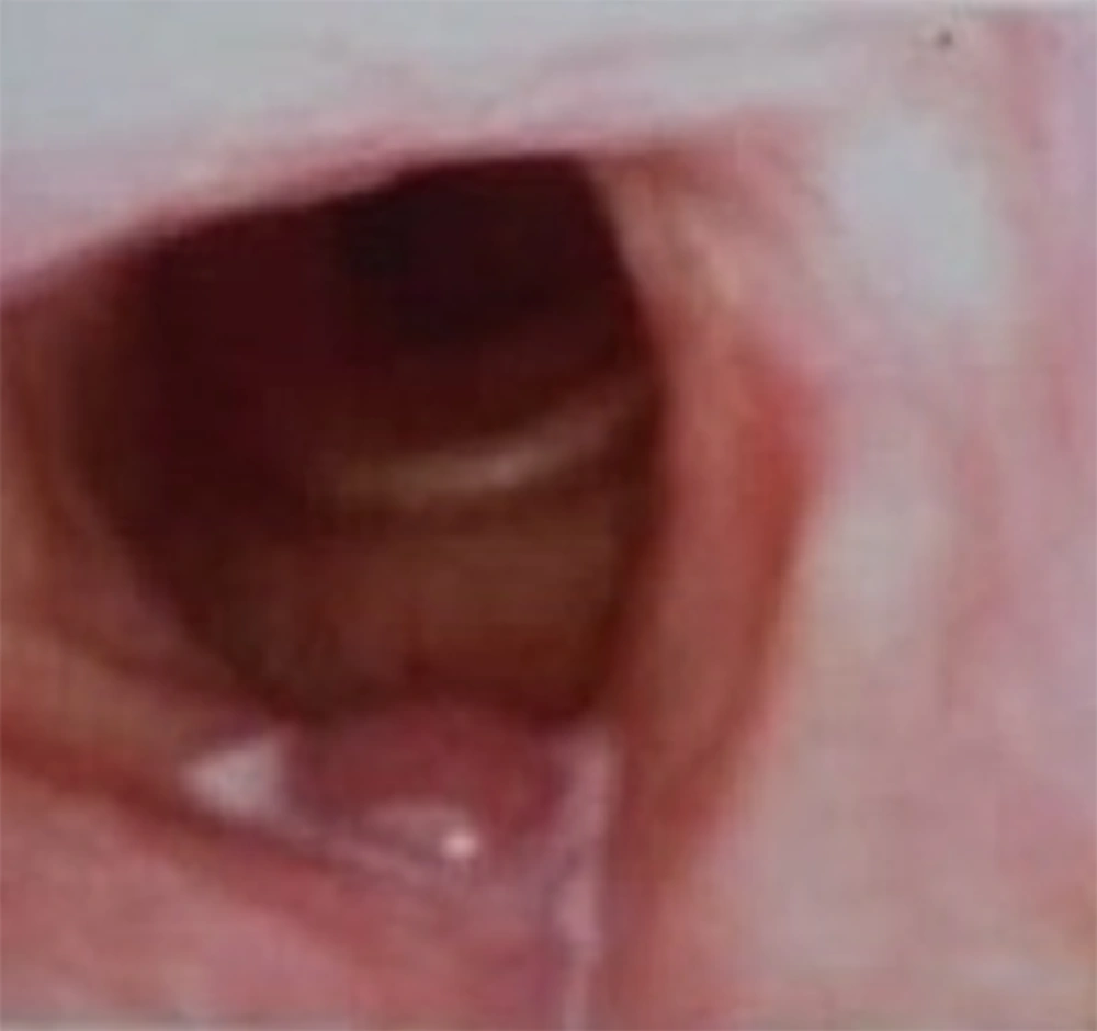
The first patient was a 61-year-old man with the chief complaint of voice hoarseness from six months prior to referral. He was a heavy smoker (150 packs per year) but did not report any history of other respiratory symptoms, medical illnesses, or surgery. Video laryngoscopy revealed a laryngeal mass in the right false vocal cord (Figure 1). Laryngeal Computerized Tomography Scan (CT scan) showed a 14 * 26 * 28 mm mass with a density equivalent to cystic lesions in the right false vocal cord, extending beyond the supraglottis to the level of the hyoid.
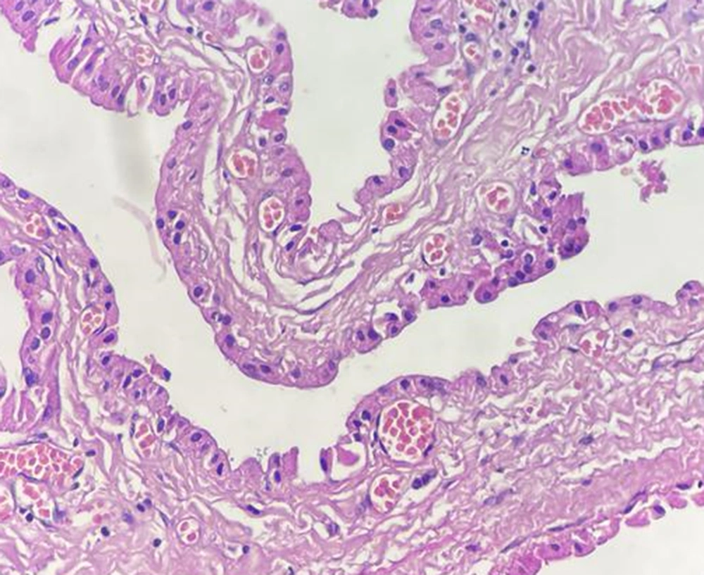
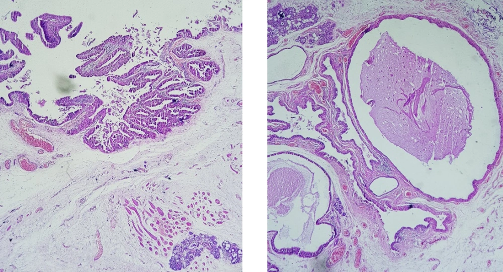
After excision, the mass appeared as a creamy-brown tissue with dimensions of 2.1 * 1.2 * 1.1 cm, a diameter of 3.2 cm, and a thickness of 1.5 cm covered by mucosa. The pathological assessment revealed the false vocal cord epithelium with seromucinous glands and multiple cysts lined by bland oncocytic columnar cells. The final diagnosis was a benign oncocytic cyst. The patient had no hoarseness or other clinical symptoms six months after the procedure (Figure 2).
2.2. Case 2
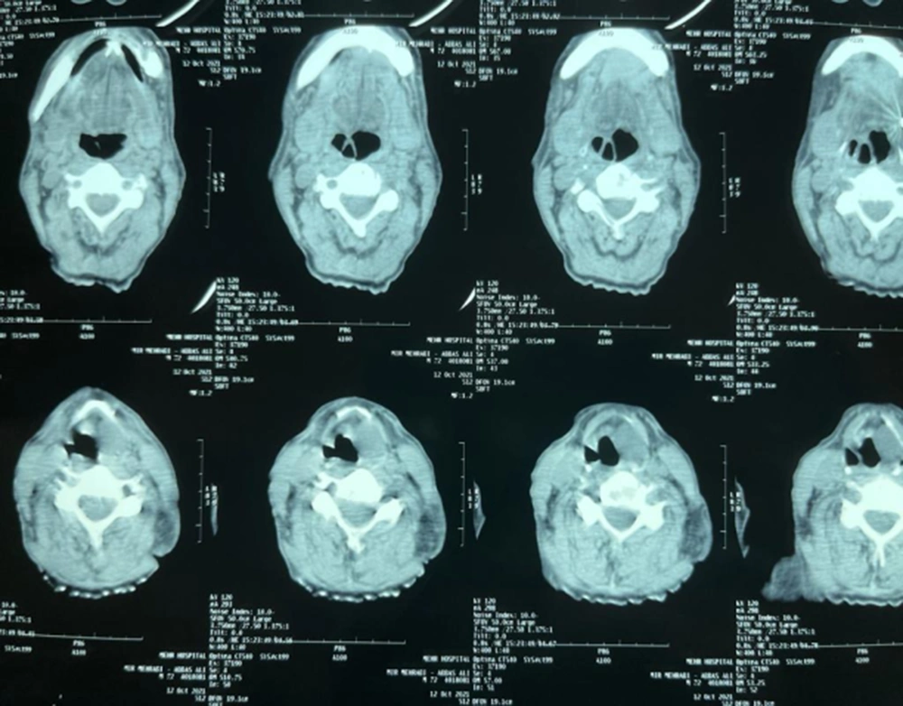
The second patient was a 76-year-old man referred due to voice hoarseness for three months. He did not report any other respiratory symptoms, past medical history, undergoing surgery, or smoking. A neck CT scan without contrast showed hypodense and homogenous foci with a dimension of 37 * 20 * 17 mm and a mass in the lateral side of the left false vocal cord. So, the radiologic diagnosis of a cyst or laryngocele was made (Figure 3). After the surgical removing the mass, the patient had a good condition with no voice hoarseness after three months of follow-up.
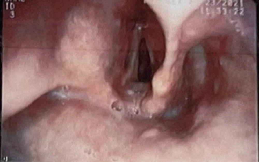
Video laryngoscopy showed three masses on the laryngeal surface of the epiglottis, the right true vocal cord, and the right false vocal cord. A biopsy was performed (Figure 4), and the histopathologic examination showed a cystic structure with papillary growth lined by bland oncocytic cells (Figure 5).
3. Discussion
Oncocytes consist of polygonal epithelial cells found in salivary glands and the lining epithelium of the respiratory tract (11). Oncocytic metaplasia in the larynx is uncommon, and oncocytic laryngeal cysts are rare, comprising less than 1% of all benign masses of the larynx (12). The oncocytic lesions of the larynx usually present as solitary lesions; however, some multifocal lesions have been described so far (3), including the second case presented in this paper.
Oncocytic metaplasia is a senile process that commonly appears in the epithelial endocrine cells possessing a high metabolic activity (13). Oncocytic cysts usually occur in ages higher than 50 years, unlike other laryngeal cysts that involve younger individuals (3).
Smoking predisposes to oncocytic lesions of the larynx due to increased oxidative stress and laryngeal inflammation. A long history of heavy smoking is reported in most patients with these lesions (14, 15), including the first case presented here. Consistently, Mettias et al. reported an oncocytic cyst in the vocal cord of a 62-year-old man with a history of smoking 20 cigarettes per day for 50 years (16). Nevertheless, the second patient described in the present paper declared no history of smoking.
The common clinical presentation of these lesions is voice hoarseness; however, lesions of different sizes and locations can present with various symptoms (17). Erfanian et al. reported a case of severe respiratory distress due to two large masses on the false vocal cord, which was identified via video laryngoscopy. This patient had aggravating dyspnea for two years prior to diagnosis and reported dysphonia for more than one year. In physical examination, inspiratory stridor was obvious, and the masses were disclosed by fiberoptic nasotracheal intubation (18).
The management of these lesions is conservative and local excision, where endoscopic removal is considered the treatment of choice (1). Oncocytic laryngeal cysts are benign lesions, but follow-up is recommended due to the risk of recurrence, especially in patients with multiple lesions (2).
3.1. Conclusions
In this paper, we reported two men referring with prolonged voice hoarseness. Medical assessments led to the diagnosis of oncocytic cysts in their vocal cords, which were removed by video laryngoscopy. Both patients were followed up after removing their laryngeal cysts and had no clinical symptoms since then.
