1. Introduction
Sarcoidosis is a multisystem inflammatory disorder characterized by non-caseating granulomatous inflammation in affected tissues. Presentation may vary with age. In adults over 90% of subjects will have lung involvement. Skin, lymph node, ocular, liver, neurologic and cardiac involvement occur in decreasing frequency .Children may present with non-specific symptoms such as fever, weight loss and general malaise. This is a rare case of genital sarcoidosis in a child presenting as a nodule in clitoris. To our knowledge this is a very rare presentation of this entity. Sarcoidosis of the female genital tract is rare and most commonly involves the uterus but there have been also cases reported in the ovaries, fallopian tubes, cervix and even placenta. Almost all of the cases reported before, were in adult females.
2. Case Presentation
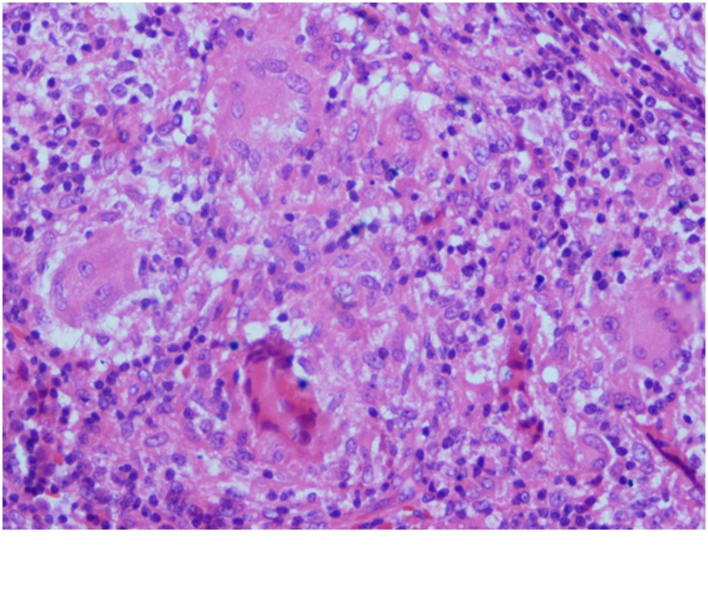
A 12 year old girl was admitted because of a large nodule in her clitoris which had been gradually developed during the last three months before admission. At the time of admission, she had a fever of 39°c, dysuria, frequency, dribbling accompanied by vaginal itching and irritation. She was not well during the last three months and she complained of weight loss, fatigue and non-productive cough. Cultures from the lesion, blood and urine were obtained and antimicrobial therapy was started for her at the day of admission. Two days later, during the course of hospitalization, her parotid glands enlarged bilaterally. Chest X-Ray was normal. Complete Blood Count indices were within normal limits for age but erythrocyte sedimentation rate was significantly elevated (71mm/ hr). A Spiral CT scan of the neck showed diffuse parotid and submandibular glands enlargement accompanied by enlarged lymph nodes in submental and posterior triangle bilaterally. Spiral CT scan of the chest revealed bilateral hilar lymphadenopathy with a non-infectious pattern of pneumonitis. Meanwhile, tissue specimens were obtained from both lesions of the genital and parotid gland. Pathologic reports were indicative of “groups of epithelial cells and epithelioid histiocytes in favor of granulomatous inflammation most likely sarcoidosis” (Figure 1). The patient was also evaluated for other conditions associated with granulomatous inflammation including tuberculosis, chronic granulomatous disease, fungal infections in addition to sarcoidosis. A complete work-up for tuberculosis including PPD, PCR and culture of sputum, broncho –alveolar lavage and gastric lavage fluids were performed to detect acid fast bacilli, which all were negative. Dihydrorhodamine dye test was within normal limits, which was against the diagnosis of chronic granulomatous disorder. Absence of primary disorders of cellular immunity, and eosinophilia, and the lack of any positive culture and histopathologic findings in favor of fungal infections ruled out fungal infections such as coccidioidomycosis.
Serum and urine calcium levels were normal. Angiotensin converting enzyme was significantly increased (125 IU/ ml with an upper limit of normal of 65 IU/ ml). Pulmonary function tests were normal.
The patient was treated with prednisolone with an impression of sarcoidosis. A favorable response was observed in follow-up visits with disappearance of the clitoris nodule, normalization of erythrocyte sedimentation rate and angiotensin converting enzyme level and improvement of respiratory symptoms.
3. Discussion
Sarcoidosis is a chronic multisystem disorder of unknown etiology affecting mostly young adults and rarely children. Pediatric sarcoidosis encompasses a spectrum of childhood granulomatous inflammatory conditions with a hallmark of non-caseating epithelioid giant cell granulomas in a variety of tissues and organ systems (1). Sarcoidosis presents in patients between 10 and 40 years of age in 70-90 percent of cases. Symptomatic sarcoidosis is rare in children (2). Children between the ages of 8 to 15 develop multisystem disorder similar to adults (3). Granulomatous lesions may occur in any organ but the lungs, lymph nodes, eyes, skin and liver are the most commonly involved. Occasionally joints, bone, spleen, central nervous system, heart or kidneys are involved. Sarcoidosis can involve the reproductive system in male and female. Testes involvement may be seen and must be differentiated from testicular cancer and tuberculosis. Also, recurrent epididymitis due to sarcoidosis rarely can occur. Sarcoidosis rarely affects the female genital tract (3-5) with majority of cases involving ovaries, fallopian tubes, cervix and vagina (6). It is very crucial that before diagnosing the patient with genital tract sarcoidosis, more obscure causes of granulomatous inflammation are ruled out including coccidioidomycosis, lymphogranuloma inguinale, foreign body reaction and tuberculosis (6). Genital sarcoidosis may cause urinary problems and in this regard may represent a therapeutic challenge (7) as was seen in our patient. We reviewed the literature to find the rate of the occurrence of the disease in female genital tract. Most of the cases were in adults. The first reported case of vaginal involvement with a proven biopsy-confirmed sarcoidosis was in a woman whose symptoms of gynecologic sarcoidosis occurred at the time of initial presentation of her pulmonary sarcoidosis (8). There were also reports of sarcoidosis presenting as an intraperitoneal mass (9) and recurrent tubo-ovarian abscesses (6).
