1. Background
Multiple sclerosis (MS) is a disorder in which the central nervous system of the body is affected. In MS, the myelin sheath is assaulted by the T cells of the body’s own immune system and the nerve cell fibers are not protected (1). Multiple patches of hard and scarred tissue, the plaques or lesions, are the last symptoms of this process which are called demyelination (2). The common sings of MS include spasticity, pain, fatigue, and sexual, bladder, and cognitive dysfunctions (3).
The most momentous purpose for which MS is treated, is the prevention of permanent neurologic disability. Different ways are introduced for MS therapy. The symptomatic therapy takes advantage of corticosteroids which can treat acute relapses of MS (4). Beta-interferons including Avonex, Betaseron, Extavia, and Rebif is a new way (5). Natalizumab and alemtuzumab are monoclonal antibodies (mAbs) which can be used for the treatment of MS. Monoclonal antibodies are beneficial for MS treatment, but have limited applications in clinics because of their immunogenicity and induction of human anti-mouse antibody (HAMA) reactions (6). High-dose intravenous steroid infusion has some contrary effects such as hyperglycemia, gastrointestinal intolerance, psychiatric manifestations, and euphoria or depression (7). Therapy with beta-interferon can expose multiple adverse reactions. So, the use of this drug is restricted due to its side effects including anaphylactic shock, thrombotic-thrombocytopenic purpura, and capillary leak syndrome (8). New treatments like targeted therapy have better results. The evaluation of the targeted therapy of NgR1 indicates some promising ways for MS treatment. NgR1 is a mediator of myelin-associated inhibition (9). Binding to the neuronal Nogo-66 receptor (NgR1) and preventing the regeneration of the “neuronal” after injury are the functions of three proteins in CNS, ie, Oligodendrocyte Myelin glycoprotein (OMgp), Myelin-Associated Glycoprotein (MAG), and Nogo-A. Lessening the prohibition and improving the recovery of the injury may be the effects of neutralizing the interactions between the inhibitory ligands and NgR1. Antibodies that neutralize ligand/receptor interactions might have therapeutic value (10).
Monoclonal antibodies, due to the fact that they are very specific and potent immunosuppressive parts, are considerably more advantageous than the other MS treatments. In the last decade, the first verified mAb for relapsing MS therapy was Anti-a4 integrin natalizumab (11). The mAbs are used to change the treatment strategies from non-specific to specific. The mAbs have the ability to revolutionize therapeutic concepts of the treatments of immune-mediated diseases, thus they are useful in this area (12). Looking for new therapeutic agents is still a crucial mission, because the difficulties of the production and the use of monoclonal antibodies have limited their applications (13).
Smaller single-gene encoded recombinant antibody fragments are produced, thus the main regions which take part in the recognition and binding of antigen include the variable regions of heavy (VH) and light (VL) chains which are linked by a short peptide linker. The obtained antibody, named scFv, can be selected against a proper epitope and the mono-specific scFvs can be isolated (14-19). They can be a good alternative to the common therapeutic drugs in human disease therapy because of their unique properties like human origin, high connection and better tissue penetration, low molecular weight, and high physical and chemical stability (20-25).
In this study, a novel immunodominant epitope of Ngr1 is designed and phage display technology is utilized to select specific scFvs against the epitope. The evaluation of the selected antibodies is performed in ELISA.
2. Objectives
The objectives of this study were to select an immunodominant epitope of NgR1, the mediator of myelin associated inhibition, using insilico method, to select specific scFvs against NgR1, and to evaluate the reactivity of the selected scFvs by ELISA.
3. Methods
3.1. The Determination of the Ngr1 Immuno-Dominant Peptide
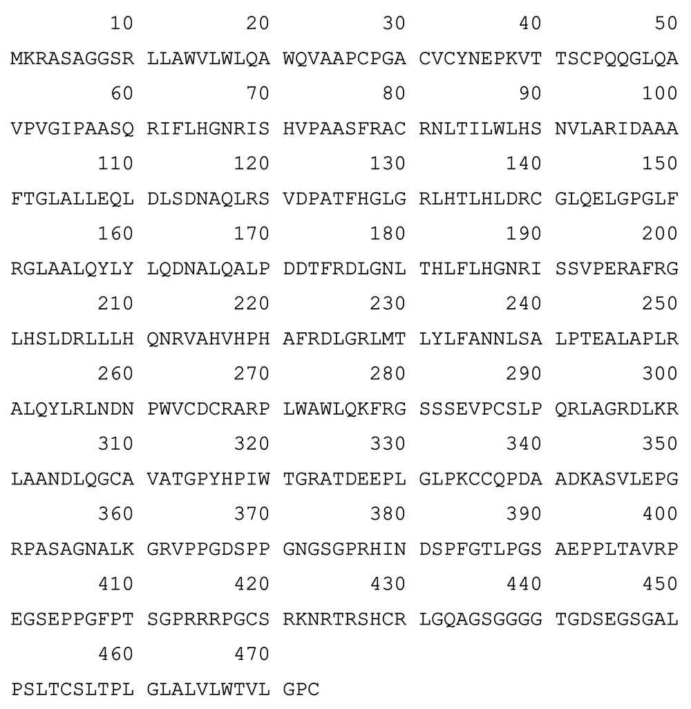
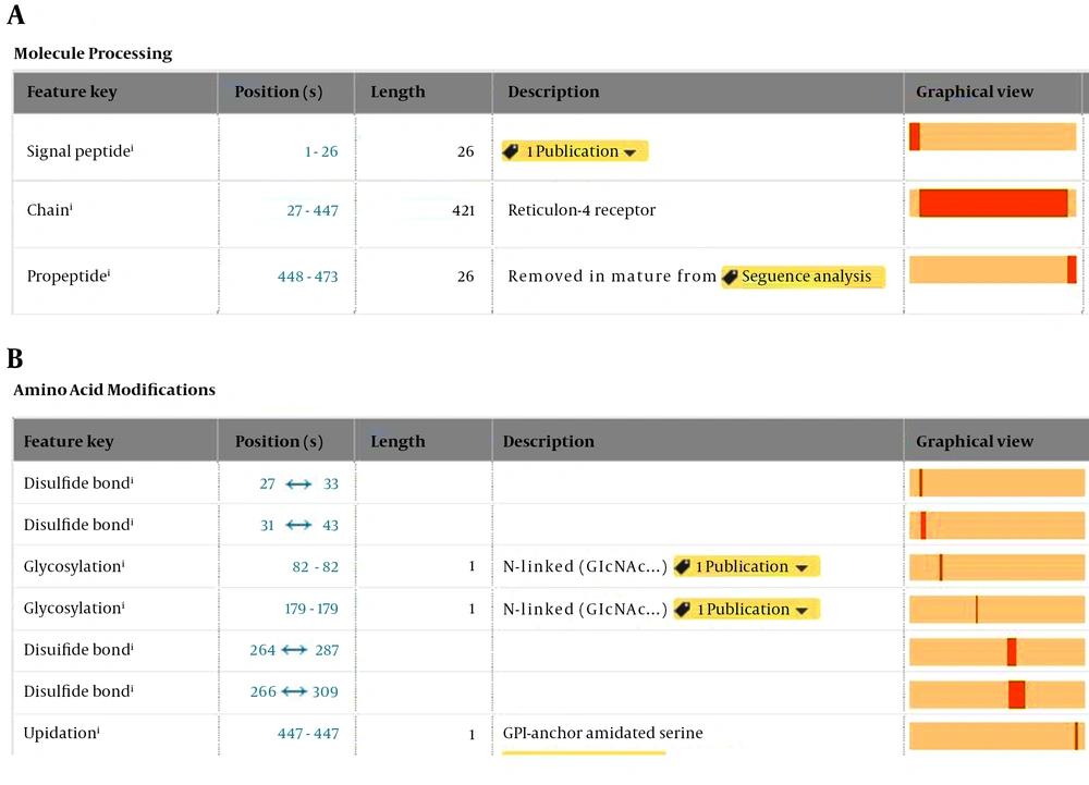
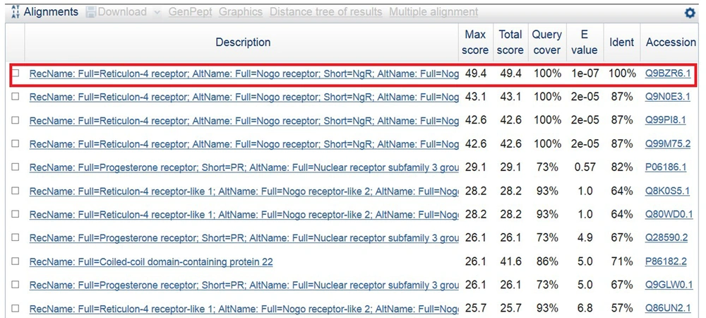
The initial molecule structure of the NgR1 is recruited using the UniProt server (26) (Figure 1). It is obvious in Figure 1 that in 473 amino acids, residues 1 to 26 can be introduced as signal sequence and cases 27 to 447 represent the main form of the cell surface NgR1 protein and are presented in Figure 2. The EpiC web server took the amino acid sequence of the extracellular region of the NgR1 receptor (27) and found the antigenic regions in the submitted sequence. Next, Protein Data Base (PDB) (28) was the source from which the three-dimensional structure of the target molecule was retrieved. Afterward, Chimera served as the evaluator of the tertiary structure of the modeled receptor (29). Then, a region of 15-amino acid was picked among the proposed immune-dominant sequences. The peptide sequence was blast with National Center for Biotechnology Information (NCBI) server (30). Human NgR1 (Nogo receptor or Reticulon-4 receptor) with accession number Q9BZR6, is 100% identical and 100% covers among all submitted sequences (See Figure 3).
3.2. Phage Rescue
As mentioned in a previous study, a phage antibody display library of scFv was produced (23). Culturing the Escherichia coli (E.coli) bacteria containing phagmid was performed on plates of 2TYG agar / ampicillin (ie, agar and ampicillin, yeast extract, tryptone, glucose) (Merck, Germany) at 30°C overnight. For a 60-minute period and a temperature of 37°C, the bacteria scraping and incubating was processed in 2TYG broth. After that, adding the helper phage of M13KO7 and incubating it at 37°C was the mission for 30 minutes. The culture was shaken for 30 minutes and it was centrifuged. The pellet containing bacteria was moved to a broth of 50µl/mL-kanamycin and 100µg/mL-2TY-ampicillin, then shaken at 30°C all night long. The culture was centrifuged and stored after passing the supernatant through some 0.2 µm filters (at 4°C).
3.3. Panning
Polystyrene immune-tubes (Nunc, Denmark) were coated at 4°C by peptide (QAVPVGIPAASQRIF) with a concentration of 10µg/ml overnight. Phosphate buffered saline (PBS) was used to wash the tubes for four times. After that, 2% skimmed milk as a blocking solution was added at 37°C and incubated for 120 minutes. The next level was a four-time washing of tubes with PBS/Tween20 and then PBS. The blocking solution’s phage supernatant was thinned (1/1) and poured on the tubes and incubated for 60 minutes with random inversions at 25°C. The logarithmic phase E. coli was added to the tube after it was washed, then it was incubated and centrifuged at 37°C for one hour. Re-suspending the bacterial pellet in a broth of 2TY (yeast extract, tryptone) of Merck, Germany, plating it on a plate of 2TY agarose/ampicillin and incubating it overnight at 30°C was the next level. Selecting the specific scFv antibodies against the epitope lasted for four rounds of panning.
3.4. DNA Fingerprinting of the Selected Clones
PCR is used to amplify the insertion of the selected clones (first minute 94°C, 55°C for the second minute, and 72°C for the next two minutes afterwards- a 30-cycle performance-). The evaluation of the fingerprinting patterns was performed by mixing the restriction enzyme (1µL of Mva-I) and restriction enzyme buffer with (2µL) PCR product (17µL). For two hours, the final mixtures were put in a dry block heater at 37°C and were run on a 2% agarose gel.
3.5. Phage ELISA
96 wells of a polystyrene plate were coated by peptide (100 µg/ mL) as an epitope and were incubated at 4°C overnight. Each well was enriched with a 2% skimmed milk as blocking solution and was incubated for two hours (in 37°C) after washing with PBS. PBS/Tween20 and PBS was used to wash the wells. Phage rescue supernatant was thinned by blocking solution. Next, it was added to each well and incubated at room for two hours at 25°C. Triple washing by PBS/Tween20 and another triple by PBS was done in order to take the non-bound phages out. At the next level, 1:100 Anti-Fd bacteriophage (Sigma, Germany) was the material which was added to each well and then the incubation was performed at 25°C for 90 minutes. Each well was enriched by secondary antibody, HRP-conjugated anti-rabbit (1:1000) (Sigma, Germany) which was incubated at 25°C for sixty minutes. After washing the wells, a solution of 10 mg of Azino-bis-3-ethylbenzothiazoline-6-sulfonic acid (ABTS) (Sigma, Germany) mixed with 20 mL citrate buffer PH4 and 6 mL H2O2 was added to each well. After 30 minutes, an ELISA reader was used to read the amount of the absorbance at 405 nm. The negative controls of this test include the wells without peptide, with unrelated scFv or scFv to HER2, M13KO7 and unrelated peptide -named prostate stem cell antigen peptide. In order to obtain more accurate results, the test was established three times and the average amounts of the absorbance and controls were calculated.
4. Results
4.1. The Selection of NgR1 - Immunodominant Epitope
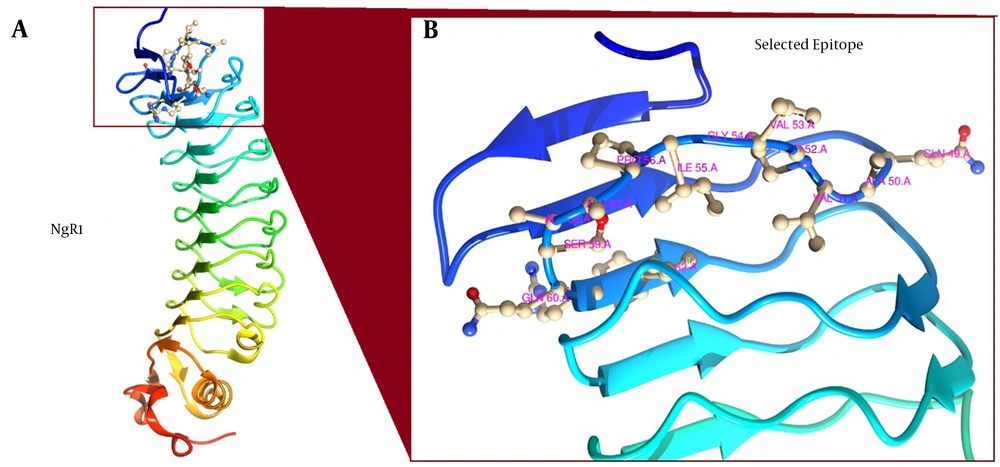
In this work, the sequence of Gln49 Ala50 Val51 Pro52 Val53 Gly54 Ile55 Pro56 Ala57 Ala58 Ser59 Gln60 Arg61 Ile62 Phe63 was selected as an immunodominant epitope and was used for the isolation of specific scFv antibody (Figure 4).
4.2. PCR and DNA Fingerprinting
The PCR and DNA Fingerprinting of 20 clones after the panning process are presented in Figures 5 and 6, respectively. For all the clones, the 950 bp band (VH-linker-VL) was obtained (Figure 5). Two Prevailing fingerprinting patterns were detected, ie, Pattern 1 (Lanes 1, 4, 6, 7, 8, 10, 11, 13, 15, 17, 20), scFv1 with frequency of 55% and pattern 2 (lanes 2, 3, 5, 9, 12, 14, 16, 18, 19), scFv2 with frequency of 45%. One clone from each pattern was selected to use in further investigations.
4.3. Phage Enzyme Linked Immunosorbent Assay
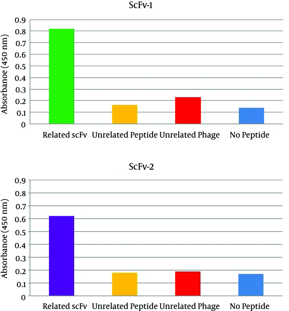
In order to observe the specific bindings of the anti-Ngr1 scFv antibodies to the related peptide, the phage ELISA was used. The absorbances obtained from the reaction of scFv1 and scFv2 with the corresponding peptide were 0.81 and 0.62 versus no peptide wells absorbances which were 0.10 and 0.14, respectively. The ODs at 405 nm are shown in Figure 7.
5. Discussion
The methods to treat the MS include modalities that want to decrease the regulation of the different immune elements involved in the immunologic cascade. The immunotherapeutic modalities are divided into two groups: the long-term treatments which want to stop the appearance of the relapses and the disease improvement in disability and those influencing the disease relapses (31). The immunotherapy aims differ from the disease clinical stage. It include (1) stopping the chronic progressive disease development from a relapsing-remitting course; (2) diminishing the relapses’ harshness or their number; (3) for patients who have chronic progressive diseases, decreasing further progression of the disorder; and (4) improving the recovery. Interferon beta-1b is a more specific immunotherapy since it diminishes the number and the roughness of the relapse (32). New era in Multiple Sclerosis therapy was performed as “targeted therapy” in which the molecular pathways of demyelination are handled by linking highly specific antibodies to the selected antigens. Multiple monoclonal antibodies have been made to target NgR1. The monoclonal antibody 7E11 prevents oligodendrocyte myelin glycoprotein myelin-associated glycoprotein and Nogo to bind with NgR1 (33). The applications of monoclonal antibodies are restricted in practice since they have many drawbacks such as having high expenses, being time consuming in production, not making the highest affinity against the target, having big size and less penetration, and having non-human parts. Yet, HAMA response takes place, even though some humanized monoclonal antibodies are made, and this is in relation to the complementarity defining the unchangeable regions part of the antibody. Single chain antibodies are very advantageous over the monoclonal antibodies. Thus, they are used in plenty of studies in order to block cancer targets such as: CEACAM1, PSCA, HER2, HER3, IL25 receptor, and MUC18 (18, 34-37). These antibodies are also recommended for some neurodegenerative diseases such as Huntington’s, Parkinson’s, Alzheimer’s, and prion disease to aim to unusual proteins or different protein aggregate forms such as SNCA, Htt, PrP proteins and Aβ (38).
In this study two specific single-chain antibodies were selected against an immunodominant portion of NgR1 antigen, an antigenic epitope (QAVPVGIPAASQRIF). The selection of this antigenic region on NgR1 receptor was performed by utilizing Insilco techniques. After the investigation of the NgR1 structure, the EpiC program was used to get a suitable sequence as epitope. Lots of parameters were assumed for defining the epitope including the experiment type, the state of the protein (Native or Denature), subcellular localization, and antibody type. A linear immune dominant epitope with 15-long residues was selected among all the presented sequences, according to accessibility, lack of any glycosylation or any other hetero atom derivatives and other parameters. After that, finding the analogy between the desired sequence and other sequences of the unrelated proteins, the blasting of the selected sequence against the protein reference sequences was performed on NCBI web server. Unlike the original protein- human NgR1 receptor- the selected epitope showed the least identity to the other proteins, but there are two exceptions, ie, mouse and rat NgR receptors (87% identity). This identity among human, mouse, and rat sequences would be very useful in future due to the importance of in vivo studies.
The results of the panning process demonstrated two specific antibodies, scFv 1 and scFv 2 which were selected with frequencies of 55% and 45%, respectively. Plenty of studies have shown the selection of specific scFvs against different targets in neurodegenerative disorders using panning process. In the most neurodegenerative diseases like Huntington’s, Parkinson’s, Creutzfeldt-Jakob, and Alzheimer’s (AD) disease, the insoluble filamentous mass is aggregated. There are general pathways suggested by these pathologies which implicate protein accumulation and could be the reason for the formation of fibril and amyloid plaques. A verified aim for AD treatment is a derived peptide from the proteolysis of the amyloid precursor protein called 4 kDa Aβ (39 - 43 amino acids). Using panning process led to the selection of scFvs against 4 kDa Aβ peptide (39 - 43 amino acids) (39). Prion diseases are introduced by PrPSc deposition, an unusual shape of the PrPC. An increasing piece of testimony declares that the antibodies to PrPC can oppose PrPSc. ScFvs against PrPC have been selected to interfere with PrPSc deposits (40). Parkinson’s and Huntington’s disease are both the disorders of neurodegeneration which occur, at least in part, because of the accumulation and misfolding of a-synuclein and Huntingtin (htt). ScFv against oligomeric a-synuclein was suggested to verify the differences and the similarities of the toxic mechanisms of a-synuclein and htt to accumulation. Panning process was used to select scFv against oligomeric a-synuclein (41). Using sequential proteolytic cleavage of APP -firstly β-secretase, secondly γ-secretase- the amyloid precursor protein (APP) is the originator of the Amyloid-β (Aβ) peptide. The supreme enzyme - a therapeutic target for the treatment of Alzheimer’s disease - which causes β-secretase processing of APP a β-Site APP is the cleaving enzyme-1 (BACE-1). Stopping the activity of BACE-1 is very beneficial in therapeutic fields. With its important physiological activity, it also separates many other substrates. Panning process suggested ScFv, since it forbids the activity of BACE-1 toward APP by means of binding the APP substrate at the proteolytic site (42). The panning results were verified by ELISA in this study. In ELISA results, it is shown that the reactivity of the two selected scFvs was significantly higher than the negative control, no peptide well. Besides the antibody controls, unrelated scFv and M13KO7 had no reactivity with the epitope. The unrelated scFv did not react with the NgR1 peptide. The absorbance of peptide-coated wells for the selected scFv 1 and scFv 2 were 8.1 and 4.4 folds higher than those of the non-coated wells. Thathaisong et al. (43) reported a positive ELISA when the optical density (OD) of specific scFvs against H5N1 influenza virus was two times higher than the assay’s negative controls. The selected scFvs did not react with the unrelated peptide which shows the specific reaction of the selected scFvs with the corresponding peptide. Phage ELISA was previously used to investigate the specificity of the antibodies for the targeted epitopes. Utilizing panning of a phage library, Esra et al. isolated phage clones against the staphylococcal enterotoxin B (SEB). The specificity of the selected clones is verified by phage-ELISA method (44). Phage ELISA showed the specificity of scFv antibodies in using human scFv antibodies against hepatocellular carcinoma (45). The selected specific anti-NgR1 scFvs, recommend new parts with high impression in targeted therapy. These antibodies not only stop producing HAMA response, but also can be manipulated and used for drug-delivery systems, radioisotopes, toxins, and enzymes. Moreover, they could also interfere with the axonal growth inhibition by binding to extracellular portion of NgR1 and would have an effect in immunotherapy of Multiple Sclerosis treatment. Much more evaluations are needed to examine these antibodies’ effects in vivo and in vitro to develop these functions.
