1. Background
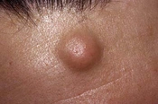
Epidermal cysts are mostly benign tumors with a dome-like appearance that are usually observed in the body, behind the ears, and in the cervical area (1). Operative management is mostly adapted to treat the affection that usually ends up scar formation (2-5). Attachment of the apical part of an epidermal cyst is to the dermal layer of cutaneous and the rest of the cyst is placed immediately under the skin with a loose attachment to the subcutaneous.
This affection is also called sebaceous cyst and includes epidermal cyst, keratin cyst, epithelial cyst, and epidermoid cyst. These cysts originate from a ruptured pilosebaceous follicle associated with acne. The obstructed duct of the sebaceous gland in the hair follicle is turned into a narrow and lengthened canal that finds a way to the surface of the cutaneous (1). These cysts also originate from exertion of trauma to the surface of cutaneous or a developmental defect of the sebaceous duct. The contents of the cysts include keratin and lipids and because of the decomposition and bacterial infection of these contents, they become odorous. They are ruptured spontaneously and a doughy discharge appears on the cutaneous (1). This affection ends up a severe inflammation and subsequent scar formation results in a complication in the surgical management of the affection (1).
To the best of the authors’ knowledge, the literature is poor regarding the comparison of the long-term surgical management of the minimal excision technique and elliptical excision technique in a prospective, randomized study.
2. Objectives
The aim of the study was to compare the results of surgery of minimal excision technique and elliptical excision technique in the surgical management of epidermal cysts.
3. Methods
Spontaneous rupture of the epidermal cysts could be due to an infection. Surgical removal of the cyst takes time and suturing is needed to close the defect (6). One of the successful and less invasive methods is the minimal excision technique. In this method, a 2- to 3-mm incision is made and the cyst contents and wall are expelled out. In this method, fingers are pressed downward a side of the lesion to make the wall loose and then the sac is removed easily. The resultant cutaneous defect could be closed using one stitch. After application of compression, a sterile tampon is placed on the resulted wound (1). Adoption of a minimal excision operation as a technique to expel out the cysts has been reported by others (1, 7). Overall, this technique is accompanied by advantages like the simplicity of the method, less scar formation, and accelerated wound healing time.
3.1. Patients
During June 2012 to September 2015, 356 patients (18 to 78 years of age) with non-infected epidermal cysts were enrolled in the present investigation. The patients had informed consent to enter the study. Exclusion criteria included patients with cysts larger than 3 cm, infected or inflamed cysts, recurrent cysts, cysts suspected to malignancy, uncertain diagnosis, cysts located in the forehead and not followed up patients. The patients were randomized into two odd and even numbers. Those with the odd number received minimal excision operation and those with the even number received the conventional method.
3.2. Surgical Procedures
All the surgical operations were done by the first author. The minimal excision operation was done based on a method described by others (1). Briefly, the skin overlying the site was prepped and anesthetized with 1 percent lidocaine without epinephrine. A stab incision (3 - 5 mm) was made on the central part of the cyst. A hemostat was inserted into the cyst and the tips of the hemostat were opened. Then, with the application of compression, the cyst contents were expelled out via the opening. Following the removal of the hemostat, the surgeon used his thumbs to expel out the cyst contents. If required, the hemostat could again be inserted to help with the discharge of the materials. After forceful and complete discharge of the contents, the capsule of the bottom of the cyst was expelled out using hemostat. The whole membrane of the cyst was taken out via the opening. Finally, the surgeon inspected the wound to make sure that the whole wall of the cyst was taken out. Using a sterile tampon and with direct pressure, the wound was compressed. Then, a topical antibiotic ointment was put on the wound and the patient was asked to hold direct pressure for a while along with tampon.
The conventional elliptical excision was also done based on a method described by others (6). The surgical preparation and anesthesia were performed as the same as minimal excision technique. However, the wound was closed using sutures. Based on the volume of the cyst and cutaneous tension line, an elliptical excision was made. The major axis of the excision was as small as possible to achieve the optimum cosmetic result. Patient’s data records and follow-up, age and gender of the patient, time of operation, date of the surgical procedures, place and the original volume of the cyst, and the length of the sutured wound were recorded. After a 24-month follow-up, all 356 patients were contacted by a phone call. Data gathered by phone call were recurrence and presence of any complications.
3.3. Statistical Analysis
Shapiro-Wilk test was used to check the normality of data. SPSS software (version 11.0, SPSS, Chicago, IL. USA) was used for statistical analyses. Two-sided p values were taken by Student’s t-tests to reveal the difference in the original volume of the cyst, length of the wound, and operative time between the groups. P values less than 0.05 were considered significant.
4. Results
354 randomized patients with age range of 18 to 78 were assigned to two equal groups of the minimal and elliptical excision groups (n = 178). The minimal excision group included 80 males and 98 females. The elliptical excision group included 85 males and 93 females. If the cyst was not ruptured or inflamed, the place of the cyst did not affect the selection of the case. Table 1 shows our findings when both groups were compared. The mean original size of the cysts in the minimal excision group was 1.5 ± 0.70. The mean original size in the elliptical excision group was 1.7 ± 0.60. There was no statistically significant difference in the original sizes between both groups. The mean length of wounds in the minimal excision group was 2.4 ± 0.50 cm, and the wound length in the elliptical excision group was 2.6 ± 0.40 cm (P = 0.001). The mean time of operation for minimal excision was 6.0 ± 2.00 minutes that was significantly shorter than that of the elliptical excision group (11 ± 3.00 minutes, P = 0.001).
| Patients Data | Minimal Excision Technique | Elliptical Excision Technique |
|---|---|---|
| Location of cysts | Head | Head |
| Number of patients | 178 | 178 |
| Age of patients | 45.0 ± 27.00 | 46 ± 29.00 |
| Mean size of cysts (cm) | 1.5 ± 0.70 | 1.7 ± 0.60 |
| Mean length of wound (cm) | 2.4 ± 0.05b | 2.6 ± 0.04 |
| Procedure time (min) | 6.0 ± 2.00b | 11 ± 3.00 |
| Recurrence (%) | 2.8 | 3.3 |
aData are expressed as Mean ± SD.
bResults were statistically significantly different from those obtained from elliptical excision technique (P < 0.05), Student’s t-test.
The incidence of recurrence in both techniques is shown in Table 1. The overall recurrence rate in the elliptical group was 3.3%. The recurrence rate in the minimal excision group was 2.8 % that was not significantly different from that of the elliptical group (P = 0.065).
5. Discussion
Some particular situations need to be considered in epidermal cysts that are simple lesions with multiple aspects. These cysts could be associated with cutaneous lipomas or fibromas and osteomas (1). There may be some confusions between dermoid cysts of the head and epidermoid cysts and excision of a dermoid cyst can end up a wound with intracranial communication (1). Sometimes, epidermal cysts could be considered complicated due to the association with some malignancies like basal cell and squamous cell carcinoma. Whenever solid tumors or unusual findings are encountered, standard histologic assessments should be taken into consideration (1).
Since epidermal cysts may interfere with cosmetic concerns and/or be very troublesome, the affected patients ask for surgical management of the case. It is a regular affection in daily practice and surgeons hardly ever search for novel surgical management. Nonetheless, cosmetic concerns of the patients are being increased nowadays. Therefore, minimally invasive surgical techniques for the removal of these cysts have been introduced in several kinds of literature (8-14).
The rationale to adopt minimally invasive surgical techniques is simplicity, less invasiveness, fewer bleeding, reduced scarring, and decreased healing time. However, objective measurements associated with these advantages are missing.
In the present randomized study, it was demonstrated that the minimal excision technique for the removal of epidermal cysts actually reduces the length of the wound, resulting in the improved cosmetic result, shorter time of the procedure, and decreased complication rate. The minimal excision technique is a satisfactory alternative method to excise non-infected epidermal cysts. Reduced surgical wound length could be mentioned as one of the greatest advantages of the minimal excision technique. In the present study, the mean value for the length of the wound in the minimal excision group was only 2.4 ± 0.50 cm with the greatest result not exceeding 3 cm. In the present study, regardless of the original size of the cyst, the resultant wound length from the minimal excision method did not exceed 3 cm. This is considered as a great benefit of the minimal excision technique when dealing with cysts on the areas of cosmetic concern. The surgeon in the present study did his best to minimize the size of the wounds treated by conventional excision. However, the wounds created by the conventional method were still larger than those of minimal excision, especially when excising cysts larger than 1 cm, because the long axis should be kept about two to three times the length of the short axis. The minimal excision procedure may seem more difficult and time-consuming when managing large cysts (larger than 2 cm in size). However, the procedure can still be performed smoothly with patience. In the present study, the size of the cyst did not make a difference in case selection and no conversion to conventional excision was required. When the surgical removal of 1 to 2 cm sized cysts in an area of cosmetic concern is the case, the privilege of minimal excision becomes significant. Other minimally invasive methods could also improve cosmetic results when compared to conventional excision. Carbon dioxide laser is adapted to create several openings and expel out the cystic content; however, the basis of this technique has not been well investigated. Others have reported making 2 to 3 mm openings over the cyst (13). The minimal excision method could result in a round to oval-shaped puncture for facilitated manipulation. Another advantage of the minimal excision technique in our investigation was the reduced surgery time. The required mean time for operation in the minimal excision method was significantly shorter than that of the conventional method. For those surgical interventions in which only a simple equipment is available, the minimal excision method is a very rapid procedure. Sometimes, expelling out the contents and wall of the cyst was time-consuming. However, the surgeon could save time because hemostasis and wound closure were needed. Since small openings in wounds are created in the minimal excision approach, no closure of the wound is required. The place of the cyst did not impact the selection of our cases if the cyst was not ruptured or inflamed. The recurrence rate of the minimal excision technique was 2.8 %, which was considered low. Compared to previous reports, no significant difference was observed in the recurrence rates. A recurrence rate of 0.66% by minimal excision within an18-month follow-up has been reported (13). It has also been reported that the recurrence rate using punch incision method was 3.6% by chart review and 8.3% by the further survey (14). It was reported that cysts excised from the back and/or ear had the highest recurrence rates compared to those excised from other places. It is believed that all surgical methods for the removal of cysts bear a significant risk of recurrence when the cyst wall is not completely removed.
In a few studies, less scar formation and comprehensive clinical evaluations incorporating MRI and CT imaging (due to a potential for intracranial and/or intradural extension associated with some scalp dermoids) have been proposed that need to be taken into consideration (15-17).
It should be taken into account that we included only non-ruptured and non-inflamed cysts in the present investigation. The findings of the present investigation revealed that the minimal excision method was more pleased for the excision of non-inflamed cysts. However, the application of this method to ruptured cysts remains to be further investigated.
5.1. Conclusion
This is the first randomized prospective study to statistically compare the results between conventional and minimal excision techniques for surgical management of epidermal cysts. The findings of the present study showed that the minimal excision technique resulted in superior cosmetic results while keeping less scarring. The minimal excision technique reduced the length of the postoperative scar wound regardless of the original size of the cyst. The patients with an epidermal cyst in the areas of cosmetic concern are the best candidates for this method. When properly performed, minimal excision was a satisfactory method to remove non-infected epidermal cysts.
