1. Background
Paraquat (PQT), a bipyridyl compound, is concentrated in many cells following absorption, where redox cycling, involving repetitive enzyme-mediated cycling between PQT and PQT radicals, occurs. Superoxide radical, a highly reactive oxygen species (ROS), is a by-product of this process, involved in direct cellular damage or further reaction to form other ROS and nitrite radicals (1, 2). Redox cycling uses NADPH as a major antioxidant defense. Oxidative stress induced by free radical production and NADPH depletion directly result in cell damage (via lipid peroxidation, mitochondrial dysfunction, necrosis, and apoptosis) and induce a significant secondary inflammatory response (3).
Although PQT is a neurotoxic agent in vitro, its neurotoxic effects are still unclear in humans. Exposure to PQT in laboratory animals reduces the level of brain neurotransmitters (4). In a study by Chen et al. (5), PQT destroyed immature and mature cerebellar granule neurons in rats. Moreover, Kriscenski-Perry et al. (6), reported that thermal stress and PQT in laboratory animals trigger synergistic effects in damaging spinal motor neurons. Furthermore, Niso-Santano et al. (7), showed that low concentrations of PQT (25 μM) lead to early and rapid activation of intracellular signaling cascades, which is associated with neural cell death induced by PQT.
An etiological cause of Parkinson’s disease is chronic exposure to PQT. Evidence suggests that the structure of PQT is very similar to the Parkinsonian toxin, known as MPTP (8). Dopamine transporters facilitate the access of PQT to dopaminergic neurons. Repeated doses of MPTP, similar to repeated doses of PQT, result in the reduced population of TH-positive neurons in the substantia nigra of mice (9).
The hippocampus is majorly involved in learning and memory processes. According to previous studies, PQT injection in the hippocampus leads to hippocampal cell damage and limbic seizures in rats (10). Some studies suggest that PQT causes selective neuronal damage in the hippocampal CA1 region through organotypic cultures. Irregular hippocampal cells and condensed cytoplasm are attributed to PQT treatment. In addition, the Nissl bodies reduce, while necrotic/apoptotic neurons may be identified. As a result, the Morris water maze test suggests that the response latency is prolonged significantly following PQT treatment. Moreover, ROS formation and malondialdehyde (MDA) content in the hippocampus increase significantly in mice (10, 11).
Based on previous studies, total superoxide dismutase (SOD) activity in the hippocampus decreases significantly due to PQT treatment in mice. Moreover, the energy metabolism analysis after PQT treatment showed that the level of adenosine triphosphate (ATP) reduced significantly in the hippocampus; therefore, the mitochondrial energy synthesis with neurocytes decreased. On the other hand, the level of 8-OHdG was found to increase significantly in mitochondrial DNA (mtDNA) following PQT treatment, which represents an increase in oxidative damage of mtDNA (10, 11).
Diosmin (DM), a naturally occurring flavonoid in different plants, exhibits significant antioxidant activities, besides potential anticancer, anti-inflammatory, and antiulcer activities. Some studies suggest that DM has antiapoptotic effects in injuries, such as renal damage and varicose veins (12, 13).
2. Objectives
In this study, we aimed at assessing the effects of DM on learning and memory following oral PQT administration in a rat model.
3. Methods
All chemical reagents were available with a relatively high quality. PQT and DM (purity ≥ 98%) were supplied by Sigma-Aldrich Co. (USA). MDA and total antioxidant capacity (TAC) kits were also obtained from Zellbio, Germany. Ketamine and xylazine were obtained from Alfasan, Netherland. Diosmin was purchased from Sigma St. Louis, MO, USA (PubChem Substance ID: 329756466).
3.1. Animals
Thirty-two male Wistar rats (300 ± 20 g) were supplied by the animal care facility of Zahedan University of Medical Sciences (ZAUMS) and kept in plexiglas cages (two rats per cage). The animals were housed in a windowless air-conditioned room (22 ± 2ºC; humidity, 55 ± 5%) with access to food and water ad libitum in a 12:12 light/dark cycle (lights on at 06:00 am). The Ethics Committee of ZAUMS approved this study (IR. ZAUMS. REC.1397.170).
3.2. Experimental Design
After one-week of acclimation, the rats were divided randomly into four groups: (1) control group, rats receiving normal saline as PQT solvent (4 mL/kg, i.p.) three times a day for one week; (2) PQT group, rats receiving normal saline similar to the control group, followed by PQT treatment; (3) DM group, control rats receiving DM at 100 mg/kg for one week; and (4) DM + PQT group, rats receiving DM at 100 mg/kg for one week prior to PQT treatment.
DM and PQT were dissolved in normal saline. DM was prepared in a way that the target dose could be administrated at 10 mL/kg. Regarding the bioavailability of DM, it was administered three times a day (once every eight hours) (14). Figure 1 presents the experimental scheme and measurement intervals of parameters. After drug treatment, changes were evaluated in the firing activities of HDG pyramidal neurons.
3.3. Passive Avoidance Memory
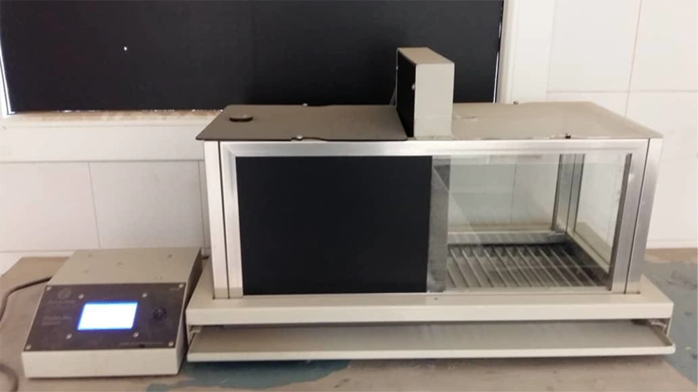
To investigate long-term memory in animals, the commonest paradigm is step-through passive avoidance. To perform this test, a protocol described previously was employed, and for this purpose an automatic commercial passive avoidance apparatus (Shuttle-box Cage, Borj Sannat- Iran) was used (Figure 2). The apparatus had two chambers with the same size (51 × 25 × 24 cm) and grid floors connected to electric current. The first chambers walls were white, and the second black. Between the chambers, a guillotine door was embedded. The test was performed in two phases as training and testing with 24 hours interval. First, an animal while facing the guillotine door was placed in the white chamber while the guillotine raised automatically; the animal was given 30 seconds time for reconciliation. Then, considering the cut-off time of 60 seconds, the animal was given the chance to enter the dark chamber. The rat was excluded on the second day if it could not move during the considered period. The guillotine door was closed and the electric current flow was connected to deliver a scrambled foot shock of 0.4 mA (20 Hz, 8.3 ms) for three seconds suddenly after the rat placed the paw inside the dark chamber. The shock intensity was determined in a way to produce the lowest jumping/vocalization response through establishing a threshold. The rat was returned to the home cage just after delivering the shock; the chambers were also cleaned. About 30 minutes before each training session held in three days, normal saline was injected intraperitoneally into the rat. The rat was returned to the home cage after each session. The procedure followed on days two and three was the same protocol used on the first day, however, a 300-second cut-off latency to enter the dark chamber was considered.
3.4. Spatial Memory Test
3.4.1. Apparatus
Spatial and learning memory test was evaluated by MWM apparatus (Technic Azma, Tabriz, Iran). A digital camera was located above the center of the water maze apparatus; thus, the animal motion is recorded. The pathway of swimming each animal was automatically recorded and calculated some parameters including path length, latency time to find the hidden platform, and time spent in goal quarter (15).
3.4.2. MWM Procedure
Briefly, animals were taught in a protocol (one session in four consecutive days and four trials in each session) with four dissimilar starting sites. Each animal was placed randomly in the water facing the wall of the tank at one of the four starting points (N, E, S, and W). During training trials, animals were permitted to swim freely until they found the hidden platform during 60 s, then, each animal remained on the platform for 30 s pending the start of the next trial. On day five, the platform was removed from the pool and the percent of time that each animal remained in the goal quadrant was recorded as a probe trial test (15, 16).
3.5. Rotarod Test
Motor performance and coordination in all groups were evaluated in the rotarod apparatus (M.T 6800, Borj Sanat Co., Tehran, Iran). The apparatus automatically recorded the time that each rat remained on the spinning wheel (75 mm diameter, 40 cm height). This procedure was performed for two days. On the first day, the rats were located on a rod (at 5 rpm for three minutes) to adapt with the apparatus. On the next day, the animals were placed on the rod with an initial constant rod speed of 5 rpm for three minutes. Afterwards, speed was increased to 40 rpm automatically (5 - 10 rpm/next 3 min, 10 - 20 rpm/next 3 min, 20 - 30 rpm/next 3 min and 30 - 40 rpm/end 3 min). The cut-off time was 15 minutes. The test session included three trials during one day. Also, the inter-trial interval was 45 minutes. Data are presented as mean retaining time on the rotating bar over three trials (16).
3.6. Determination of Hippocampal MDA Concentration
The rats were anesthetized at the end of the research with 100 mg/kg ketamine and then beheaded. The removed hippocampi were isolated and dried, weighed and then, a 5% tissue homogenate was prepared in ice-cold 0.9% NaCl solution. After centrifugation at 1000 ×g at 4ºC for 10 minutes, the supernatant was collected and stored at −70ºC until using. The MDA concentration was measured (17).
3.7. Electrophysiological Study
In anesthetized rats, SUR was determined in hippocampal pyramidal neurons in vivo. Spontaneously active pyramidal neurons were identified extracellularly in the dorsal hippocampal DG region within almost 15 minutes.
3.8. Electrical Recording from Hippocampus Dentate Gyrus
Herein, we used a single unit recording method to evaluate neuronal firing in the dentate gyrus (DG) area of hippocampus formation. Briefly, rats will be anesthetized with intraperitoneal injection of 1.5 g/kg of urethane, and an extracellular recording of a neuron is then taken by the Tungsten Microelectrode (WPI; with an extra fine tip; 1 MΩ impedance tip). The microelectrode is implanted using a manual microdrive into the DG cells according to the Paxinos stereotactic coordinates until the maximum spike activity of neurons with a signal/noise ratio of more than two isolated from the contextual noise (18). Band pass was filtered at 0.3 - 3 kHz, and digitized at 50 kHz sampling rate and 12-bit voltage resolution via a data acquisition system. The action potentials are evaluated by a window discriminator software based on the spike amplitude.
3.9. Data Analysis
Data are presented as mean ± SEM. Normal distribution of data was examined using the Kolmogorov-Smirnov test. The VCS measure was determined at different intervals using repeated measures ANOVA, while cytokine and electrophysiological data were evaluated using one-way ANOVA and Tukey’s test. The significance level was set at P < 0.05.
4. Results
4.1. DM Administration Improved Motor Coordination in Rats Treated with PQT
According to the rotarod tests, descent latencies reduced significantly in the PQT group, compared with the controls (P < 0.001); however, it increased significantly due to DM administration before PQT treatment (P < 0.001). No significant difference was found between the DM and control groups (Figure 3).
4.2. DM Increased Spatial Learning and Memory in Rats Treated with PQT
4.2.1. Probe Trial
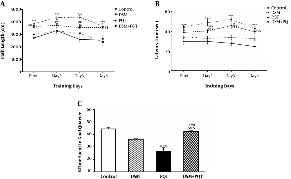
The mean latency to find the hidden platform was found to decrease during the test trial in all groups. The PQT-treated animals spent a significantly less amount of time in the platform quadrant in comparison with the controls, as shown in Figure 4 (P < 0.001). The DM treatment could not reverse this effect. Each animal’s swimming speed was recorded in order to control any differences in MWM performance. The swimming speed of animals revealed that there were differences between the groups during the MWM trial. Figure 4 presents the latency data of the experimental groups during five days of MWM test. Comparisons between the groups indicated that the PQT group took longer to find the platform than the other groups on days 3, 4, and 6 (P < 0.05).
The Effect of diosmin on A: path length, B: escape latency, C: percent of total time spent by each one of the rats at goal quarter in probe trial in the paraquat-induced memory impairments in control, diosmin, paraquat, and diosmin + paraquat groups, during spatial memory test. (***P < 0.001 vs. control, #P < 0.05, ##P < 0.01 ###P < 0.001 vs. diosmin, and $$P < 0.01, $$$P < 0.001 vs paraquat, mean ± SEM (n = 8) two-way ANOVA repeated measurements, followed by Tukey’s post hoc tests).
4.3. Effect of DM on Neuronal Firing Rate in HDG
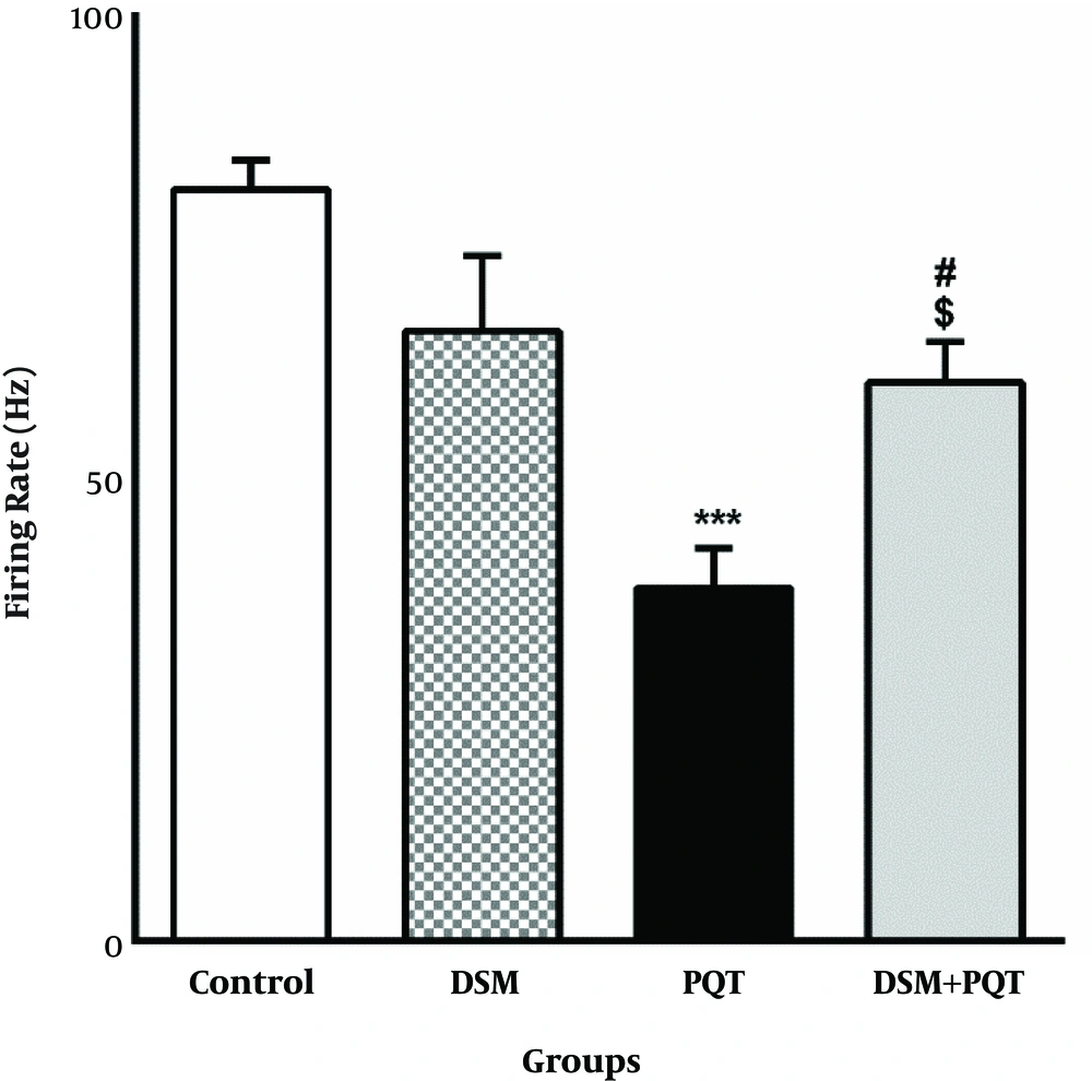
To determine the neuronal firing frequency in HDG, single-unit recordings were used. The number of spikes/bin was calculated in 1200 seconds for all the groups. Representative tracing and magnification of the sample spike of neuronal activity in granular cells are shown in Figure 5. The spike/bin count reduced in the PQT group in comparison with the control group. Based on the findings, PQT + DM treatment increased the spike rate in comparison with the PQT group. The average spike/bin count significantly reduced in the PQT group versus the controls (P < 0.001) (Figure 5). In addition, DM pretreatment significantly increased the spike rate in comparison with the PQT group (P < 0.001).
4.4. The Administration of Diosmin Decreased MDA Concentration in the PQT–induced Memory Impairment
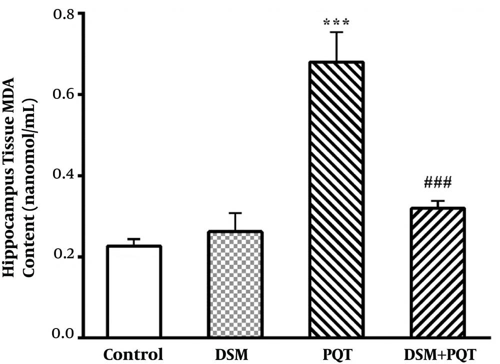
AS shown in Figure 6, hippocampal tissue MDA concentration in the PQT group was significantly increased than the control group (P < 0.001). DM treatment caused a significant decrease in MDA concentration as compared DSM + PQT with PQT group (P < 0.001).
5. Discussion
The present findings showed that intraperitoneal injection of PQT at 4 mg/kg (three times a day for one week) impaired the rats’ memory, balance, and motor performance. On the other hand, administration of DM before PQT injection improved the memory, balance, and motor coordination of rats, while reducing the MDA content of hippocampal tissues in comparison with the PQT group.
PQT preferentially affects the nigrostriatal pathway, although it is known to accumulate in the cerebral cortex and hippocampus (19). A study by Chen et al. (5), revealed that the hippocampus was damaged by PQT through oxidative stress. In addition, damage was associated with the mitochondrial dysfunction of hippocampal neurons, which is attributed to mtDNA oxidative damage; consequently, learning and memory might be affected in rats (4). In the present study, spatial memory test and passive avoidance test (PAT) were carried out for evaluating memory function. We initially used the rotarod test for evaluating motor function and determining DM potential to prevent changes in memory and learning due to PQT treatment.
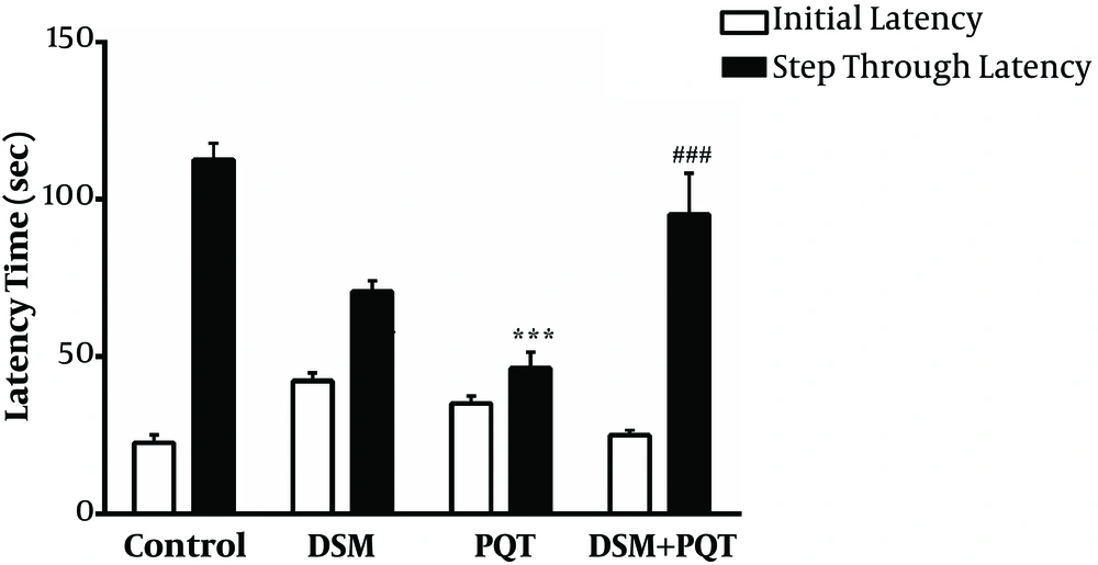
Based on our results, administration of PQT induced memory impairment in rats, which is in accordance with previous findings (4). Moreover, cognitive disorders were assessed in rats using PAT. The animals received electric shocks in the first trial as soon as they entered the dark compartment. Generally, avoidance of entrance into the dark chamber indicated the learning and memory ability of animals (20). In our study, intraperitoneal administration of PQT at 4 mg/kg three times a day for one week before the acquisition trial caused a significant reduction in retention latency and impaired learning and memory.
In order to examine memory deficits in cases of hippocampal dysfunction, spatial and learning memory tests were applied. Overall, latency time to find the hidden platform is related to the application of information stored during trail training. Consistent with previous findings, PQT treatment prior to the acquisition trial increased the latency time and caused cognitive deficits in our study (14). On the other hand, DM pretreatment caused a significant reduction in the latency time of the retention trial in the mentioned tests.
DM, with potential antioxidant, anti-inflammatory, and anti-lipid peroxidation activities, is isolated from different plants (21, 22). In previous reports, the effective role of DM in neuroprotection has been confirmed. In this regard, Sawmiller et al., reported that DM significantly decreased the level of Aβ and Aβ soluble oligomers, besides tau hyperphosphorylation in Alzheimer’s disease. Moreover, they reported that DM decreased neuroinflammation and cognitive damage in AD (23).
Furthermore, in ischemia/reperfusion damage, DM exhibits neuroprotective and antiapoptotic effects through activation of JAK2/STAT3 signaling pathway. In multiple studies, this signaling pathway has been suggested to account for the neuroprotective effects of DM (at least partly). In addition, it is involved in various neuron-specific CNS functions, including cell inflammation, proliferation, survival, and differentiation (24).
Additionally, DM may exhibit potential antiapoptotic activities in cerebral I/R rats by reducing DNA fragmentation and improving cell survival via Bax downregulation and Bcl-2 upregulation (24). Based on previous studies, DM prevents cerebral I/R by ameliorating neurological deficits, infarcted area, and brain edema by preventing venular protein leakage and post-ischemic adhesion/migration of leukocytes in mice (25). In our study, behavioral findings indicated that different DM doses could alleviate memory disorders, which is in line with previous research (13).
In conclusion, DM injection prior to PQT administration could improve the motor functions and memory (spatial and passive avoidance memory) or rats and reduce the hippocampal MDA concentration induced by PQT. However, further research is necessary to determine the neuroprotective mechanism of DM.

