1. Background
Septic arthritis is a painful condition demonstrating a microorganism invasion to the synovium. Pediatrics and geriatrics are more at risk and the knee is the most common site (1). Its incidence is more probable due to trauma (such as intra-articular injection), immunosuppressive conditions, and inflammatory disorders, including rheumatoid arthritis and tenosynovitis (2, 3). Increased prevalence could be seen in joint implantations (4). Staphylococcus aureus is the most common cause of acute bacterial arthritis, because this facultative anaerobic gram-positive coccus binds to bone sialoproteins, which is a glycoprotein found in joints (3). As antibiotic-resistant strains of S. aureus are increasing (5), choosing a suitable antibiotic to start a therapeutic protocol is challenging, especially while waiting for results of antibiogram tests (6). Also, recently increased bacterial resistance to antibiotics plus their irrefutable side effects has led worldwide health care to an alternative herbal medicine (7).
Pistacia atlantica or Báneh is a native Iranian nut, which is found more in Zagrossian area; and has considerable medical effects such as anti-atherogenic, antidiabetic, antioxidant, anti-inflammatory, anticholinesterase, antimicrobial, and antifungal activities. Antimicrobial activity of P. atlantica for some bacteria has been assessed by the disc diffusion method, and S. aureus has admissible sensitivity to it (8). In vivo and in vitro evaluations demonstrated that phenolic compounds of P. atlantica cause microorganisms’ death by damaging their memberane (9). Also, biosynthesis of silver nanoparticles (Ag-NPs) with P. atlantica illustrated considerable inhibitory effects on S. aureus (10). Gas chromatography/mass spectrometry (GC-MS) analysis has shown α-Pinene as the most ingredient of P. atlantica by significant antibacterial activity, which its minimum inhibitory concentration (MIC) and minimum bactericidal concentration (MBC) was assessed for gram-positive and gram-negative bacteria (11).
Unknown onset and rapid joint destruction in septic arthritis make it difficult to design controlled studies on human septic arthritis, but experimental studies on animal models make the possibility for critical evaluations. Mostly rabbit and intra-articular (IA) bacterial injection models have been used for septic arthritis but also rat and blood-borne models have been described, as well (12-15). Paraclinical assessments have not yet been performed using this herbal in cases of septic arthritis.
2. Objectives
Considering S. aureus as the most important etiology of nosocomial infections and septic arthritis, this experimental study was designed to evaluate the clinical and paraclinical effects of oral and IA administration of P. atlantica on septic arthritis.
3. Methods
In this experimental study, 80 adult white male Wistar rats (weighing 300 ± 50 gr) were used. The rats were housed (22ºC, 12 h light/12 h darkness) and had free access to food and water. The study was approved by the Research Ethics Committee of Shiraz University of Medical Sciences (SUMS) (IR.SUMS.REC.1397.441). Practical steps complied with the ARRIVE guidelines (16). The numbers of rats in each experimental group was determined to be 10, considering animal rights and the statistician’s consult.
3.1. Extraction of P. atlantica Nut
The P. atlantica fresh nuts (voucher herbarium specimen No. 2817) were provided from Fasa city, Fars province of Iran and dried in the shade. All the parts of P. atlantica nut, including outer soft shell, hard shell, and the kernel were powdered and then hydroalcoholic extract was catered from its powder (8). Syringe filters (0.45 μm pore size and 30 mm diameter) were used for providing sterile injectable solutions.
3.2. LD50
For LD50 determination, groups of six male rats received three doses of both forms of extract (1000, 2000, and 3000 mg/kg) and then were monitored for 48 hours in terms of behavioral changes and mortality. As the P. atlantica is a commonly used nut, the LD50 could not be determined since the rats showed no behavioral changes even mortality. Thus, it was decided to use the 1000 mg/kg dose because of its possibility to be filtered for sterilization.
3.3. Preparation of S. aureus Suspension
Staphylococcus aureus strain (PTCC1112) was purchased from the Burn Research Center of SUMS; then prepared according to the standard methods. Also, MIC and MBC values of P. atlantica extract were determined for this microorganism, and both values were compatible with previous studies (11, 17, 18).
3.4. Experiment
Eighty rats were randomly (using random number table) categorized into eight groups of ten as follows:
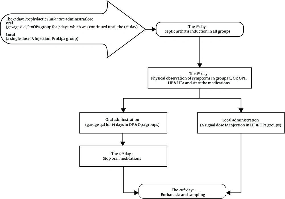
A. Septic arthritis was induced by IA injection of 100 µL of S. aureus suspension of 105 CFU/mL (104 CFU) (19, 20) into the left stifle joint of fifty rats after anesthesia and aseptic preparation. After observing the joint inflammation and lameness, the rats were categorized into five groups of ten, based on the medications:
1. Control (C): The rats received no treatment.
2. Oral Placebo (OP): The rats received daily per oral (P.O) normal saline (0.1 mL) for 14 days.
3. Local Injected Placebo (LIP): The rats received a single dose (0.1 mL) IA injection of normal saline.
4. Oral P. atlantica (OPa): The rats received daily P.O. P. atlantica extract solution (0.1 mL) for 14 days.
5. Local Injected P. atlantica (LIPa): The rats received a single dose (0.1 mL) IA injection of P. atlantica hydroalcoholic extract.
B. In order to evaluate the prophylactic effects of P. atlantica, the P.O. and IA medications started one week prior to septic arthritis induction in two groups consisting of 10 rats each:
6. Prophylactic Oral P. atlantica (ProOPa): Rats received daily P.O. P. atlantica extract solution (0.1 ml) one week prior to septic arthritis induction, and then continued receiving it for the next 14 days (totally, 21 days).
7. Prophylactic Local Injected P. atlantica (ProLIPa): The rats received a single dose (0.1 ml) IA injection of P. atlantica hydroalcoholic extract one week prior to septic arthritis induction.
Three days following the last oral treatment, blood samples were obtained from the rats on Ethylenediaminetetraacetic acid (EDTA) tubes and then they were euthanized to harvest samples of the whole stifle joint for histopathological assessments. The duration of the animal monitoring period was similar in all groups (20 days post septic arthritis induction).
C. In addition, ten normal blood samples and histopathological specimens were obtained to compare the experiment with the normal joint values:
8. Normal (N): The rats with no experimental procedure.
3.5. Complete Blood Count (CBC)
Blood samples in EDTA-containing tubes were sent to the laboratory and were analyzed by URIT-2900Vet Plus CBC counter calibrated for rat hematological parameters.
3.6. Histopathology
The hematoxylin and eosin (H&E) stained stifle samples (21) were studied by Olympus microscope BX41 in a single-blind manner by a pathologist. The following details have been surveyed (22, 23):
1. Hypertrophy and proliferation of synovial tissue and pannus formation (synovial tissue overlaying the cartilage).
2. The presence of inflammatory cells in the joint cavity and the cell types.
3. Degree of synovitis (scales 0 - 3):
0 = no sign of inflammation; 1 = mild synovial hypertrophy; 2 = moderate inflammation (hyperplasia of synovial membrane and influx of inflammatory cells to synovial tissue); 3 = marked synovial hypertrophy and inflammatory infiltrations in synovial tissue)
4. Alternative categorization of degree of sub-chondral bone and cartilage erosion (scales 0 - 3):
0 = no bone erosion; 1 = mild bone erosion; 2 = moderate bone erosion; 3 = severe bone erosion
3.7. Statistical Analysis
For CBC data, the Kruskal Wallis test (and post hoc Mann-Whitney test) and analysis of variance (and post hoc Duncan’s multiple range test) were performed for non-parametric and parametric data, respectively, based on Levene statistic test. For ordinal histopathological data, the Kruskal Wallis test (and post hoc Mann-Witney test) was done. The analyses were performed using an SPSS package (SPSS 24 for Windows, SPSS Inc, Chicago, IL, USA). P values (P < 0.05) were considered statistically significant.
4. Results
4.1. Clinical Observation
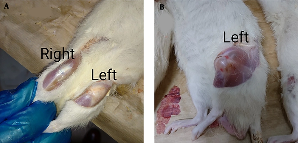
Symptoms of inflammation in the joint were observed just three days after septic arthritis induction, including swelling, pain, warmth, erythema, joint tenderness, stiffness, and lameness of the left stifle joint of rats in the experimental groups compared to the right stifle joint of the same animal as the negative control. These signs confirmed septic arthritis; thus medical treatments were initiated (Figure 1).
During twenty days of the monitoring period, only two cases of mortality were recorded in the control group. Physical examination of the left stifle joint at the end of the period revealed that medication by P.O. P. atlantica (OPa group) reduced the aforementioned symptoms (Figure 2).
Rat model of septic arthritis. (A) The two stifle joints of an animal of the OPa group, which received oral P. atlantica once a day for 14 days. The right stifle joint is the normal one without any experimental intervention (negative control); the left stifle joint is the experimentally induced septic arthritis, showing alleviation of symptoms at the end of the study with a nearly the same appearance as the right one. (B) The left stifle joint of an animal of LIPa group, which received a single dose of intra-articular injection of P. atlantica, showing no alleviation of symptoms at the end of the study with severe swelling, inflammation, and erythema in the joints.
4.2. CBC
The white blood cells (WBC) count in the control group was reduced non-significantly compared to the normal group (P = 0.172). The WBC count in all medicated groups increased compared to the normal and control groups. The results were significant compared to the control group, and non-significant as compared to the normal group, except for the OP group (P = 0.004). The highest WBC count was seen in the OP group. The closest WBC count to the normal group was observed in the OPa group (P = 0.073).
In differential WBC count, lymphocyte, monocyte, and granulocyte counts of the control group showed a non-significant decrease in comparison to the normal group (P = 0.173, P = 0.121, and P = 0.051, respectively). The lymphocyte values increased non-significantly in all experimental groups compared to the control group, except the OP group (P = 0.024). The OP group had the highest lymphocyte value. The monocyte and granulocyte values of all experimental groups significantly increased compared to the control group, except for the ProOPa group’s monocytes (P = 0.121) and the OPa group’s granulocytes (P = 0.073). These two cells in the experimental groups indicated no significant difference compared to the normal group. The ProOPa group showed the closest monocyte value to the normal group (P = 0.118). The highest monocyte value was seen in the OP group. The closest granulocytes value to the normal group was observed in the OPa (P = 0.051), LIP, and LIPa groups, which showed a non-significant difference with the normal group (P = 0.053 and P = 0.135; respectively). The highest granulocyte value was seen in the ProOPa group.
The red blood cell (RBC) count analysis dropped significantly in the control group compared to the normal group (P = 0.04). The count increased in all experimental groups was significant compared to the control group, but not significant with the normal group, except for the LIPa group (P = 0.01). LIPa group had the highest RBC count. ProLIPa group had the closest RBC count to the normal group (P = 0.806).
The same trend was observed in hemoglobin and hematocrit values, which showed a non-significant decrease in the control group compared to the normal group (P = 0.143). The increase in all experimental groups was significant compared to the control, except for the LIP and ProLIPa groups’ hemoglobin (P = 0.09 and P = 0.120; respectively). However, this difference was not significant compared with the normal group, except for the LIPa group with the highest hemoglobin and hematocrit values.
The platelets also showed a non-significant decline in the control group compared to the normal group (P = 0.306). The increased platelets in all experimental groups had significant differences with the control group. The highest platelet count was seen in the ProLIPa group. Table 1 shows the hematological values of this assay.
| Factor | Group | |||||||
|---|---|---|---|---|---|---|---|---|
| Normal | Control | OP | LIP | OPa | LIPa | ProOPa | ProLIPa | |
| WBC (× 109/L) | 7.96 ± 3.99 | 5.61 ± 3.12 | 12.75 ± 4.10b | 9.66 ± 1.42 | 9.46 ± 4.12 | 10.93 ± 2.40 | 11.47 ± 4.71 | 11.40 ± 2.59 |
| Lymphocyte | 2.24 ± 1.03 | 1.55 ± 0.98 | 3.15 ± 1.97b | 2.09 ± 0.51 | 2.04 ± 1.35 | 2.61 ± 1.46 | 2.49 ± 1.68 | 2.35 ± 0.80 |
| Monocyte | 3.66 ± 1.97 | 2.60 ± 1.46 | 5.64 ± 1.74b | 4.38 ± 0.67 | 4.48 ± 1.99 | 4.75 ± 1.15 | 3.83 ± 1.61b | 4.91 ± 1.20 |
| Granulocyte | 2.06 ± 1.20 | 1.46 ± 0.76 | 3.96 ± 1.29 | 3.19 ± 0.78 | 2.94 ± 1.01b | 3.57 ± 1.53 | 5.15 ± 2.50b | 4.14 ± 1.22 |
| RBC (× 1012/L) | 5.53 ± 2.37 | 3.61 ± 2.40 | 6.91 ± 1.01 | 6.38 ± 0.24 | 6.53 ± 0.48 | 7.74 ± 0.99b | 6.79 ± 1.07 | 6.26 ± 0.37b |
| Hemoglobin (g/dL) | 12.56 ± 5.28 | 8.12 ± 5.65 | 16.03 ± 2.10 | 14.04 ± 0.45 | 14.23 ± 1.32 | 17.02 ± 2.26b | 14.83 ± 1.92 | 13.79 ± 0.77 |
| Hematocrit (%) | 28.94 ± 12.09 | 18.80 ± 12.67 | 36.75 ± 4.84 | 32.59 ± 1.13 | 33.13 ± 2.97 | 38.66 ± 4.41b | 34.89 ± 5.04 | 32.33 ± 2.02 |
| Platelet | 202.40 ± 91.19 | 129.09 ± 97.67 | 243.80 ± 37.03 | 254.80 ± 36.71 | 239.80 ± 41.89 | 247.60 ± 39.24 | 257.70 ± 48.76 | 281.20 ± 17.84b |
Hematological Effect of P. atlantica Nut Extract on Staphylococcal Septic Arthritisa
4.3. Histopathology
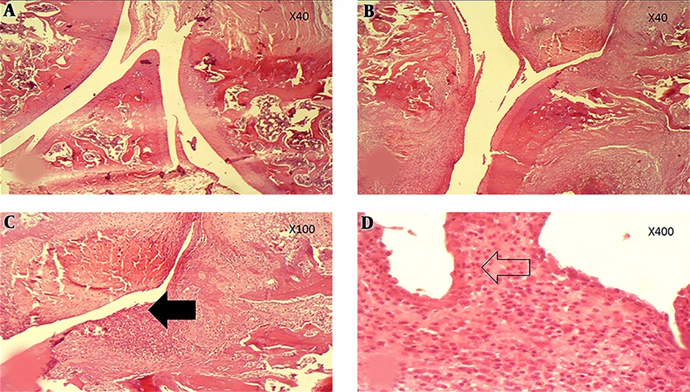
All the ordinal histopathological parameters had significantly higher scores in the control group compared to the normal group (P < 0.001). Further, placebo-receiving groups (OP and LIP) showed a significant decline in the scores compared to the control group, except for synovitis degree (P = 0.201 and P = 0.699, respectively). While all P. atlantica-receiving groups did not a significant increase in scores compared to the control group, they non-significantly differed from each other (Figure 3).
Histopathological evaluation of articular space. (A) Section shows normal articular space without any erosion or destruction in the controls (H&E, ×40). (B & C) Articular surface destruction in conjunction with cartilage loss pointed by filled arrow (H&E, ×40 & ×100) associated with acute synovial inflammation. (D) The arrow is representative of inflammatory cells involved in arthritis (H&E, ×400).
The ProOPa and ProLIPa groups revealed higher non-significant hypertrophy and proliferation of synovial tissue and pannus formation scores compared to the OPa and LIPa groups. The ProLIPa group had the highest score. Measuring the degree of synovitis, the OPa group demonstrated a non-significant reduction in comparison to the control group (P = 0.690), while other groups had a significant increase, except for the LIP group, which had a slight difference with the control group (P = 0.699).
The infiltration scores of inflammatory cells into the synovial cavity differed slightly between P. atlantica-receiving groups. These differences were not significant. Lymphocytes were one type of these cells, which decreased non-significantly in the OPa group in comparison to the control group. The ProLIPa group showed the highest significantly lymphocyte counts (P = 0.021) and then the LIPa group had the highest lymphocyte infiltration. Other cell types, such as plasma cell and polymorphonuclear granulocytes (PMN) showed non-significant minor changes compared to the control group. The ProLIPa demonstrated the highest plasma cell infiltration. Similar to the normal group, the LIPa group had the lowest non-significant PMN counts compared to the other groups (P = 0.118).
Reporting on synovial cartilage/bone erosion degree, all P. atlantica-receiving groups increased significantly compared to the control group, except the OPa group (P = 0.065). Although local injections revealed higher erosion scores, changes were not significant among P. atlantica-receiving groups. Meanwhile, placebo-receiving groups did not show a significant reduction compared to the control group. Table 2 shows the histopathological values of this assay.
| Factor | Group | |||||||
|---|---|---|---|---|---|---|---|---|
| Normal | Control | OP | LIP | OPa | LIPa | ProOPa | ProLIPa | |
| Hypertrophy of ST & Pannus Formation | 0.00 | 1.62 ± 0.51 | 0.51 ± 0.40 | 0.90 ± 0.73 | 1.80 ± 0.63 | 1.80 ± 0.63 | 2.20 ± 0.78 | 2.40 ± 0.84b |
| Inflammatory Cells Infiltration | 0.00 | 2.62 ± 0.51 | 1.00 ± 0.81 | 1.90 ± 0.87 | 2.70 ± 0.48 | 2.80 ± 0.63 | 2.80 ± 0.42 | 2.80 ± 0.42 |
| Lymphocyte | 0.00 | 1.62 ± 0.51 | 0.60 ± 0.51 | 1.10 ± 0.56 | 1.60 ± 0.51 | 1.90 ± 0.31 | 1.80 ± 0.42 | 2.50 ± 0.84b |
| Plasma cell | 0.00 | 2.00 ± 0.00 | 1.70 ± 0.48 | 1.90 ± 0.31 | 2.10 ± 0.31 | 2.00 ± 0.00 | 2.00 ± 0.00 | 2.70 ± 0.48b |
| PMN | 0.00 | 0.37 ± 0.51 | 0.30 ± 0.48 | 0.50 ± 0.52 | 0.30 ± 0.48 | 0.00 ± 0.00 | 0.20 ± 0.42 | 0.20 ± 0.42 |
| Synovitis | 0.00 | 1.87 ± 0.83 | 1.70 ± 0.48 | 2.00 ± 0.66 | 1.40 ± 0.51 | 2.80 ± 0.63b | 2.80 ± 0.42b | 2.70 ± 0.67b |
| Cartilage/Bone Erosion | 0.00 | 2.25 ± 0.46 | 2.00 ± 0.66 | 1.60 ± 0.84 | 2.40 ± 0.96 | 2.80 ± 0.63b | 2.60 ± 0.84 | 2.70 ± 0.48b |
Histopathological Effect of P. atlantica Nut Extract on Staphylococcal Septic Arthritisa
5. Discussion
In this study, synovitis appeared just three days after the IA injection of S. aureus. Evaluating clinical and paraclinical effects of P. atlantica on S. aureus septic arthritis showed that joint inflammation symptoms were present so obviously in the control group to the end of the study revealing two cases of mortality, but no mortality was recorded in the experimental rats. The final physical examination revealed that P.O. P. atlantica (OPa group) reduced gross symptoms of inflammation and lameness impressively, while inflamed joints in local injections were the same as or even worse than the control (Figure 2). The WBC and platelet counts decreased in the control group, whereas RBC count, hemoglobin, and hematocrit increased in experimental groups. Histopathological findings showed synovitis and cartilage/bone destruction, which was ameliorated by P.O. administration of P. atlantica extract.
The WBC counts illustrated a leukopenia in the control group and significant reflective leukocytosis in all experimental groups compared to the control group. Induced septic arthritis released chemotactic factors to activate leukocyte migration toward the infected/inflamed site (22, 24). The leukopenia of the control group might show the microorganisms overcome the body and depletion of reservoir leukocytes. In reflective leukocytosis, WBCs egress from bone marrow reservoir to blood flow by stress, fever, trauma, a medical agent administration, or even an edible (25). Phagocytic cells of innate immunity, as well as mast cells and platelets, will be affected by chemotaxis (24). Here, the effect of stress and synovial trauma could not be ignored since no significant differences were detected between experimental groups. Stress leukogram is due to increased endogenous/exogenous corticosteroids (25, 26). In this study, the OPa group had the most closed WBC count to the normal that may be probably attributed to the benefits of P.O. P. atlantica. Similar to WBC counts, differential counts showed lymphopenia, monocytopenia and granulocytopenia in the control group and reflective lymphocytosis, monocytosis and granulocytes in the experimental groups. Lymphocytosis indicates the involvement of acquired immunity as well as the recovery stage of an infectious disease (24, 25). So, close lymphocyte counts to the normal in the experimental groups might present improvement, especially in the OPa group ascribing to the benefits of P.O. P. atlantica. This improvement could not be achieved by oral normal saline, as the highest lymphocytosis belonged to the OP group. Mononuclear phagocytic cells eliminate pathogens via specific receptors of pathogen-associated molecular patterns (PAMPs) (24). Significant increases in monocyte count in the experimental groups indicated body reflection to medications and monocyte egression from bone marrow reservoirs to blood fellow. Strong stimulatory effect of daily gavage stress plus infected synovium was defeated by benefits of P.O. P. atlantica in the OPa and ProOPa groups; compared to the OP group which had the highest monocyte count. Oral prophylactic P. atlantica had the closest monocyte count to the normal group, possibly explaining accumulative effects of long-term P.O. P. atlantica. Peripheral neutrophils will be attracted to the infected site by chemotaxis (24). Here, all medications caused reflective agranulocytosis but P.O. P. atlantica had the closest count to the normal group, whereas the ProOPa group had the highest granulocyte count probably due to stress dominancy prior to benefits of P. atlantica on the granulocytes.
Intra-articular septic arthritis induction caused a significant RBC fall compared to the normal state, which means that the impact of the local infection was systemic and RBCs immigrated and settled down in the spleen. The RBC increment in all experimental groups explained the secretion of epinephrine/nor-epinephrine, affecting the spleen to release RBCs; thus stress might stimulate their secretion (24, 25). With this respect, RBC alterations in the experimental groups seem to be due to daily gavage stress and painful stimulation of IA injected agent. The highest RBC count in the local injection of P. atlantica after septic arthritis diagnosis (LIPa group) may be as a result of higher adrenal release from painful and erosive synovium, which remained high after 17 days, but it was alleviated after 25 days in the ProLIPa group with the closest RBC count to the normal group. Daily gavage stress was alleviated by P.O. P. atlantica benefits in the OPa group. The same trend was seen in the hemoglobin and hematocrit values.
Platelets contribute to the inflammatory response as well as other innate immune cells (24). The release of their granule ingredients results in excessive endothelial permeability and complement system activation (24). The observed thrombocytopenia in the control group suggests the consumption of platelets beside the influence of catecholamines on the clearance of platelets by the spleen. Although it was not significant, the slightly higher platelet values in IA injected groups may be due to more damage than oral groups since P.O. P. atlantica had the closest count to the normal group.
Pathological study illustrated that septic arthritis could significantly affect the histopathologic pattern of synovium, leading to synovitis followed by cartilage and bone destruction. it seems that wash out effect by normal saline had caused significant alleviation of the increased factors in placebo groups; the P. atlantica-receiving groups exacerbated the condition by affecting erosive and stimulatory conditions. This is more obvious in hypertrophy of synovial tissue/pannus formation and cartilage/bone erosion that had the highest score in the ProLIPa group. Considering the arthritogenic properties of bacterial debris and released soluble components such as lipoteichoic acid and peptidoglycan (22), it can correlate with P. atlantica antibacterial activity (11). Elimination of bacterial components, inflammatory mediators, and infiltrated immune cells and their harmful effects from synovium by normal saline, can encourage the idea. The observed synovitis indicates the involvement of innate immunity. Here, the degree of synovitis was ameliorated by P.O. P. atlantica, confirming the least macroscopic symptoms. The therapeutic P.O. and IA P. atlantica differed significantly from each other, but prophylactic P. atlantica and local therapeutic P. atlantica showed no significant differences. Increased infiltrated inflammatory cells into synovial cavity were also affected by washout effect of normal saline, while P. atlantica extract slightly deteriorated it. Acquired immunity is needed by lymphocyte infiltration for the destruction of infected cells at the site (24). Here, despite the lowest peripheral lymphocytes level of the control group, its pickup in histopathology of synovium explained the immigration of these cells apparently. Both infiltrated lymphocytes and degree of synovitis were reduced by P.O. P. atlantica, but the local IA P. atlantica showed the highest lymphocyte infiltration possibly due to more stimulation and destruction of synovium manifested by erosion scoring. In lymph nodes, B lymphocytes would change to plasma cells to form a specific immunoglobulin isotype and then leave the node (24). Here, alterations can be attributed to the same reason. The PMNs as innate immune phagocytic cells destroy pathogens via PAMPs (24). Although these infiltrated cells did not show significant alterations at any level, P. atlantica medications showed lower values, indicating the effect of P. atlantica on PAMPs.
Increased cartilage/bone erosion scoring in P. atlantica-receiving groups reinforced in mind the subsequent arthritogenic outcome of P. atlantica extract antibacterial activity, especially by a direct effect on local administration, in addition to painful stimulation and the amount of induced stress it causes. The first step of septic arthritis treatment is joint lavage or drainage to eliminate bacterial component (1, 22). In this study, the painful and stressful needle arthrotomy was avoided, but the effects of diluting and washing out of microorganisms were found in the local injection of normal saline. To find erosive nature of P. atlantica on synovial contents and other unexpectable effects (27), future studies on detecting the erosive action of P. atlantica in a non-infectious condition are suggested to differentiate possible arthritogenic properties of bacterial contents. Also, it is better to evaluate the effects of P. atlantica on serum levels of immune mediators, and biopsy of spleen and lymph nodes plus the synovial tissues for more advanced serological and immunohistochemical tests.
5.1. Conclusion
In conclusion, P.O. P. atlantica could alleviate clinical symptoms of septic arthritis.
Systemic benefits of P.O. P. atlantica could improve the body condition, but local P. atlantica could not show good systemic effects.
Histopathological findings confirmed the cellular immunity involvement and antibacterial activity of P. atlantica, encouraged by more erosion of synovial cartilage and bone due to more bacterial debris and contents release with arthritogenic properties. Meanwhile, possible stimulatory effects of P. atlantica extract on synovial content should not be ignored. The P.O. P. atlantica showed reasonable reduction in the inflammation of the synovial capsule.


