
1. Introduction
The pulmonary artery (PA) catheter is usually inserted while the pressure waveform detected from the catheter tip is continuously monitored (1). The detection of the PA wedge pressure (PAWP) indicates when the catheter tip is located in the PA. The PA blood sample is then collected through the catheter tip. The inserted length of the PA catheter varies according to parameters including the size of the cardiac chambers and the course of the PA catheter. Blood must not be sampled while the PA branch is obstructed with an inflated balloon because this may cause the PA branch to rupture (2-4). Here, we report our experience of a case in which pulmonary venous blood was unexpectedly collected via the PA catheter tip, even though the blood pressure and waveform at the PA catheter tip were typical of those in the PA.
2. Case Presentation
A 74-year-old male with the height of 162 cm and weight of 68 kg was scheduled for mitral valve plasty for cardiac failure caused by mitral valve regurgitation. An atrial septal defect had been surgically closed when the patient was 56 years old. Preoperative oral medications were digoxin, furosemide, aspirin, and warfarin. The patient also had chronic atrial fibrillation. The cardiothoracic ratio on a preoperative chest radiograph was 72%. Preoperative transthoracic echocardiography showed severe mitral regurgitation with an annulus enlargement and decreased posterior leaflet mobility, ejection fraction of 70%, estimated PA pressure of 65 mmHg, moderate pulmonary hypertension, and tricuspid regurgitation with a peak pressure gradient of 46 mmHg. There was no residual shunt at the atrial septum. Renal function was mildly reduced without electrolyte disturbance. Preoperative percutaneous oxygen saturation was 98%.
Anesthesia was induced via intravenous administration of propofol, remifentanil, rocuronium, and sevoflurane inhalation. Volume-controlled ventilation was started with a fraction of inspired oxygen (FiO2) of 0.6, tidal volume of 0.8 mL/kg, and a positive end-expiratory pressure of 0 cmH2O. A transesophageal echocardiography probe was inserted. General anesthesia was maintained with inhalation of sevoflurane, and intravenous administration of remifentanil and rocuronium.
After endotracheal intubation, a central venous catheter was inserted from the right internal jugular vein at the level of the thyroid cartilage; the inserted length was 10 cm from the site where the catheter was inserted transcutaneously. A PA catheter sheath introducer® (Edwards Lifesciences, CA, USA) was then inserted into the right internal jugular vein at the level of the cricoid cartilage. The PA was catheterized with a Swan Ganz thermodilution catheter® (Edwards Lifesciences) of size 8 Fr and length 110 cm. After a 15-cm PA catheter inserted through the sheath, the balloon was inflated with 1.5 mL of air. We slowly advanced the tip of the PA catheter forwards, while monitoring the pressure and detecting its waveform from the catheter tip. When the tip of the PA catheter showed the PA pressure waveforms, systolic pressure of 32 mmHg and diastolic of 21 mmHg following right ventricular pressure, the PAWP was 21 mmHg at the end-expiration. We then deflated the balloon and pulled the tip of the PA catheter back 3 cm so that the inserted length was 58 cm from the sheath inlet. We confirmed that the tip of the PA catheter showed a waveform typical of the PA and appropriate a and v waves, and also transesophageal echocardiography showed that the tip of the PA catheter was located in the right PA.
We collected 0.5 mL of blood through the tip of the PA catheter without inflating the catheter balloon. The blood sample had an oxygen partial pressure (PaO2) of 358.8 mmHg, carbon dioxide partial pressure (PaCO2) of 20.1 mmHg, and oxygen saturation of 99% (Table 1).
Table 1.
Parameters of Blood Sampled Through the Pulmonary Artery Catheter From Various Sites
| Inserted Length of PA Catheter, cm | 58 | 58 | - | 53 | 47 |
|---|---|---|---|---|---|
| Site of blood Sampling | Tip of PA Catheter | Opening 26 cm from the Tip | Radial Artery | Tip of the PA Catheter | Tip of the PA Catheter |
| FiO2 | 0.6 | 0.6 | 0.6 | 0.6 | 0.6 |
| pH | 7.64 | 7.48 | 7.49 | 7.59 | 7.48 |
| PaO2, mmHg | 358.8 | 37.0 | 310.0 | 340.6 | 41.6 |
| PaCO2, mmHg | 20.1 | 37.1 | 32.0 | 22.0 | 36.2 |
| SpO2, % | 99.7 | 78.2 | 100.0 | 99.7 | 84.4 |
| Base excess, mEq/L | 2.1 | 3.4 | 2.2 | 0.7 | 0.7 |
Abbreviations: FiO2, fraction of inspired oxygen; PA, pulmonary artery; PaO2, oxygen partial pressure; PaCO2, carbon dioxide partial pressure; SpO2, oxygen saturation.
Alveolar PaO2 is calculated as follows:
PaO2 = (PB - 47 mmHg) × FiO2 - PaCO2 / 0.8
Where PB: atmospheric pressure, FiO2: fraction of inspired oxygen, and PaCO2: partial pressure of carbon dioxide in arterial blood. In the present case, PaO2 was calculated as 374.5 mmHg after PB of 760 mmHg, FiO2 of 0.6, and PaCO2 of 32.0 mmHg applied to the equation. The blood sample collected from the tip of the PA catheter showed a PaO2 of 358.8 mmHg; therefore, the sample must have either been from the pulmonary vein or alveolar blood. We have never performed blood sampling from the pulmonary veins, because sampling from pulmonary veins requires obstruction of the PA branch. We thought that the tip of the PA catheter had been over-inserted into a branch of the PA and pulled the tip of the PA catheter back 5 cm, but the second blood sample showed the same results (Table 1). We then pulled the PA catheter back until the inserted length of the PA catheter was 47 cm, while watching the location of the catheter tip on X-ray fluoroscopy (Figure 1A and 1B). The blood gas testing on a sample collected through the catheter tip showed an oxygen saturation of 84.4 % and PaO2 of 41.6 mmHg (Table 1). The surgical procedure was performed without problems.
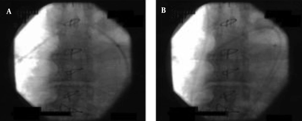
Figure 1.
A, The inserted catheter length was 53 cm; B, the inserted catheter length was 47 cm.
Postoperative chest radiography showed proper placement of the PA catheter, but a chest radiograph taken the next day showed over-insertion, although the insertion length was unchanged (Figure 2A and 2B). The PA catheter was removed from the patient. The postoperative course was uneventful, but long-term rehabilitation was required. The patient was discharged from the hospital 2 months after the surgery.
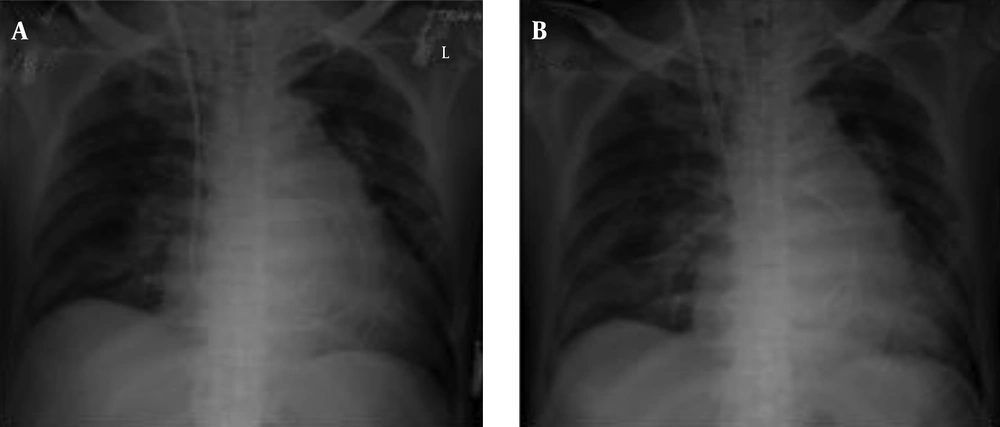
Figure 2.
A, The postoperative image showed appropriate placement of the pulmonary artery catheter; B, the image taken on postoperative day 1 showed that the catheter tip had moved peripherally.
2.1. Consent
Written informed consent was obtained from the patient for publication of this case report and any accompanying images. A copy of the written consent is available for review by the Editor-in-Chief of this journal.
3. Discussion
The PA catheter is usually inserted while continuously monitoring the pressure and its waveform from the catheter tip. After the waveform and pressures of the right atrium, right ventricle, and PA confirmed on the monitor, the PA branch can be wedged and the PAWP is shown on the monitor. After about 2 - 3 cm of the PA catheter pulled back with the balloon deflated in order to avoid obstruction of the PA branch, the PA catheter is fastened to the skin with thread and tape. Because the tip of the PA catheter is advanced under the guidance of monitoring of intracardiac blood pressures and their waveforms, the exact location of the catheter tip cannot be detected until transesophageal echocardiography or chest radiography is performed (1). In the present case, the PA catheter was inserted quickly, but over-insertion was first detected after blood testing and X-ray fluoroscopy. Blood testing could provide enough information to determine over-insertion.
Major and minor complications caused by PA catheterization include bleeding from PA, pneumothorax, ventricular arrhythmia, complete heart block, carotid and subclavian arterial puncture, catheter sepsis, endocarditis, pulmonary embolism, difficulty in catheter removal, pseudoaneurysm, thrombosis, pulmonary infarction, balloon rupture, catheter knotting, and cardiac tamponade (2-5). Factors associated with increased risk of PA injury include female sex, age above 60 years, pulmonary hypertension, improper balloon inflation, and active anticoagulation treatment (6, 7).
In the present case, the tip of the PA catheter showed PA pressure and its waveform just before blood sampling, and the balloon was not inflated when the blood was taken. The unexpected sampling results may be attributable to the followings: movement of the catheter tip to a peripheral branch of the PA because of negative pressure created when taking the blood sample, increase in left atrial pressure and backward flow of pulmonary venous blood due to severe mitral regurgitation, and/or insertion of the catheter tip into an intrapulmonary shunt (Figure 3). Although there were no obvious intrapulmonary shunts in the pre- and post-operative computed tomography images in this case, small shunts may have been presented.
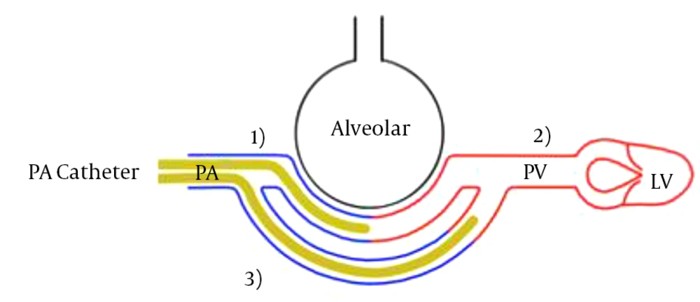
Figure 3.
1) Movement of the catheter tip to a peripheral branch of the pulmonary artery caused by negative pressure during blood sampling, 2) increase in left atrium pressure caused by severe mitral regurgitation and backflow from the pulmonary veins, and 3) insertion of the catheter into an intrapulmonary shunt. PA, pulmonary artery; PV, pulmonary vein; LV, left ventricle.
When using cardiopulmonary bypass for cardiothoracic surgery, the catheter tip may accidentally show the PAWP when weaning from cardiopulmonary bypass, because the course of the PA catheter varies and the catheter tip can easily move to a peripheral branch of the PA because of the collapse of the cardiac chambers (8). The appropriate site of the catheter tip is the hilum of the lung, and if the PAWP must be measured intra- and post-operatively, the catheter tip can be easily advanced using a cover for the PA catheter.
The use of PA catheterization has decreased recently (9); however, PA catheterization is still needed for patients with shock and patients undergoing cardiovascular surgery because it gives important information on accurate PA pressure and PAWP in patients with pulmonary hypertension (10, 11). Although the physician who inserted the PA catheter in the present case was not a pulmonary and critical care fellow, she was an anesthetic fellow with catheterization experience in over 50 cases, and also an expert cardiovascular anesthesiologist observed the catheterization. The shape of the loop of the PA catheter can vary, especially in dilated cardiac chambers. If the loop is small, the tip can be over-inserted. Thus, the catheter tip can move easily, and over-insertion must be avoided by pulling the catheter back several centimeters at catheterization.
3.1. Conclusions
We experienced a case in which pulmonary venous blood was unexpectedly sampled through the PA catheter tip, even though the blood pressure at the PA catheter tip showed PA pressure. This indicates that the PA catheter tip can easily advance into a peripheral branch of the PA, and that the over-inserted catheter should be pulled back immediately with careful monitoring to ensure correct catheter placement in order to avoid PA rupture.