
Comparison of Esophagography and Chest Computed Tomography and the Prognostic Value of Inflammatory Markers for Detecting Anastomotic Leakage After Esophageal Resection

Author(s):
Seyed Hosein MirjavadiSeyed Hosein Mirjavadi1, Mohadese EstajiMohadese Estaji2, Soheila KoopaeeSoheila KoopaeeSoheila Koopaee ORCID1, Farideh PakFarideh Pak3, Amirmohsen JalaeefarAmirmohsen Jalaeefar4, Elahe Jazayeri GharehbaghElahe Jazayeri Gharehbagh5,*
1Department of Cancer Radiology, Imam Khomeini Complex Hospital, Tehran University of Medical Scinece, Tehran, Iran
2Department of Medical Physics and Biomedical, School of Medicine, Tehran University of Medical Sciences, Tehran, Iran
3Department of Radiation Oncology, Cedars Sinai Medical Center, Los Angeles, California, USA
4Department of Surgical Oncology, Imam Khomeini Complex Hospital, Tehran University of Medical Sciences, Tehran, Iran
5Department of Radiation Twchnology, Faculty of Allied Medicine, Tehran University of Medical Science, Tehran, Iran

Annals of Military and Health Sciences Research:Vol. 24, issue 1; e156851
Published online:Mar 31, 2026
Article type:Research Article
Received:Oct 15, 2024
Accepted:Feb 21, 2026
How to Cite:Mirjavadi SH, Estaji M, Koopaee S, Pak F, Jalaeefar A, et al. Comparison of Esophagography and Chest Computed Tomography and the Prognostic Value of Inflammatory Markers for Detecting Anastomotic Leakage After Esophageal Resection. Ann Mil Health Sci Res. 2026;24(1):e156851. doi: https://doi.org/10.69107/amh-156851

Abstract

Background:

Esophageal cancer poses substantial treatment challenges, particularly when esophagectomy is required. Despite advances in multimodality therapy, esophagectomy remains the primary curative approach. Anastomotic leakage (AL) after esophagectomy is associated with mortality rates of up to 40%–60%. Therefore, timely detection and intervention are essential to mitigate adverse outcomes.

Objectives:

This study aimed to identify the gold standard for assessing AL after esophageal resection.

Methods:

This prospective study was conducted at Imam Khomeini Complex Hospital, Tehran, Iran. From May to December 2022, a total of 46 patients who underwent esophageal or gastric resection for cancer were enrolled. Patients were evaluated using esophagography, chest computed tomography (CT), C-reactive protein (CRP) levels on postoperative days (within 30 days, as the gold standard of care), and clinical signs. Thirty patients underwent postoperative radiographic examination using a water-soluble contrast medium, and 16 underwent chest CT with oral and intravenous contrast medium during the same period. CRP levels were measured in all patients on the day of esophagography or chest CT.

Results:

Ten patients exhibited clinical signs of leakage. Among the 30 patients who underwent radiographic examination, 27 (90%) had normal findings and 3 (10%) had AL, whereas clinical signs of AL were present in 10 patients (33.3%). Among the 16 patients who underwent chest CT, 9 (56.3%) had normal findings and 7 (43.8%) had AL. The sensitivity, specificity, and accuracy of radiographic examination were 30%, 100%, and 71.43%, respectively, compared with 71.43%, 66.67%, and 68.75%, respectively, for chest CT. The chi-square test showed no association between CRP levels and clinical signs of leakage (P = 0.771).

Conclusions:

Esophagography demonstrated lower sensitivity than CT, although its accuracy was higher, indicating that both methods can be used to evaluate patients with clinical signs of AL. C-reactive protein levels were not associated with AL.

1. Background

Esophageal cancer poses significant challenges because of its high mortality rate and the complexity of treatments such as esophagectomy (1). Although advances in multimodality treatment have improved long-term survival, esophagectomy remains the primary curative approach (2). Nevertheless, esophagogastric resection is a high-risk surgical procedure and accounts for nearly 40% of postoperative deaths (3). Anastomotic leakage (AL) after esophagectomy is associated with substantial morbidity, prolonged hospitalization, high health care resource utilization (4), and mortality rates of up to 40%-60% (5). Timely detection and intervention are therefore necessary to mitigate adverse outcomes. The clinically silent nature of some leaks underscores the need for vigilant diagnostic strategies.
Esophagography has traditionally been a mainstay for diagnosing leaks and facilitating prompt therapeutic intervention. However, the limited sensitivity of this method and the occurrence of clinically silent leaks, as reported by Sauvanet et al. (3), underscore the need for alternative diagnostic approaches.
Helical computed tomography (CT) has emerged as a valuable tool for identifying leaks after esophagectomy (6). Compared with esophagography, CT is easier to perform, particularly in patients with restricted mobility. Although some studies have examined the role of CT in detecting esophageal perforation (7-9), few have directly compared esophagography and CT for detecting leakage after esophagectomy.
The rapid response of C-reactive protein (CRP) to inflammation and tissue damage suggests that it may serve as an early indicator of leakage after esophagectomy. C-reactive protein is an acute-phase protein synthesized by hepatocytes in response to proinflammatory cytokines (10). Its level increases rapidly in the presence of infection, tissue damage, or ischemia and declines rapidly when these conditions resolve (11). In colorectal surgery, CRP has shown utility as a reliable marker of AL in multiple prospective studies (12-14) and 2 comprehensive meta-analyses. When CRP levels remain elevated or increase again after surgery, the priority is to control sepsis and secure the anastomosis rather than attempt to lower the CRP level directly.

2. Objectives

By assessing the diagnostic performance of esophagography and CT and examining the correlation between CRP levels and AL, both individually and in combination, this study aimed to improve the diagnostic approach for identifying AL after esophagectomy.

3. Methods

3.1. Study Design and Participants

This prospective study was approved by the review board of Tehran University of Medical Sciences. The sample size for each method was determined based on previous studies in this field. Alvandipour et al. reported that 2.8% of 72 patients experienced AL. Accordingly, this estimated proportion was used in the Cochran formula to calculate the required sample size. In this formula, n represents the sample size, p is the estimated proportion, d is the allowable error (usually 0.05 - 0.08), and Z is obtained from the standard normal distribution for the desired confidence level (type I error). With an allowable error of d = 0.06, the required sample size for each method was n = 29. Therefore, 30 samples were used for each modality.
A total of 46 patients who underwent esophageal or gastric resection for cancer at Imam Khomeini Complex Hospital, Tehran, Iran, from May to December 2022 were enrolled. All patients provided written informed consent to participate.
Patients had no history of esophageal surgery before study enrollment. Medical histories unrelated to the patients’ current conditions were not considered.
Age is a recognized risk factor for AL after esophagectomy. Both very young and older patients may be at increased risk, although this relationship is complex and interacts with other comorbidities and technical factors (15). Therefore, no age criterion was applied.
Patients who were unable to swallow the contrast medium, experienced contrast aspiration, died, or declined to participate were excluded from the analysis.

3.2. Clinical Monitoring and Management

During the 30-day postoperative period (16), patients were monitored for nausea, vomiting, early satiety, dysphagia, and elevated CRP levels. Recorded data included sex, age, medical history, surgical method, primary cancer, and surgical indication. When a patient’s symptoms suggested an abnormality, the patient was recalled for further evaluation. Patients without abnormalities within 5 days were discharged and instructed to monitor symptoms closely and report any suspicious symptoms.
If the CRP level was high (a CRP level of 170 mg/L on postoperative day 3 was considered indicative of leakage) or did not decline as expected, and clinical signs were present, including fever, tachycardia, pain, ileus, sepsis, or dysphagia, the usual steps were as follows:
1) Prompt imaging to confirm or exclude AL or an intra-abdominal collection.
2) Source control tailored to the findings, including percutaneous drainage of collections, endoscopic stenting or endoscopic vacuum therapy for contained leaks, or reoperation for uncontrolled sepsis or major disruption.
3) Broad-spectrum antibiotics, hemodynamic stabilization, and organ support as needed for sepsis.
Patients were assessed using esophagography and chest CT to rule out AL.

3.3. Esophagography

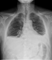
Thirty patients underwent postoperative radiographic examination using a water-soluble contrast medium (Visipaque-320). The examinations included a chest radiograph without contrast as a scout view, followed by radiography with contrast medium to detect leakage, fistula, or stricture (Figure 1).
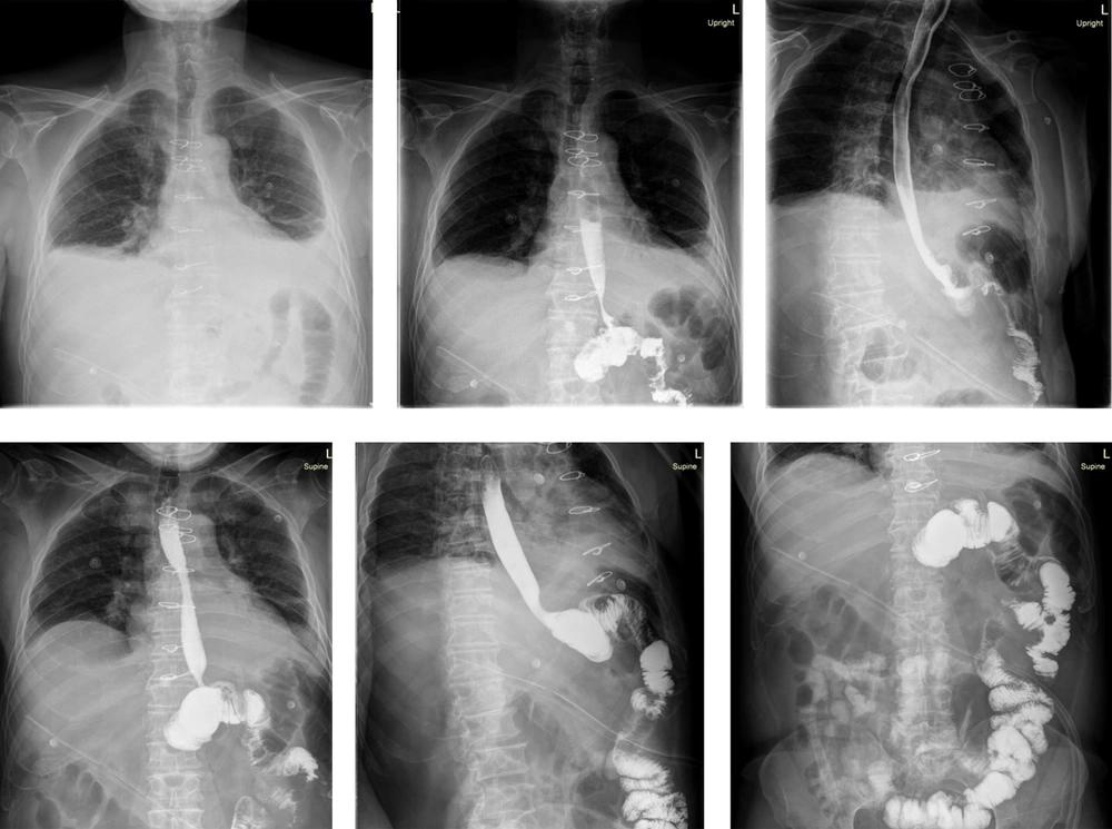
A 56-year-old man after total gastrectomy and partial esophagostomy, with an elevated C-reactive protein (CRP) level of 58 mg/L but no evidence of anastomotic leakage on radiographic evaluation
Figure 1.

A 56-year-old man after total gastrectomy and partial esophagostomy, with an elevated C-reactive protein (CRP) level of 58 mg/L but no evidence of anastomotic leakage on radiographic evaluation

3.4. Computed Tomography

Sixteen patients underwent postoperative chest CT both with and without oral and intravenous (IV) contrast medium. The examinations were performed in the Imaging Department of Imam Khomeini Complex Hospital using a 16-slice Philips CT system. Scans were obtained in the portal phase, 55 seconds after injection, at a rate of 2.5 mL/s, using 1.5 mL/kg of Visipaque-320. Three-dimensional reconstructions were created in the sagittal, coronal, and axial planes; the axial and coronal views were the most informative for diagnosing leakage (Figure 2).
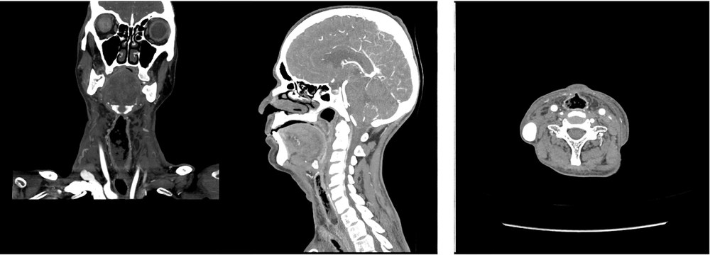
A 51-year-old woman with clinical signs of leakage, an elevated C-reactive protein (CRP) level of 50 mg/L, and computed tomography-confirmed anastomotic leakage after total gastrectomy and partial esophagostomy
Figure 2.

A 51-year-old woman with clinical signs of leakage, an elevated C-reactive protein (CRP) level of 50 mg/L, and computed tomography-confirmed anastomotic leakage after total gastrectomy and partial esophagostomy

3.5. C-Reactive Protein Measurement

C-reactive protein levels were measured in all patients using blood samples and a Hitachi 917 analyzer (Japan). The laboratory reported CRP levels in mg/L on the day of esophagography or chest CT.

3.6. Statistical Analysis

Descriptive analyses and cross-tabulation were used to calculate the sensitivity, specificity, and accuracy of the 2 diagnostic methods. These measures were used to assess the effectiveness of the diagnostic tests in detecting postoperative leakage.
Quantitative data were analyzed using the chi-square test. Statistical analyses were performed using SPSS, version 20. A P value of less than 0.05 was considered statistically significant.

4. Results

A total of 46 patients were recruited, including 32 men and 14 women. The mean age was 62.8 ± 9.2 years (range, 34 - 90 years).
Fourteen patients were excluded from the analysis because of an inability to swallow the contrast medium, contrast aspiration, death, or refusal to participate.
Patients underwent diagnostic procedures during the 30-day postoperative period, including esophagography, chest CT, clinical assessment, and measurement of CRP levels.
Of the 46 patients, 10 had clinical signs of leakage, including elevated CRP levels and other significant clinical findings.
Among the 30 patients who underwent radiographic examination, 27 (90%) had normal findings and 3 (10%) had AL. Clinical signs of AL were present in 10 patients (33.3%).
Among the 16 patients who underwent chest CT, 9 (56.3%) had normal findings and 7 (43.8%) had AL.
The sensitivity, specificity, and accuracy of radiographic examination were 30%, 100%, and 71.43%, respectively. The corresponding values for CT were 71.43%, 66.67%, and 68.75%.
The mean CRP level among all patients was 43.31 ± 26.07 mg/L. During postoperative days 2 - 10, the mean CRP level was 43.31 ± 26.07 mg/L. The mean CRP levels in patients with and without AL were 53.33 ± 15.2 mg/L and 38.35 ± 25.79 mg/L, respectively.
Among patients with clinical signs of leakage, CRP levels ranged from 35 - 85 mg/L, with a mean of 53.33 ± 15.2 mg/L. Only 1 patient had a CRP level of 105 mg/L; however, this patient showed no leakage on esophagography or CT.
Chi-square analysis showed no correlation between CRP levels and clinical leakage (P = 0.771).

5. Discussion

Anastomotic leakage is one of the most serious complications after esophagectomy (17-19), with a high mortality rate of 35% and a substantial contribution to post-esophagectomy deaths (25% - 50%) (5). Diagnosing AL before resuming an oral diet is essential (17).
Although esophagography is routinely used to diagnose leaks (3), it demonstrated relatively low sensitivity (30%), a high number of false-negative results (8 patients), and high specificity (100%) in this study. These results are consistent with a literature review by Fabbi et al. (20), which reported sensitivity values ranging from 33% - 52% and a specificity of 100%. Other authors have reported similar findings. Mejia-Rivera et al. reported a sensitivity of 43% (21), and Boone et al. reported a sensitivity of 52% for detecting AL using a contrast-enhanced swallow study (22). The accuracy of this method for identifying leaks has therefore been questioned because of false-negative results and the need for interpretation by experienced radiologists (23). Accordingly, discontinuation of its routine use has been recommended, although it may remain useful when leakage is clinically suspected and its extent must be assessed (22, 24).
Computed tomography is a noninvasive, safe, and widely available technique for investigating leakage (25). In this study, CT had higher sensitivity than esophagography (71.43% vs 30%). Upponi et al. reported CT sensitivity and specificity of 100% and 80%, respectively (6), compared with fluoroscopy. Although CT was compared with esophagography in the present study, both fluoroscopy and esophagography have lower sensitivity, and CT is better tolerated by severely ill patients. Helical CT can more readily detect small periesophageal air collections indicative of esophageal perforation. Such collections may be the most useful finding for suggesting esophageal rupture (6).
In this study, CRP levels were not correlated with AL. The mean interval between surgery and postoperative CRP measurement was 5.73 ± 1.68 days, and the mean CRP level was 43.31 ± 26.07 mg/L. The mean CRP levels in patients with and without AL were 53.33 ± 15.2 mg/L and 38.35 ± 25.79 mg/L, respectively. Although the value was slightly higher in patients with AL, the difference was not significant. In contrast to some studies of colorectal surgery (26), the present study found no significant correlation between CRP levels and AL after esophageal resection (27). Although some research has supported CRP as an early biomarker of leakage, these findings are consistent with conflicting results from previous studies (28-30).
These findings suggest that esophagography remains valuable for detecting leakage because of its high specificity, although its sensitivity is limited. Computed tomography appears to be a more sensitive alternative but has lower diagnostic accuracy and specificity for detecting leakage after esophagectomy.

5.1. Conclusions

This study provides insight into the diagnosis of AL after esophagectomy, highlighting the limitations of esophagography and the potential benefit of CT as a more sensitive diagnostic tool. Further research and clinical efforts are needed to optimize diagnostic strategies and improve patient outcomes associated with this serious postoperative complication.

Footnotes

References

  • 1.
    Ferlay J, Colombet M, Soerjomataram I, Mathers C, Parkin DM, Piñeros M, et al. Estimating the global cancer incidence and mortality in 2018: GLOBOCAN sources and methods. International journal of cancer. 2019;144(8):1941-53. [PubMed ID: 30350310]. [PubMed Central ID: PMC8791452]. https://doi.org/10.1002/ijc.31937.
  • 2.
    Schmidt HM, Gisbertz SS, Moons J, Rouvelas I, Kauppi J, Brown A, et al. Defining benchmarks for transthoracic esophagectomy: a multicenter analysis of total minimally invasive esophagectomy in low risk patients. Annals of surgery. 2017;266(5):814-21. [PubMed ID: 28796646]. https://doi.org/10.1097/SLA.0000000000002445.
  • 3.
    Sauvanet A, Baltar J, Le Mee J, Belghiti J. Diagnosis and conservative management of intrathoracic leakage after oesophagectomy. British journal of surgery. 1998;85(10):1446-9. [PubMed ID: 9782035]. https://doi.org/10.1046/j.1365-2168.1998.00869.x.
  • 4.
    Kassis ES, Kosinski AS, Ross P, Koppes KE, Donahue JM, Daniel VC. Predictors of anastomotic leak after esophagectomy: an analysis of the society of thoracic surgeons general thoracic database. The Annals of thoracic surgery. 2013;96(6):1919-26. [PubMed ID: 24075499]. https://doi.org/10.1016/j.athoracsur.2013.07.119.
  • 5.
    Alanezi K, Urschel JD. Mortality secondary to esophageal anastomotic leak. Ann Thorac Cardiovasc Surg. 2004;10(2):71-75. [PubMed ID: 15209546].
  • 6.
    Upponi S, Ganeshan A, D'costa H, Betts M, Maynard N, Bungay H, et al. Radiological detection of post-oesophagectomy anastomotic leak-a comparison between multidetector CT and fluoroscopy. The British Journal of Radiology. 2008;81(967):545-8. [PubMed ID: 18559902]. https://doi.org/10.1259/bjr/30515892.
  • 7.
    Backer CL, LoCicero J, Hartz RS, Donaldson JS, Shields T. Computed tomography in patients with esophageal perforation. Chest. 1990;98(5):1078-80. [PubMed ID: 2225947]. https://doi.org/10.1378/chest.98.5.1078.
  • 8.
    Lantos JE, Levine MS, Rubesin SE, Lau CT, Torigian DA. Comparison between esophagography and chest computed tomography for evaluation of leaks after esophagectomy and gastric pull-through. Journal of Thoracic Imaging. 2013;28(2):121-8. [PubMed ID: 23059739]. https://doi.org/10.1097/RTI.0b013e31826ff062.
  • 9.
    Fadoo F, Ruiz DE, Dawn SK, Webb WR, Gotway MB. Helical CT esophagography for the evaluation of suspected esophageal perforation or rupture. American Journal of Roentgenology. 2004;182(5):1177-9. [PubMed ID: 15100114]. https://doi.org/10.2214/ajr.182.5.1821177.
  • 10.
    Du Clos TW. Function of C-reactive protein. Annals of medicine. 2000;32(4):274-8. [PubMed ID: 10852144]. https://doi.org/10.3109/07853890009011772.
  • 11.
    Pepys MB, Hirschfield GM. C-reactive protein: a critical update. The Journal of clinical investigation. 2003;111(12):1805-12. https://doi.org/10.1172/JCI200318921.
  • 12.
    Garcia-Granero A, Frasson M, Flor-Lorente B, Blanco F, Puga R, Carratalá A, et al. Procalcitonin and C-reactive protein as early predictors of anastomotic leak in colorectal surgery: a prospective observational study. Diseases of the colon & rectum. 2013;56(4):475-83. [PubMed ID: 23478615]. https://doi.org/10.1097/DCR.0b013e31826ce825.
  • 13.
    Lagoutte N, Facy O, Ravoire A, Chalumeau C, Jonval L, Rat P, et al. C-reactive protein and procalcitonin for the early detection of anastomotic leakage after elective colorectal surgery: pilot study in 100 patients. Journal of visceral surgery. 2012;149(5):e345-e9. [PubMed ID: 23102916]. https://doi.org/10.1016/j.jviscsurg.2012.09.003.
  • 14.
    Ortega-Deballon P, Facy O, Binquet C. C-reactive protein and procalcitonin as predictors of anastomotic leak. Diseases of the Colon & Rectum. 2013;56(10). e395. [PubMed ID: 24022538]. https://doi.org/10.1097/DCR.0b013e31829eb9a7.
  • 15.
    Bracale U, Peltrini R, De Luca M, Ilardi M, Di Nuzzo MM, Sartori A, et al. Predictive factors for anastomotic leakage after laparoscopic and open total gastrectomy: a systematic review. Journal of Clinical Medicine. 2022;11(17):5022. [PubMed ID: 36078954]. [PubMed Central ID: PMC9457286]. https://doi.org/10.3390/jcm11175022.
  • 16.
    McOwan M, Johnson MA, Ward S, Read M, Chong L, Taylor L, et al. C-reactive protein is a predictor of severe infective complications following gastrectomy-a retrospective analysis. Journal of Gastrointestinal Oncology. 2022;14(1):64-72. [PubMed ID: 36915451]. [PubMed Central ID: PMC10007915]. https://doi.org/10.21037/jgo-22-675.
  • 17.
    Solomon DG, Sasaki CT, Salem RR. An evaluation of the routine use of contrast radiography as a screening test for cervical anastomotic integrity after esophagectomy. The American journal of surgery. 2012;203(4):467-71. [PubMed ID: 21906717]. https://doi.org/10.1016/j.amjsurg.2011.04.005.
  • 18.
    Jones CM, Heah R, Clarke B, Griffiths EA. Should routine radiological assessment of anastomotic integrity be performed after oesophagectomy with cervical anastomosis? Best evidence topic (BET). International Journal of Surgery. 2015;15:90-4. [PubMed ID: 25667064]. https://doi.org/10.1016/j.ijsu.2015.01.034.
  • 19.
    Markar SR, Arya S, Karthikesalingam A, Hanna GB. Technical factors that affect anastomotic integrity following esophagectomy: systematic review and meta-analysis. Annals of surgical oncology. 2013;20(13):4274-81. [PubMed ID: 23943033]. https://doi.org/10.1245/s10434-013-3189-x.
  • 20.
    Fabbi M, Hagens ERC, van Berge Henegouwen MI, Gisbertz SS. Anastomotic leakage after esophagectomy for esophageal cancer: definitions, diagnostics, and treatment. Diseases of the Esophagus. 2021;34(1). doaa039. [PubMed ID: 32476017]. [PubMed Central ID: PMC7801633]. https://doi.org/10.1093/dote/doaa039.
  • 21.
    Mejía-Rivera S, Pérez-Marroquín SA, Cortés-González R, Medina-Franco H. Contrast-enhanced swallow study sensitivity for anastomotic leak detection in post-esophagectomy patients. Revista de Gastroenterología de México (English Edition). 2018;83(4):400-4. https://doi.org/10.1016/j.rgmxen.2018.05.006.
  • 22.
    Boone J, Rinkes IB, Van Leeuwen M, Van Hillegersberg R. Diagnostic value of routine aqueous contrast swallow examination after oesophagectomy for detecting leakage of the cervical oesophagogastric anastomosis. ANZ Journal of Surgery. 2008;78(9):784-90. [PubMed ID: 18844909]. https://doi.org/10.1111/j.1445-2197.2008.04650.x.
  • 23.
    Page RD, Asmat A, McShane J, Russell GN, Pennefather SH. Routine endoscopy to detect anastomotic leakage after esophagectomy. The Annals of Thoracic Surgery. 2013;95(1):292-8. [PubMed ID: 23200235]. https://doi.org/10.1016/j.athoracsur.2012.09.048.
  • 24.
    Hu Z, Wang X, An X, Li W, Feng Y, You Z. The diagnostic value of routine contrast esophagram in anastomotic leaks after esophagectomy. World Journal of Surgery. 2017;41(8):2062-7. [PubMed ID: 28255626]. https://doi.org/10.1007/s00268-017-3923-9.
  • 25.
    Kim TH, Kim JH, Shin CI, Kim SH, Han JK, Choi BI. CT findings suggesting anastomotic leak and predicting the recovery period following gastric surgery. European Radiology. 2015;25(7):1958-66. [PubMed ID: 25708962]. https://doi.org/10.1007/s00330-015-3608-4.
  • 26.
    Gans SL, Atema JJ, van Dieren S, Koerkamp BG, Boermeester MA. Diagnostic value of C-reactive protein to rule out infectious complications after major abdominal surgery: a systematic review and meta-analysis. International journal of colorectal disease. 2015;30(7):861-73. [PubMed ID: 25935447]. [PubMed Central ID: PMC4471323]. https://doi.org/10.1007/s00384-015-2205-y.
  • 27.
    Liesenfeld LF, Sauer P, Diener MK, Hinz U, Schmidt T, Müller-Stich BP, et al. Prognostic value of inflammatory markers for detecting anastomotic leakage after esophageal resection. BMC surgery. 2020;20(1). 324. [PubMed ID: 33298038]. [PubMed Central ID: PMC7726907]. https://doi.org/10.1186/s12893-020-00995-2.
  • 28.
    Dutta S, Fullarton GM, Forshaw MJ, Horgan PG, McMillan DC. Persistent elevation of C-reactive protein following esophagogastric cancer resection as a predictor of postoperative surgical site infectious complications. World J Surg. 2011;35(5):1017-1025. [PubMed ID: 21167776]. https://doi.org/10.1007/s00268-011-1002-1.
  • 29.
    Gordon AC, Cross AJ, Foo EW, Roberts RH. C-reactive protein is a useful negative predictor of anastomotic leak in oesophago-gastric resection. ANZ journal of surgery. 2018;88(3):223-7. [PubMed ID: 27457697]. https://doi.org/10.1111/ans.13681.
  • 30.
    Noble F, Curtis N, Harris S, Kelly JJ, Bailey IS, Byrne JP, et al. Risk assessment using a novel score to predict anastomotic leak and major complications after oesophageal resection. Journal of Gastrointestinal Surgery. 2012;16(6):1083-95. [PubMed ID: 22419007]. https://doi.org/10.1007/s11605-012-1867-9.

Similar Articles

30
Jun
2009

Predicting Factors for Anastomotic Leakage after Esophageal Cancer Resection

SA Tabatabaee,
SM Hashemi,
Mohammad Eidy,
AH Davarpanah Jazi

Tabatabaee S, Hashemi S, Eidy M, Davarpanah Jazi A. Predicting Factors for Anastomotic Leakage after Esophageal Cancer Resection. Int J Cancer Manag. 2009;2(2):e80545. doi:

30
Jun
2017

Comparison of Early and Late Complications in Three Esophagectomy Techniques

Seyed Reza Mousavi,
Mohammad Esmaeil Akbari

Mousavi SR, Akbari ME. Comparison of Early and Late Complications in Three Esophagectomy Techniques. Int J Cancer Manag. 2017;10(6):e7644. doi: https://doi.org/10.5812/ijcm.7644

25
Jul
2022

The Risk Factors of Anastomotic Leakage After Rectal Cancer Surgery

Seyed Vahid Hosseini,
Ahmad Kashif Alghataa,
Alimohammad Bananzadeh,
Faranak Bahrami,
Hajar Khazraei,
Seyed Mohammad Kazem Tadayon
,et al.

Hosseini SV, Kashif Alghataa A, Bananzadeh A, Bahrami F, Khazraei H, et al. The Risk Factors of Anastomotic Leakage After Rectal Cancer Surgery. Int J Cancer Manag. 2022;15(6):e120889. doi: https://doi.org/10.5812/ijcm-120889

30
Jun
2010

Lateral esophagostomy with thoracic drainage in treatment of esophageal perforation

H Parsa,
P Fallah,
M Rezaei,
S Shahmoradi

Parsa H, Fallah P, Rezaei M, Shahmoradi S. Lateral esophagostomy with thoracic drainage in treatment of esophageal perforation. J Inflamm Dis. 2024;14(1):e155523. doi:

28
Feb
2007

Reinforced Primary Repair in Delayed Thoracic Esophageal Perforation

M Vaziri,
H Kadkhodaii

Vaziri M, Kadkhodaii H. Reinforced Primary Repair in Delayed Thoracic Esophageal Perforation. Shiraz E-Med J. 2007;8(1):20431. doi:


Crossmark
Crossmark
Checking
Share on
Metrics

Ordering Reprints

Articles are published under the Creative Commons license stated on each article. No permission or royalty fee is required for uses permitted by that license. CCC handles optional bulk and customized reprint orders. Any quotation covers production and delivery services only, not copyright permission. > Request Reprints from CCC 

Search Relations

Author(s):

Related Articles