
1. Background
Mucormycosis (zygomycosis) is an opportunistic fungal infection caused by various molds classified in the order Mucorales of the phylum Glomerulomycota and subphylum Mucormycotina (1). Mucorales fungi belong to seven families, all of which are capable of causing mucormycosis. Among these, Rhizopus oryzae (from the Mucoraceae family) is the most common infectious agent in the Western Hemisphere. Less common species in the Mucoraceae family that cause a similar range of infections include Rhizopus microsporus, Rhizomucor pusillus, Lichtheimia corymbifera (formerly Absidia corymbifera), Apophysomyces elegans, and Mucor species (which, despite their name, rarely cause mucormycosis) (2).
Conditions that increase patient risk include acidosis—especially in diabetes, leukemia, lymphoma, corticosteroid therapy, severe burns, immunodeficiencies, other debilitating diseases, and dialysis with iron chelators such as deferoxamine (3).
The primary clinical form of mucormycosis is rhino-orbito-cerebral, resulting from the germination of sporangiospores in the nasal passages and the invasion of hyphae into blood vessels, leading to thrombosis, infarction, and necrosis. This disease can progress rapidly by attacking the sinuses, eyes, skull bones, and brain. It damages blood vessels and nerves, causing facial edema, bloody nasal discharge, and orbital cellulitis, an acute infection of the eye tissue (4). Pulmonary mucormycosis occurs after inhalation of sporangiospores, with invasion of the lung parenchyma and vascular system. In both cases, ischemic necrosis leads to extensive tissue destruction (3). Direct examination of cultures from nasal secretions, tissue, or sputum reveals broad hyphae (15 - 10 μm) with uneven thickness, irregular branching, and few septa. These fungi grow rapidly in laboratory settings and produce numerous fluffy colonies. Identification is based on sporangium structures (3).
Although mucormycosis is rare, its global incidence has increased in the last two decades, particularly in France, Belgium, Switzerland, and India (5-9). In developed countries, nearly all mucormycosis cases occur in immunocompromised patients. In France, a national hospital database identified 35,876 invasive fungal infections from 2001 to 2010, of which 1.5% were associated with mucormycosis (7). Additionally, in France, the incidence of mucormycosis increased from 0.7 per million in 1997 to 1 per million in 2006 (6). In a Spanish hospital study, 19 cases of mucormycosis were diagnosed from 2007 to 2015 (incidence 2.3 per 100,000), compared to 1.2 per 100,000 from 1998 to 2006 (10).
In a Geneva, Switzerland hospital, only three mucormycosis cases were diagnosed from 1989 to 2003, compared to 16 cases from 2003 to 2008—an incidence increase that appears linked to a rise in immunosuppressed patients (9).
Treatment includes surgical excision of nonviable tissue, rapid administration of amphotericin B, and disease control. Many patients survive; however, partial facial paralysis or eye loss may occur (3). The most important risk factors for developing mucormycosis include uncontrolled diabetes, hematological malignancies leading to neutropenia, organ transplantation, autoimmune diseases, immunosuppressive treatments, HIV infection, iron overload, burns, trauma requiring surgery, peritoneal dialysis, and malnutrition (11-17).
Based on anatomical location, mucormycosis is classified into six categories: Rhino-orbito-cerebral, pulmonary, cutaneous, gastrointestinal, disseminated, and rare sites (18).
Rhino-orbito-cerebral mucormycosis is the most common form of the disease. The initial symptoms of rhino-orbital-cerebral mucormycosis are non-specific and include eye or facial pain and facial numbness, followed by conjunctival congestion and blurred vision. Fever is absent in more than half of cases (19). Without proper treatment, the infection often spreads from the ethmoid sinus to the orbit, resulting in dysfunction of the extraocular muscles and causing proptosis, primarily due to inflammation of the conjunctiva (chemosis) (19).
Infected tissues may appear normal initially, then progress to an erythematous stage, which may or may not include edema, followed by a purplish appearance, and ultimately leading to a black ulcer with necrosis. The infection can spread from the sinuses to the mouth, causing a painful necrotic ulcer on the hard palate; however, this is a late finding and indicates widespread infection (19). Diagnosing mucormycosis requires a high degree of clinical suspicion. Unfortunately, serial autopsies show that up to 50% of cases are diagnosed only post-mortem. Since mucorales can be isolated from the environment, an accurate diagnosis requires a positive culture from a sterile area (such as a needle aspiration specimen, tissue biopsy, or pleural fluid) or histopathological evidence of invasive mucormycosis. A presumptive diagnosis can be made based on a culture from a non-sterile area (such as sputum or bronchoalveolar lavage) when the patient has sufficient risk factors and clinical and radiographic evidence of the disease (20). Other fungal infections, including aspergillosis, pseudallescheriasis, and fusariosis, cause symptoms similar to mucormycosis. Histopathological examination typically distinguishes these infections from mucormycosis, and a positive culture confirms the specific organism type (20).
There are four key factors for successful treatment of mucormycosis: Early diagnosis, removal of predisposing factors if possible, surgical debridement, and timely and extensive antifungal treatment. Early diagnosis allows treatment to begin sooner, improving survival rates. Host defense deficiencies must be addressed or prevented during treatment, which includes stopping or reducing immunosuppressive medications and correcting blood sugar and acid-base levels. In patients with active mucormycosis, iron should be avoided, as it has been shown to cause flare-ups in animal models (20).
In recent decades, there has been a significant increase in mucormycosis rates among patients who are at classical risk for other opportunistic fungal infections. This rising incidence highlights the importance of investigating this invasive fungal infection, which is associated with high mortality if not diagnosed promptly (11).
Late diagnosis of mucormycosis results in prolonged hospitalization, the need for debridement, and repeated surgeries, leading to a higher probability of morbidity and mortality as well as increased hospital costs. Mucormycosis can be successfully treated with early diagnosis, elimination of predisposing factors, surgical debridement, and systemic antifungal medication. Therefore, early and accurate diagnosis and treatment protocols reduce hospitalization and improve patient outcomes (11).
In recent years, researchers have conducted studies in Iran and other parts of the world to investigate the prevalence of mucormycosis. In a study by Skiada et al. from 2005 to 2007 in Europe, patients with mucormycosis were analyzed. This study group identified proven and probable cases of mycosis across thirteen European countries. The average patient age was 50 years, and 60% of the patients were male. Background conditions included hematological malignancies, trauma, transplantation, and diabetes mellitus. Overall mortality was 47% (21).
Ruping et al. evaluated 41 patients with mucormycosis in a 2009 study in Germany. The most common underlying conditions were malignancy, diabetes mellitus, and transplantation. Diagnosis was made by culture in 28 patients and by histology in 26 patients. The main infection site was the lung in 58% of cases, followed by soft tissue, rhino-sino-orbital, and brain. Disseminated infection was observed in six patients. The overall survival rate was 51% (21 cases) (22).
Between 1998 and 2012, Tavanaee Sani et al. conducted a study in Mashhad, where 123 patients were examined, and 92 patients were confirmed cases of mucormycosis. Diabetes and immunodeficiency were the most common contributing factors (23).
2. Objectives
Given the rise in mucormycosis cases globally, including in Iran, and considering the high mortality rate and the need for timely diagnosis to initiate mucormycosis treatment, this study investigated the clinical manifestations and treatment outcomes of hospitalized mucormycosis patients in two centers in northeastern Iran.
3. Methods
This retrospective study was conducted among mucormycosis patients hospitalized in two educational-research and medical referral centers, Imam Reza and Ghaem Hospitals of Mashhad University of Medical Sciences, between 2011 and 2017. The selection criteria included a positive pathologic specimen or mycologic culture, file completeness, and clarity of the patient’s outcome. These patients were examined for variables such as demographic characteristics, underlying diseases, clinical symptoms, treatments performed, and disease outcome.
By referring to the registered pathology information, files, and graphs of hospitalized patients diagnosed with mucormycosis, information was collected from the files, and the designed checklist was completed. The data were then entered into the statistical software SPSS version 26. Eligible patients were those admitted to Imam Reza and Ghaem Hospitals in Mashhad between June 1, 2011, and the end of September 2017 with a clinical and pathological diagnosis of mucormycosis and who did not meet the criteria for exclusion from the study.
Due to the rare occurrence of this disease and the limited population of affected patients, all patients referred to the project implementation site from 2011 to 2017 were selected if they met the study entry criteria. Patients were classified into proven, possible, and probable cases based on the guidelines of the Mycoses Study Group Education and Research Consortium (18). Descriptive statistics, including the frequency and percentage of data, as well as central indicators such as mean and median, and measures of dispersion such as standard deviation and interquartile range, were calculated and displayed in tables.
To compare qualitative data between the deceased and surviving patient groups, the chi-square test was used, with Fisher’s exact test applied when necessary. For comparing quantitative data between these two groups, normality was first assessed using the Shapiro-Wilk test. If the data were normally distributed, the independent t-test was used; otherwise, the Mann-Whitney U test was applied. Finally, the Cox regression test was employed to investigate patient mortality risk factors, considering the length of hospitalization. The hazard ratio and 95% confidence interval were calculated, and a Kaplan-Meier curve was drawn. P-values less than 0.05 were considered significant.
4. Results
A total of 97 patients were included during the study period, of whom 53 were men (54.6%). The average age of patients was 40.82 ± 19.45 years, with an age range from 13 to 83 years. Additionally, 64 patients (66.0%) were from urban areas, while 33 patients (34.0%) were from rural areas.
Table 1 shows the frequency and percentage of underlying diseases and contributing factors. The three most common risk factors were malignancy with 58 cases (59.8%), chemotherapy with 48 cases (49.5%), and diabetes with 34 cases (35.1%). Additionally, three cases (3.1%) of mucormycosis had no underlying disease (Figure 1).
Table 1.
Frequency and Percentage of Underlying Diseases and Contributing Factors
| Underlying Diseases and Risk Factors | Frequency (%) |
|---|---|
| Malignancy | 58 (59.8) |
| Chemotherapy | 48 (49.5) |
| Diabetes | 34 (35.1) |
| Corticosteroid use | 10 (10.3) |
| Kidney failure | 9 (9.3) |
| Pulled tooth | 5 (5.2) |
| Dialysis | 4 (4.1) |
| No underlying cause | 3 (3.1) |
| Head and face trauma | 1 (1) |
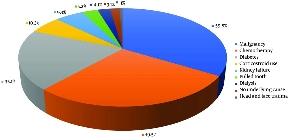
Figure 1.
Frequency and percentage of underlying diseases and contributing factors shown in pie chart.
The most common symptom was fever, observed in 61 cases (62.9%), followed by facial swelling in 52 cases (53.6%) and facial pain in 48 patients (49.5%). Table 2 presents the frequency and percentage of various symptoms in patients diagnosed with mucormycosis (Figure 2).
Table 2.
Frequency and Percentage of Symptoms of Patients Diagnosed with Mucormycosis
| Symptoms and Signs | Frequency (%) |
|---|---|
| Fever | 61 (62.9) |
| Headache | 46 (47.4) |
| Nasopharyngeal necrosis | 38 (39.2) |
| Symptoms of orbital involvement | 42 (43.3) |
| Ptosis | 33 (34) |
| Proptosis | 27 (27.8) |
| Decreased vision | 42 (43.3) |
| Decreased eye movements | 30 (30.9) |
| Symptoms of nervous system involvement | 28 (28.9) |
| Facial nerve involvement | 23 (23.7) |
| Facial swelling | 52 (53.6) |
| Facial pain | 48 (49.5) |
| Decreased facial sensation | 34 (35.1) |
| Nasal discharge | 43 (44.3) |
| Nasal congestion | 44 (45.4) |
| Nausea and vomiting | 15 (15.5) |
| Stomachache | 2 (2.1) |
| Toothache | 6 (6.2) |
| Epistaxis | 2 (2.1) |
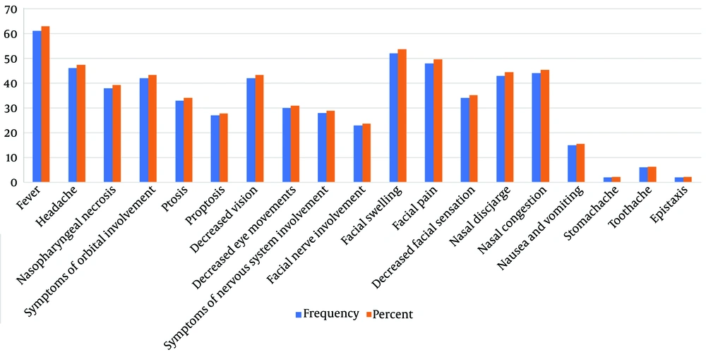
Figure 2.
Frequency and percentage of symptoms of patients diagnosed with mucormycosis displayed by chart.
A paranasal CT scan was requested for 95 patients (94.8%), a brain MRI for 40 patients (41.2%), and a brain CT for 33 patients (34.0%).
Overall, 88 patients (90.7%) showed maxillary involvement, 83 patients (85.6%) had ethmoid involvement, and 66 patients (68.0%) had sphenoid involvement. Additionally, 18 patients (18.6%) presented with orbital involvement, and only one patient had CNS involvement on imaging. Pansinusitis was observed in 31 patients (32.0%), while simultaneous involvement of the ethmoid, sphenoid, and maxillary sinuses was found in 62 patients (63.9%).
In total, a direct fungal scope was requested for 18 patients, yielding a positive result in only 1 patient (1.0%). Additionally, a fungal culture was requested for 6 patients, with positive results in 4 patients (4.1%).
Table 3 presents the frequency and percentage of medical and surgical treatments, while Table 4 displays the frequency and percentage of medical treatments, considering therapeutic combinations. The most commonly administered drug was intravenous (IV) Amphotericin B Deoxycholate (1 mg/kg once daily), given to 78 patients (80.4%), whereas the least frequently administered drug was IV voriconazole (4 mg/kg twice daily), given to 3 patients (3.1%). Additionally, 94 patients (96.9%) underwent surgery. Twenty-two patients (22.7%) were admitted to the ICU, and 40 patients (41.2%) expired.
Table 3.
Frequency and Percentage of Medical and Surgical Treatments Regardless of the Therapeutic Combination
| Surgical and Drug Treatments/(Drug Dosage) | Frequency (%) |
|---|---|
| Liposomal amphotericin B (10 mg/kg/day) | |
| Taken | 18 (18.6) |
| Amphotericin B deoxycalate (1 mg/kg/day) | |
| Taken | 78 (80.4) |
| Caspofungin (50 mg/day) | |
| Taken | 14 (14.4) |
| Posaconazole (200 mg four times a day) | |
| Taken | 14 (14.4) |
| Voriconazole (1 mg/kg/day) | |
| Taken | 3 (3.1) |
Table 4.
Frequency and Percentage of Medical Treatments Considering the Therapeutic Combination
| Drug Treatments and Combinations | Frequency (%) |
|---|---|
| Liposomal amphotericin B (10 mg/kg/day) | |
| Alone | 12 (12.4) |
| + Caspofungin (50 mg/day) | 1 (1.0) |
| + Posaconazole (200 mg four times a day) | 2 (2.1) |
| + Caspofungin (50 mg/day) + Posaconazole (200 mg four times a day) | 3 (3.1) |
| Amphotericin B deoxycalate (1 mg/kg/day) | |
| Alone | 57 (58.8) |
| + Caspofungin (50 mg/day) | 8 (8.2) |
| + Posaconazole (200 mg four times a day) | 8 (8.2) |
| + Voriconazole (1 mg/kg/day) | 3 (3.1) |
| + Caspofungin (50 mg/day) + Posaconazole (200 mg four times a day) | 2 (2.1) |
The groups of patients who expired and those who survived showed similarities in sex, residence, inpatient department, occupation, and season of illness. The frequency and percentage of underlying diseases and risk factors differed between the deceased and surviving groups only in the category of dialysis patients (P = 0.026). None of the survivors were dialysis patients, while four of the deceased patients had undergone dialysis.
Table 5 compares the frequency and percentage of symptoms in deceased and surviving mucormycosis patients. Nasopharyngeal necrosis (P = 0.034) and ptosis (P = 0.017) were significantly higher in the deceased and surviving groups, respectively.
Table 5.
Comparison of Frequency and Percentage of Symptoms of Patients Between Deceased and Survivors a
| Symptoms and Signs | Death | Alive | P-Value |
|---|---|---|---|
| Fever | 28 (70.0) | 33 (57.9) | 0.287 |
| Headache | 18 (45.0) | 28 (49.1) | 0.837 |
| Nasopharyngeal necrosis | 21 (52.5) | 17 (29.8) | 0.034 |
| Symptoms of orbital involvement | 15 (37.5) | 27 (47.4) | 0.407 |
| Ptosis | 8 (20.0) | 25 (43.9) | 0.017 |
| Proptosis | 10 (25.0) | 17 (29.8) | 0.651 |
| Decreased vision | 14 (35.0) | 28 (49.1) | 0.213 |
| Decreased eye movements | 10 (25.0) | 20 (35.1) | 0.373 |
| Symptoms of nervous system involvement | 8 (20.0) | 20 (35.1) | 0.118 |
| Facial nerve involvement | 7 (17.5) | 16 (28.1) | 0.332 |
| Facial swelling | 23 (57.5) | 29 (50.9) | 0.542 |
| Facial pain | 22 (55.0) | 26 (45.6) | 0.413 |
| Decreased facial sensation | 12 (30.0) | 22 (38.6) | 0.398 |
| Nasal discharge | 14 (35.0) | 29 (50.9) | 0.148 |
| Nasal congestion | 15 (37.5) | 29 (50.9) | 0.219 |
| Nausea and vomiting | 8 (20.0) | 7 (12.3) | 0.394 |
| Abdominal pain | 2 (5.0) | 0 (0.0) | 0.168 |
| Toothache | 3 (7.5) | 3 (5.3) | 0.688 |
| Epistaxis | 1 (2.5) | 1 (1.8) | > 0.999 |
a Values are expressed as Frequency (%).
There was no significant difference between the two groups regarding the frequency and percentage of involvement based on imaging between deceased and surviving patients. However, the relative frequency of posaconazole administration was significantly higher in surviving patients compared to deceased patients (P = 0.007). Additionally, the relative frequencies of maxillectomy (P = 0.036) and ethmoidectomy (P = 0.006) were significantly higher in surviving patients than in deceased patients.
Table 6 compares quantitative data, including the number of surgeries, duration of drug treatment, and length of hospitalization, between the deceased and surviving groups. The median number of surgeries and duration of drug treatment were significantly higher in survivors (P < 0.001). Furthermore, the average length of hospitalization was significantly longer in surviving patients compared to deceased patients (P = 0.007).
Table 6.
Comparison of Quantitative Data Between Two Groups of Deceased and Survivors
| Surgical and Drug Treatments | Death | Alive | P-Value |
|---|---|---|---|
| Hospitalization frequency-interquartile range and median | 1.0 (1.0 - 1.0) | 1.0 (1.0 - 1.0) | 0.335 |
| Treatment distance from hospitalization (day)-interquartile range and median | 8.5 (15.0 - 1.0) | 2.0 (16.5 - 1.0) | 0.712 |
| The treatment interval from the onset of symptoms (day)-interquartile range and median | 5.0 (10.0 - 3.0) | 5.0 (10.0 - 3.0) | 0.706 |
| Number of surgeries -interquartile range and median | 1.0 (2.0 - 1.0) | 3.0 (4.0 - 2.0) | < 0.001 |
| Duration of drug treatment (day)-interquartile range and median | 11.0 (19.5 - 6.0) | 30.0 (38.5 - 16.0) | < 0.001 |
| Length of hospitalization (day); mean ± SD | 24.32 ± 14.33 | 33.37 ± 16.27 | 0.007 |
The Cox regression test was used to assess risk factors. Based on the most suitable model obtained, three variables—ICU hospitalization, ethmoidectomy, and duration of drug treatment—were included. The results indicated that ethmoidectomy (OR = 0.316; 95% CI = 0.142 - 0.703; P = 0.005) and the duration of drug treatment (OR = 0.907; 95% CI = 0.874 - 0.940; P < 0.001) were identified as protective factors against patient mortality. Table 7 provides the relevant details.
Table 7.
Cox Regression Test Results
| Variables | Risk Ratio | 95% Confidence Interval | P-Value |
|---|---|---|---|
| Hospitalization in ICU | 1.87 | 3.949 - 0.892 | 0.097 |
| Ethmoidectomy | 0.316 | 0.703 - 0.142 | 0.005 |
| Duration of drug treatment (days) | 0.907 | 0.940 - 0.874 | < 0.001 |
The Kaplan-Meier curves for the two factors, ethmoidectomy and ICU hospitalization, are shown in Figures 3. and 4.
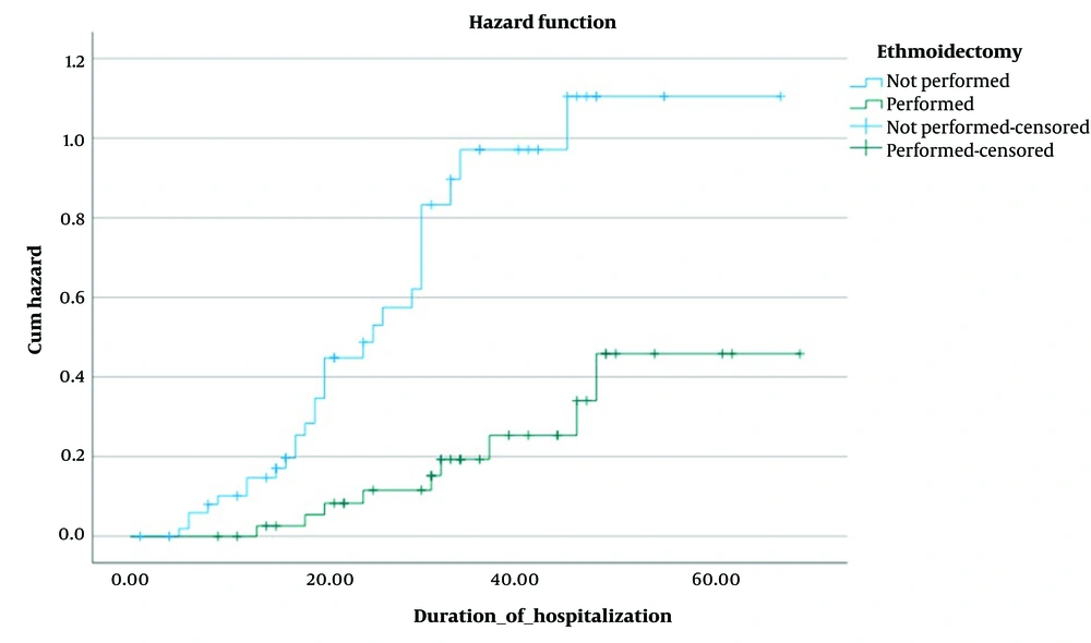
Figure 3.
Kaplan-Meier curve of the effect of ethmoidectomy as a protective factor
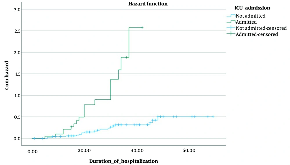
Figure 4.
Kaplan-Meier curve of the effect of ICU admission as a risk factor
5. Discussion
Among the 97 patients with mucormycosis, 57 cases were confirmed, while the remaining cases were classified as possible or probable. The mean age of patients was 41 years, with a higher prevalence observed in males. The onset of the disease showed an increase during the autumn season. Malignancy and diabetes were the most common underlying diseases. The predominant symptoms observed included fever, facial swelling, and nasal secretions. Imaging indicated that nearly all patients exhibited maxillary sinus involvement, followed by involvement of the ethmoid and sphenoid sinuses, with the frontal sinus being the least involved across all patients. All confirmed patients underwent surgical intervention. Ethmoidectomy was the most frequently performed surgical procedure, followed by maxillectomy and sphenoidectomy.
Amphotericin B deoxycholate was the primary treatment, followed by liposomal amphotericin B, posaconazole, caspofungin, and voriconazole. Patients with confirmed cases exhibited a mortality rate of 35%, with a significantly higher rate observed in those in the ICU. All dialysis patients who developed invasive fungal infections succumbed to the disease. Improved survival rates were observed in patients who received prolonged antifungal drug treatment.
The epidemiology and characteristics of mucormycosis have been widely studied worldwide, with a notable focus on India. Research indicates that males exhibit a slightly elevated incidence of the disease. In our study, men constituted 54.6% of the patients. These findings align with the sex distribution results from studies by Patel et al. (24), Manesh et al. (25), Sarvestani et al. (26), Corzo-León et al. (27), and Tavanaee Sani et al. (23), all of which reported a higher prevalence of mucormycosis in males.
The highest prevalence of the disease is seen among individuals aged 30 to 50 years. The mean age of confirmed cases in our study was 41 years.
In our study, the highest number of cases occurred in autumn. This finding corresponds with Tavanaee Sani et al. (23), who reported the most cases in October. Seasonality appears to influence disease prevalence. Al-Ajam et al. (28) found that mucormycosis prevalence in Eastern Mediterranean countries increases from the hot to cold seasons, peaking in September and October. In Aspergillus, atmospheric fungal spore concentrations vary with the seasons (29). Talmi et al. found a similar seasonal prevalence of mucormycosis in the Eastern Mediterranean, with a peak in October (30). Animal models suggest that mucormycosis arises from inhalation of fungal spores in susceptible hosts, indicating a higher likelihood of disease occurrence when fungal spore concentrations are highest (31). It is hypothesized that plant decomposition during late summer and early autumn correlates with increased mucor spore concentrations, leading to a peak in disease incidence (28).
Diabetes and hematological malignancies represent the primary risk factors for mucormycosis. Malignancies are increasingly surpassing diabetes as a risk factor, attributed to improved diabetes management and treatment outcomes. In Tavanaee Sani et al. (23), 42.4% of patients had diabetes, while in our study, 35.1% did. Sarvestani et al. (26) found diabetes in 34.9% and leukemia in 58.6% of cases. In our study, 59.6% of patients had malignancies. Each epidemiological case depends on the population studied. In India, Patel et al. found diabetes to be the main risk factor (24), with 73.5% of participants having diabetes and 9% having cancer. Notably, 81.57% of individuals with diabetes exhibited inadequate control, underscoring the importance of blood glucose management in preventing invasive fungal infections. Similarly, studies by Manesh et al. (25) and Corzo-León et al. (27) found diabetes to be more prevalent than hematologic malignancies as a risk factor.
The symptoms of mucormycosis are typically nonspecific. Fungal culture and pathology serve as diagnostic methods; however, due to limited availability, time requirements, and invasiveness, they are not suitable for all patients with suspected mucormycosis (32). Therefore, a strong clinical suspicion and consideration of risk factors can assist clinicians in diagnosing mucormycosis. Early diagnosis and treatment improve outcomes in mucormycosis, making understanding disease symptoms essential for reducing mortality rates.
In our study, fever was the most common symptom in confirmed cases (59.6%), followed by facial swelling (28 cases) and nasal discharge (49.1%). According to Tavanaee Sani et al. (23), eye symptoms were the most common (59.7%), followed by headache (55.4%) and palate necrosis (53.6%). Additionally, 41.3% of patients in Tavanaee Sani et al.'s study reported fever (23).
According to a 2020 systematic review of diabetic mucormycosis symptoms, facial swelling is the most common symptom (53.3%). Additionally, patients reported headache in 44.4%, facial pain in 35.5%, nasal discharge in 24.4%, and fever in 22.2% of cases (33). However, our study was not limited to diabetic patients.
Mucormycosis can also cause toothaches and other non-specific symptoms. This symptom was reported by 6 of all mucormycosis cases and by 3 of the 57 confirmed cases in our study. A rare case report by Singh et al. described a 17-year-old Indian woman with a toothache who subsequently developed mucormycosis (34). Prioritizing these symptoms is crucial, particularly in dental training.
Many patients experience irreversible eye complications due to orbital involvement (35). Orbital involvement symptoms were found in 43.3% of our patients, with 34.7% presenting with ptosis, 30% with reduced eye movements, and 27.8% with proptosis. Additionally, 43.3% of patients retained some sense of sight. In Tavanaee Sani et al.'s study (23), ocular symptoms also had the highest relative frequency, at 59.7%.
Maxillary sinus involvement was observed in 90.7% of cases in our study. Other similar studies investigating mucormycosis imaging report maxillary sinus involvement ranging from 80 to 100% (36-38). Fungal spores enter the upper respiratory tract through inhalation, initially affecting the maxillary sinus. Due to the fungus’s invasive nature, the infection gradually progresses to other sinuses and the orbital cavity (36). The frontal sinus consistently shows the lowest rate of involvement across all studies, including ours (36-38). Multiple sinus involvement is common due to the invasive behavior of the fungus. In our study, 63.9% of patients had involvement of the ethmoid, sphenoid, and maxillary sinuses, and 32% presented with pansinusitis.
Our investigation into mycological findings revealed that physicians at the studied clinics did not prioritize direct microscopy and culture methods. In some studies, histopathological examination is considered the gold standard for diagnosis (39, 40). In Badiee et al.'s study (40), the sensitivity, specificity, positive predictive value, and negative predictive value of culture were 70%, 100%, 100%, and 91%, respectively. Nonetheless, it is important to note that fungal culture may require 3 to 5 days, and in certain instances, cultures may yield negative results despite a mucormycosis diagnosis (41). In our study, among the 4 confirmed cases for which cultures were requested, cultures were positive in 3 cases (75.0%). Additionally, only 1 case (5.5%) was positive out of 18 smear samples taken from confirmed patients. For PCR, a sensitivity of 86%, specificity of 96%, positive predictive value of 86%, and negative predictive value of 96% have been reported (40). However, this technique is costly and lacks widespread availability. Culture and pathology findings appear to hold significant value when evaluated from various perspectives. In our study, all confirmed cases (57 patients) exhibited positive pathology for mucormycosis, consistent with the Mycoses Study Group Education and Research Consortium guidelines (18).
According to recent studies, amphotericin B, posaconazole, and isavuconazole show the highest sensitivity to mucormycosis. Intravenous amphotericin B is the preferred treatment for mucormycosis, with oral posaconazole recommended as an adjunct if fungal sensitivities are confirmed (42). In our study, 10 confirmed patients and 4 possible and probable patients received oral posaconazole at a dosage of 200 mg four times daily as adjunctive treatment. All patients with confirmed diagnoses in our study received IV amphotericin B as the primary treatment. Among possible and probable cases, only one patient had not received amphotericin B; this patient died before receiving medication or surgery and was therefore likely untreated. Drug therapy continued until biopsy results indicated a negative outcome.
Patient mortality was 41.2% across proven, possible, and probable cases, with a final death rate of 35% for confirmed cases. In a study by Tavanaee Sani et al. (23) conducted at Ghaem and Imam Reza medical centers, mortality was reported at 67.3%. It is important to note that in the previous study by Tavanaee Sani et al. (23), only 70.5% of patients with a confirmed diagnosis received both surgery and amphotericin B, whereas all confirmed cases in our study received amphotericin B and underwent surgery. Recent advancements in antifungal drug treatments and surgical interventions for mucormycosis patients may have contributed to this decrease in mortality.
5.1. Conclusions
Our study found that the mortality rate for rhino-orbito-cerebral mucormycosis has changed compared to a study conducted 10 years ago in these two referral centers. Increased availability of drug treatments, particularly amphotericin B, along with more frequent surgical interventions, may have contributed to a reduction in mortality rates. Our study indicates a 35% mortality rate for mucormycosis, suggesting that one in three patients succumbs to this condition, which remains substantial. Education, attention to clinical symptoms, timely diagnosis and clinical suspicion, prompt treatment, and management of contributing factors such as diabetes and immunodeficiency may improve outcomes in rhino-orbito-cerebral mucormycosis.