



1. Background
Nosocomial fungal infections are associated with high mortality and are therefore important in patient health management. These infections can be acquired through various routes, including inhalation of fungal spores, contact with contaminated surfaces, and exposure to infected or colonized individuals (1, 2). Nosocomial fungal infections can affect multiple organs, including the liver, lungs, skin, eyes, and the central nervous system. The etiologic agents include a variety of fungal species. Filamentous fungi, such as Aspergillus species, are causative agents of nosocomial fungal infections, and their conidia are present in various environments, including hospital settings. Other fungi responsible for nosocomial fungal infections include Candida species and Mucorales (3). The prevalence of nosocomial fungal infections varies by hospital setting and patient population. For example, the prevalence of fungal bloodstream infections among 21,098 intensive care unit (ICU) patients in China from January 2008 to December 2017 was 0.38% (81 cases) (3). The rate of these infections among hospitalized pediatric patients was reported as 2.69% (4). During COVID-19, fungal infections among ICU patients in Mexico were reported at 9.3% (78/842) (5).
Common clinical signs and symptoms of nosocomial fungal infections include fever, chills, cough, chest pain, shortness of breath, skin rash, abdominal pain, vomiting, diarrhea, hemoptysis, shock, organ failure, and death (6). Radiologic findings may include infiltrates, consolidations, nodules, or cavities in pulmonary aspergillosis (chest X-rays) and halo, crescent, and/or cavity signs in the lung, sinus opacities, and bone erosion in rhino-cerebral mucormycosis (CT, computed tomography scan) (7). Magnetic resonance imaging (MRI) can detect brain abscesses and meningitis in central nervous system fungal infections (8). Several risk factors contribute to the development of nosocomial fungal infections, including immunosuppression, prolonged hospitalization, use of broad-spectrum antibiotics, use of immunosuppressive drugs, and invasive procedures. Patients with underlying conditions such as cancer, human immunodeficiency virus, and diabetes are also at increased risk for nosocomial fungal infections (9).
2. Objectives
Fungal infections often result in prolonged hospitalizations and high treatment costs. Accordingly, this study aimed to determine the rate of nosocomial fungal infections among adult patients admitted to a university hospital in Shiraz, southern Iran, and to evaluate infection-related factors, including patient demographics, infection sites, and etiologic agents.
3. Methods
This descriptive, cross-sectional study was conducted over 10 months (01/06/2023 to 31/03/2024) at Nemazi University Hospital, a 1000-bed tertiary care hospital in Shiraz, Iran. The study was conducted in accordance with the Helsinki Rules. The study was reviewed and approved by the Research Ethics Committee of Shiraz University of Medical Sciences, Shiraz, Iran (IR.SUMS.REC.1401.671), and was conducted in accordance with Iranian national guidelines for medical research and ethical principles. Informed consent for participation was obtained from all participants.
A stepwise surveillance approach was applied during the study period. Upon hospital admission, patients’ clinical, radiological, and laboratory signs and symptoms were evaluated (n = 12,941). The inclusion criteria were the absence of symptoms or signs of infectious diseases, such as fever, at the time of hospitalization; no receipt of antifungal therapy before or at admission; and a hospital stay of ≥ 7 days (n = 3,890) (Figure 1). According to the European Organization for Research and Treatment of Cancer and the Mycoses Study Group Education and Research Consortium (EORTC-MSGERC) (7), the clinical, radiological, and laboratory examinations (culture, microscopy, and molecular testing) of these patients were evaluated for fungal infections, as well as other diseases. One hundred fifty-three patients were classified as having proven, probable, or possible fungal infections.
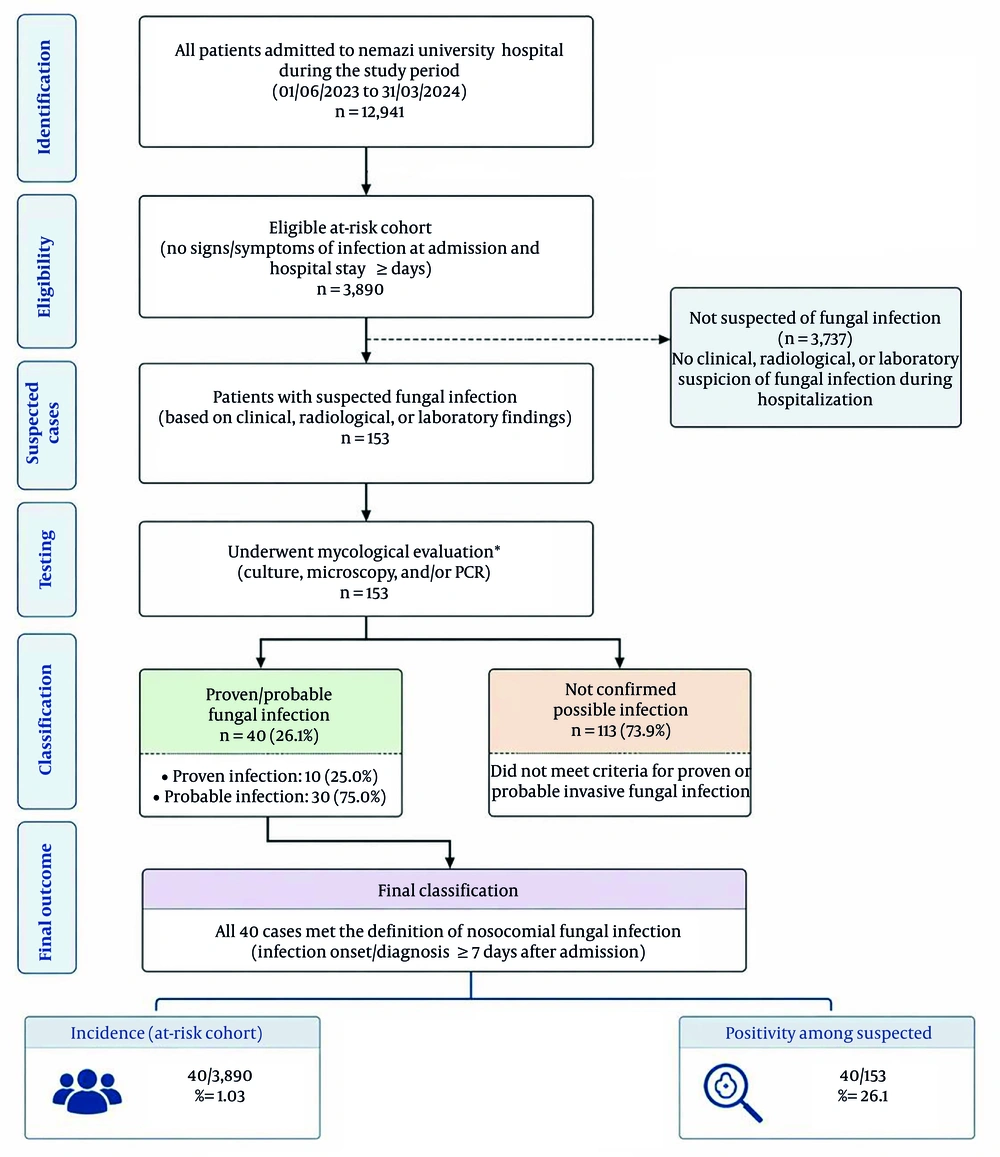
Figure 1.
Patient flow diagram of study participants
To evaluate fungal colonization in these patients (1, 9), clinical samples, including mouth, throat, and rectal swabs, as well as sputum and urine, were cultured on Sabouraud-dextrose agar (SDA, Merck, Darmstadt, Germany) with chloramphenicol (Merck, Darmstadt, Germany). Isolation of fungi from any site in patients without signs or symptoms of infection was considered colonization rather than fungal infection. To prevent contamination, laboratory tests were performed in a Class II biosafety cabinet. Demographic, radiological, and clinical data were collected from patient records.
3.1. Routine Lab Identification
In suspected patients, clinical samples, including midstream urine, wound swabs, sputum, bronchoalveolar lavage (BAL), cerebrospinal fluid (CSF), pleural fluid, abscess fluid, and tissue biopsies, were examined microscopically (10% KOH smear) and cultured on SDA with chloramphenicol. Blood samples were cultured in BACTEC medium (Becton–Dickinson, Sparks, MD, USA). Data related to pathological findings and radiological and clinical symptoms were extracted from the patient’s file, and the site of infection was determined based on all available data. The API 20C system (BioMérieux Vitek, St. Louis, MO, USA) was used to identify isolated Candida spp. Aspergillus species were identified macroscopically (colony morphology) and microscopically (lactophenol cotton blue smear).
3.2. Molecular Identification
In addition, blood samples and available clinical samples (such as CSF, sputum, BAL, tissue biopsy, and body fluids) were evaluated weekly by real-time PCR for the diagnosis of aspergillosis and candidiasis. DNA was extracted from 200 μL of clinical samples using the QIAmp DNA Minikit (Qiagen, Hilden, Germany) according to the manufacturer’s recommendations. The primers and TaqMan probes used in this study were purchased from Metabion (Martinsried, Germany). Candida DNA was detected using a real-time PCR assay designed by Shin et al. (10), and Aspergillus DNA was identified according to Kami et al. (11). Molecular laboratory procedures were performed in a laminar flow cabinet to minimize the possibility of environmental contamination.
3.3. Sensitivity Test
The susceptibility of mold isolates to antifungal agents [including amphotericin B (AMB), caspofungin (CAS), voriconazole (VOR), itraconazole (ITR), posaconazole (POS), luliconazole (LUL), and isavuconazole (ISA) (Sigma, UK)] was tested according to the Clinical and Laboratory Standards Institute (CLSI) M38-A2 and M61 guidelines. For yeast isolates, susceptibility testing was performed according to the CLSI M27, M59, and M60 guidelines. The final concentrations of the antifungal agents were as follows: AMB, CAS, VOR, ITR, and POS ranged from 8 to 0.016 µg/mL; FLU ranged from 32 to 0.064 µg/mL (for yeast isolates); and LUL and ISA ranged from 4 to 0.008 µg/mL. Two Candida species, C. parapsilosis ATCC 22019 and C. krusei ATCC 6258, were used as quality controls for the sensitivity tests in the present study.
3.4. Definition
There is no universal definition for the onset of a nosocomial fungal infection because the incubation period of the disease is unknown. However, in general, a nosocomial fungal infection usually begins after one week of hospitalization (12, 13). In this study, fungal infection was diagnosed based on the patient’s clinical and radiological symptoms. Sampling was performed immediately, and infection was confirmed by various laboratory tests, including microscopic examination, culture, and molecular tests.
Proven invasive mold infection was defined based on the identification of the etiologic agents by histopathological examination of sterile specimens (e.g., peritoneal fluid, CSF, or biopsy), in which hyphae and evidence of tissue damage were observed (7). Proven infection was also confirmed by isolating fungi from cultures of normally sterile specimens and/or amplifying fungal DNA by PCR when molds were observed in formalin-fixed paraffin-embedded tissue, together with clinical and radiological signs and symptoms (7). Proven candidiasis was defined as the isolation of Candida species from blood culture and histopathologic examination of a sterile specimen showing yeast cells, pseudohyphae, or true hyphae, with clinical or radiological evidence of infection (7). Probable invasive fungal infections were defined as the microscopic detection of fungal elements or isolation of molds (Aspergillus, Fusarium, and Mucorales) from sputum, BAL, bronchial brush, and sinus aspirate samples by culture. Probable mold infections may be defined as one positive PCR result for mold DNA in blood (plasma, serum, or whole), two or more consecutive PCR-positive results in BAL fluid, or a combination of one positive PCR result in blood and one positive result in BAL fluid (7). In clinical practice, when a patient shows symptoms of disease but the specific site cannot be determined, this is referred to as fever of unknown origin (FUO) (14). In this study, the site of infection in patients with FUO was designated as "other". These patients had two positive PCR results along with clinical symptoms.
Data were collected using the Statistical Package for the Social Sciences (SPSS) version 16 (International Business Machines Corp., USA). The distribution of fungal species and their susceptibility to antifungal agents were analyzed using descriptive statistics.
4. Results
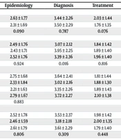
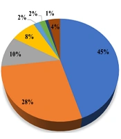
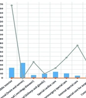
A total of 12,941 patients, with a mean hospitalization period of 7.41 days (range, 2.5 - 28.2 days), were admitted to various wards of Nemazi Hospital during the study period (Figure 2). The mean hospitalization periods and the number of mortalities in each ward are presented in Table 1. Of all admitted patients, 3,890 who had no signs or symptoms of infection at the time of admission were included in the present study. One hundred fifty-three patients were suspected of having nosocomial fungal infections based on the definition of nosocomial fungal infection (risk factor, mycological, clinical, and radiological criteria). The demographic characteristics of these patients are presented in Figure 3. The mean age of the evaluated patients was 40 years (range, 18 - 88 years), and 104 of 153 (68%) were male. Candida species were isolated from 41 patients (41/153, 26.8%) as colonization. The isolated Candida species included C. albicans, C. parapsilosis, C. glabrata, Kluyveromyces marxianus (C. kefyr), C. tropicalis, and Pichia kudriavzevii (C. krusei). Prophylactic antifungal therapy was administered to 87 of 153 (56.9%) patients (Figure 3).
Table 1.Number of Patients Admitted to Different Wards During the Study Period
| Ward | Number of Hospitalized Patients | Mean Hospitalization Period | The Number of Patients Who Died |
|---|---|---|---|
| Internal wards | |||
| Public interior | 808 | 4.9 | 167 |
| Internal (brain and nerves) | 1191 | 6.6 | 19 |
| Internal (oncology diseases) | 229 | 13.9 | 37 |
| Internal (kidneys and glands) | 434 | 8.1 | 27 |
| Special care units | |||
| Special cardiac care | 332 | 5.5 | 23 |
| Neurosurgery special care | 133 | 7.3 | 47 |
| Internal special care | 184 | 11.2 | 75 |
| Special care for surgery | 206 | 5.8 | 31 |
| Central special care | 157 | 6.3 | 19 |
| Surgery wards | |||
| Kidney and urinary tract surgery | 3655 | 3.8 | 30 |
| Bone and joint surgery | 589 | 9.3 | 23 |
| Heart surgery | 1028 | 2.5 | 25 |
| General surgery | 1037 | 5.2 | 25 |
| Surgery events | 1029 | 4.6 | 26 |
| Plastic Surgery | 569 | 5 | 20 |
| Neurosurgery | 836 | 4.1 | 14 |
| Bone marrow transplant ward | 73 | 28.2 | 15 |
| Solid organ transplant ward | 451 | 6.7 | 18 |
| Total | 12941 | 7.7 | 641 |
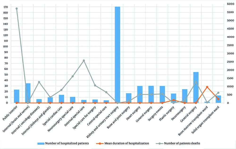
Figure 2.
Number of patients, mean hospitalization periods, and mortality rates of admitted cases in different hospital wards
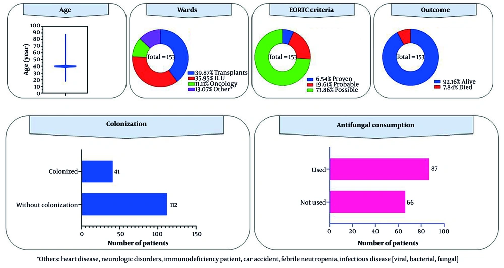
Figure 3.
Demographic characteristics of patients included in the study
A total of 612 blood samples and 405 clinical samples (including CSF, abdominal fluid, tissue, and sputum) from 153 patients suspected of having a nosocomial fungal infection were examined in the laboratory to identify infections. Forty patients were identified as having proven and probable nosocomial fungal infections. The demographic characteristics of patients with proven and probable fungal infections are shown in Table 2. Twenty-five (25/40, 62.5%) and 15 (15/40, 37.5%) patients were male and female, respectively. Ten patients (10/153, 6.54%) were diagnosed with proven nosocomial invasive fungal infections based on clinical, radiological, and mycological criteria. Candida albicans was identified in two blood cultures, one CSF sample, one abdominal fluid sample, and one liver abscess sample (five isolates in total), and C. glabrata was identified from two CSF samples. Sinusitis caused by Aspergillus species was identified in one case (based on a tissue sample). On pathological examination, Mucorales infection was confirmed in two patients. Probable cases were identified in 30 patients (30/153, 19.6%). Aspergillus flavus (2 isolates), A. fumigatus (1 isolate), and Mucorales species (1 isolate) were identified from sputum samples. Two consensus Aspergillus PCR results were positive in the blood of 18 immunocompromised patients (liver transplant recipients and patients with cancer) and in 8 BAL samples (Table 2). Patients who were clinically and radiologically suspected of having a fungal infection but whose disease was not confirmed by laboratory examination were classified as having a possible fungal infection.
Table 2.Demographic Data of Patients with Proven and Probable Nosocomial Fungal Infections a
| Variables | Values |
|---|---|
| Sex | |
| Male | 25 (62.5) |
| Female | 15 (37.5) |
| Age (y) | (18 - 85) 18.8 ± 15.1 |
| Colonization | |
| Colonized | 8 (20) |
| Without colonization | 32 (80) |
| Wards | |
| Transplant (Kidney and liver) | 18 (45) |
| Intensive care unit | 11 (27.5) |
| Oncology | 8 (20) |
| Surgery | 2 (5) |
| Trauma | 1 (2.5) |
| Background | |
| Transplant (Kidney and liver) | 21 (52.5) |
| Malignancy | 8 (20) |
| Diabetes mellitus | 3 (7.5) |
| Surgical operation | 8 (20) |
| Site infections | |
| Liver | 14 (35) |
| Lung | 12 (30) |
| Brain | 3 (7.5) |
| Sinuses | 3 (7.5) |
| Abdominal | 1 (2.5) |
| Others b | 7 (17.5) |
| Antifungal therapy | |
| Not used | 6 (15) |
| Used | 34 (85) |
| EORTC criteria | |
| Proven | 10(25) |
| Probable | 30(75) |
| Etiologic agents | |
| Positive pathology results | |
| Aspergillus species | 1 (2.5) |
| Mucor species | 2 (5) |
| Isolation from culture | |
| Candida albicans | 5 (12.5) |
| Candida glabrata | 2 (5) |
| Aspergillus flavus | 2 (5) |
| Aspergillus fumigatus | 1 (2.5) |
| Mucorales | 1 (2.5) |
| Positive PCR results | |
| Aspergillus species | 26 (65) |
| Outcome | |
| Alive | 36 (90) |
| Died | 4 (10) |
a Values are expressed as No. (%) or (min - max) mean ± SD.
b Include a patient with a fever of unknown origin (FUO) with positive Aspergillus PCR in the blood samples, with signs and symptoms of infection.
The rate of probable and proven nosocomial fungal infections in this study was 1.03% (40/3,890). All proven and probable cases (n = 40) met the predefined criterion for fungal nosocomial infection according to the EORTC criteria (7). The most common risk factors were transplantation, hematologic disorders, and prolonged ICU stays. The most common sites of fungal infection were the liver, lungs, and brain (Figure 4). The overall mortality rate during the study period was 4.95% (641/12,941). However, the mortality rate among patients with proven and probable nosocomial fungal infections was 10% (4/40 cases).
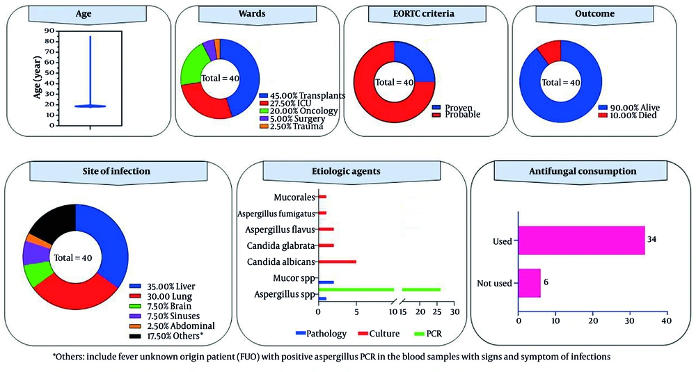
Figure 4.
Details of patients with proven and probable nosocomial infections
In the present study, 52 fungal species were isolated from patients’ clinical samples (colonization or pathogen). These included C. albicans (21/52, 40.4%), followed by C. glabrata (12/52, 23.1%), K. marxianus (2/52, 3.8%), P. kudriavzevii (5/52, 9.6%), C. parapsilosis (5/52, 9.6%), C. tropicalis (3/52, 5.8%), A. fumigatus (1/52, 1.9%), A. flavus (2/52, 3.8%), and Mucorales species (1/52, 2%). The results of antifungal susceptibility tests for the isolated species are reported in Table 3.
Table 3.In Vitro Susceptibility Patterns of Isolated Species to Antifungal Agents a
| Isolates | No. of Isolates with MIC (µg/ml) | ||||||||||
|---|---|---|---|---|---|---|---|---|---|---|---|
| 0.008 | 0.016 | 0.032 | 0.064 | 0.125 | 0.250 | 0.500 | 1 | 2 | 4 | ≥8 | |
| Aspergillus flavus / (2 isolates) | |||||||||||
| Amphotericin B | 1 | 1 | |||||||||
| Caspofungin | 1 | 1 | |||||||||
| Voriconazole | 1 | 1 | |||||||||
| Itraconazole | 1 | 1 | |||||||||
| Posaconazole | 2 | ||||||||||
| Luliconazole | 1 | 1 | |||||||||
| Isavuconazole | 1 | 1 | |||||||||
| Aspergillus fumigatus / (1 isolates) | |||||||||||
| Amphotericin B | 1 | ||||||||||
| Caspofungin | 1 | ||||||||||
| Voriconazole | 1 | ||||||||||
| Itraconazole | 1 | ||||||||||
| Posaconazole | 1 | ||||||||||
| Luliconazole | 1 | ||||||||||
| Isavuconazole | 1 | ||||||||||
| Candida albicans / (21 isolates) | |||||||||||
| Amphotericin B | 3 | 2 | 1 | 8 | 2 | 4 | 1 | ||||
| Caspofungin | 12 | 3 | 4 | 1 | 1 | ||||||
| Voriconazole | 10 | 6 | 2 | 1 | 2 | ||||||
| Fluconazole | 1 | 1 | 2 | 4 | 9 | 4 | |||||
| Itraconazole | 3 | 4 | 6 | 3 | 1 | 4 | |||||
| Posaconazole | 2 | 1 | 5 | 4 | 2 | 4 | 2 | 1 | |||
| Luliconazole | 3 | 2 | 1 | 1 | 2 | 2 | 8 | 1 | 1 | ||
| Isavuconazole | 3 | 4 | 1 | 3 | 1 | 2 | 2 | 2 | 1 | ||
| Candida glabrata / (12 isolates) | |||||||||||
| Amphotericin B | 2 | 3 | 5 | 2 | |||||||
| Caspofungin | 2 | 2 | 5 | 3 | |||||||
| Voriconazole | 1 | 5 | 2 | 1 | 2 | 1 | |||||
| Fluconazole | 1 | 1 | 3 | 7 | |||||||
| Itraconazole | 1 | 1 | 2 | 7 | 1 | ||||||
| Posaconazole | 2 | 5 | 2 | 3 | |||||||
| Luliconazole | 1 | 4 | 2 | 1 | 1 | 1 | 1 | 1 | |||
| Isavuconazole | 3 | 6 | 2 | 1 | |||||||
| Candida kefyr / (2 isolates) | |||||||||||
| Amphotericin B | 1 | 1 | |||||||||
| Caspofungin | 2 | ||||||||||
| Voriconazole | 2 | ||||||||||
| Fluconazole | 1 | 1 | |||||||||
| Itraconazole | 1 | 1 | |||||||||
| Posaconazole | 1 | 1 | |||||||||
| Luliconazole | 2 | ||||||||||
| Isavuconazole | 2 | ||||||||||
| Candida krusei / (5 isolates) | |||||||||||
| Amphotericin B | 1 | 1 | 1 | 1 | 1 | ||||||
| Caspofungin | 1 | 1 | 1 | 1 | 1 | ||||||
| Voriconazole | 2 | 2 | 1 | ||||||||
| Fluconazole | 2 | 1 | 2 | ||||||||
| Itraconazole | 3 | 1 | |||||||||
| Posaconazole | 3 | 1 | 1 | ||||||||
| Luliconazole | 1 | 1 | 1 | 2 | |||||||
| Isavuconazole | 2 | 1 | 1 | 1 | |||||||
| Candida parapsilosis (5 isolates) | |||||||||||
| Amphotericin B | 1 | 2 | 2 | ||||||||
| Caspofungin | 1 | 2 | 1 | 1 | |||||||
| Voriconazole | 1 | 2 | 2 | ||||||||
| Fluconazole | 1 | 2 | 2 | ||||||||
| Itraconazole | 1 | 4 | |||||||||
| Posaconazole | 1 | 2 | 2 | ||||||||
| Luliconazole | 1 | 3 | 1 | ||||||||
| Isavuconazole | 2 | 2 | 1 | ||||||||
| Candida tropicalis / (3 isolates) | |||||||||||
| Amphotericin B | 1 | 2 | |||||||||
| Caspofungin | 2 | 1 | |||||||||
| Voriconazole | 1 | 1 | 1 | ||||||||
| Fluconazole | 1 | 1 | 1 | ||||||||
| Itraconazole | 1 | 1 | 1 | ||||||||
| Posaconazole | 1 | 1 | 1 | ||||||||
| Luliconazole | 1 | 1 | |||||||||
| Isavuconazole | 2 | 1 | 1 | ||||||||
a Abbreviation: MIC, minimum inhibitory concentration.
Caspofungin and VOR demonstrated lower MIC values against Candida albicans isolates than other tested agents. Luliconazole and ISA showed the lowest MIC values against C. glabrata. The minimum inhibitory concentration geometric mean values (MICGM) for all Candida species isolates of AMB, CAS, FLU, VOR, ITR, POS, LUL, and ISA were 0.253, 0.213, 9.137, 0.147, 0.94, 0.487, 0.449, and 0.157 µg/mL, respectively. The geometric mean values for Aspergillus species isolates of AMB, CAS, VOR, ITR, POS, LUL, and ISA were 1.166, 0.117, 0.458, 1.166, 2.333, 0.013, and 0.211 µg/mL, respectively.
According to reliable sources, some fungi isolated from patients in this study do not have a specific breakpoint for certain drugs. Table 4 shows the breakpoints of isolated fungi relative to antifungal drugs.
Table 4.Clinical Breakpoints for Isolated Fungi According to CLSI Documents M59, M60, and M61
| Fungal Species and Antifungals | ECV for species Without a Breakpoint | ECV for Species with a Breakpoint | Breakpoints | |||
|---|---|---|---|---|---|---|
| Sensitive | Intermediate | Susceptible dose dependent | Resistant | |||
| A. flavus | ||||||
| Amphotericin B | 4 | - | - | - | - | - |
| Caspofungin | 0.5 | - | - | - | - | - |
| Voriconazole | 2 | - | - | - | - | - |
| Posaconazole | 0.5 | - | - | - | - | - |
| Itraconazole | 1 | - | - | - | - | - |
| Isavuconazole | 1 | - | - | - | - | - |
| A. fumigatus | ||||||
| Amphotericin B | 2 | - | - | - | - | - |
| Caspofungin | 0.5 | - | - | - | - | - |
| Voriconazole | 1 | - | - | - | - | - |
| Posaconazole | 2 | - | - | - | - | - |
| Itraconazole | 1 | - | - | - | - | - |
| Isavuconazole | 1 | - | - | - | - | - |
| C. albicans | ||||||
| Amphotericin B | 2 | - | - | - | - | - |
| Caspofungin | - | - | ≤ 0.25 | 0.5 | - | ≥1 |
| Voriconazole | - | 0.03 | ≤ 0.12 | 0.25 - 0.5 | - | ≥1 |
| Fluconazole | - | 0.5 | ≤ 2 | - | 4 | ≥ 8 |
| Posaconazole | 0.06 | - | - | - | - | - |
| Itraconazole | - | - | - | - | - | - |
| Isavuconazole | - | - | - | - | - | - |
| C. glabrata | ||||||
| Amphotericin B | 2 | - | - | - | - | - |
| Caspofungin | - | - | ≤ 0.12 | 0.25 | - | ≥ 0.5 |
| Voriconazole | 0.25 | - | - | - | - | - |
| Fluconazole | - | 8 | - | - | ≤ 32 | ≥ 64 |
| Posaconazole | 1 | - | - | - | - | - |
| Itraconazole | 4 | - | - | - | - | - |
| Isavuconazole | - | - | - | - | - | - |
| C. krusei | ||||||
| Amphotericin B | 2 | - | - | - | - | - |
| Caspofungin | - | - | ≤ 0.25 | 0.5 | - | ≥1 |
| Voriconazole | - | 0.5 | ≤ 0.12 | 0.25 - 0.5 | - | ≥1 |
| Fluconazole | - | - | - | - | - | - |
| Posaconazole | 0.5 | - | - | - | - | - |
| Itraconazole | 1 | - | - | - | - | - |
| Isavuconazole | - | - | - | - | - | - |
| C. parapsilosis | ||||||
| Amphotericin B | 2 | - | - | - | - | - |
| Caspofungin | - | - | ≤ 2 | 4 | - | ≥ 8 |
| Voriconazole | - | 0.03 | ≤ 0.12 | 0.25 - 0.5 | - | ≥1 |
| Fluconazole | - | 1 | ≤ 2 | 4 | ≥ 8 | |
| Posaconazole | 0.25 | - | - | - | - | - |
| Itraconazole | - | - | - | - | - | - |
| Isavuconazole | - | - | - | - | - | - |
| C. tropicalis | ||||||
| Amphotericin B | 2 | - | - | - | - | - |
| Caspofungin | - | - | ≤ 0.25 | 0.5 | - | ≥1 |
| Voriconazole | - | 0.12 | ≤ 0.12 | 0.25 - 0.5 | - | ≥1 |
| Fluconazole | - | 1 | ≤ 2 | 4 | ≥ 8 | |
| Posaconazole | 0.12 | - | - | - | - | - |
| Itraconazole | 0.5 | - | - | - | - | - |
| Isavuconazole | - | - | - | - | - | - |
5. Discussion
Invasive fungal infections are associated with high morbidity and mortality rates in hospitals. The diagnosis of these infections is challenging because the signs and symptoms often mimic those of other bacterial infections. Risk factors for nosocomial fungal infections are increasing (5). The identification of underlying host diseases, early diagnosis, and timely initiation of antifungal drugs have a significant impact on effective patient management (6). In the present study, the rate of proven and probable nosocomial fungal infections was 1.03%, and mold fungi (Aspergillus and Mucor species) were the most common etiologic agents. A study by Zingg et al. reported that, of 392 microorganisms isolated from 342 (44%) health-care-associated infections, 28 (7%) were fungi, including Aspergillus and Candida species (15). Among ICU patients in China, the prevalence of fungal septicemia from January 2008 to December 2017 was 0.38% (81/21098 cases), with a mortality rate of 36% (29 patients) (4). The incidence of these infections varies among regions and is related to healthcare management. The higher rate of proven and probable nosocomial fungal infections in our study may be attributed to the fact that Nemazi Hospital is a large, specialized hospital in the south of Iran and served as a center for oncology and liver transplants during this study. Immunocompromised patients had the highest risk of nosocomial fungal infections, and their clinical outcomes were unfavorable.
The most common risk factors for nosocomial fungal infections in the present study included prolonged ICU stays and a high prevalence of immunocompromised conditions, such as transplantation or cancer. These findings are consistent with previous studies indicating that solid organ recipients (16) and patients with viral infections, such as AIDS (acquired immunodeficiency syndrome) and COVID-19 (17, 18), are at risk for fungal infections. In the present study, 20% of infected patients (8/40) had fungal colonization; however, in Van Bang et al., 90% of burn patients were colonized with fungi, and heavy colonization with Candida species was reported to be an independent predictor of fungal infections (19).
In the current study, respiratory infections (in the lungs and sinuses) were the most common, followed by infections in the liver and brain. Wang et al. reported that the respiratory system was the most common site of infection (59.0%), followed by intra-abdominal infection (8.8%) (20). A global analysis of nosocomial fungal infections in 1,149 hospitals (29 countries) identified bloodstream infections as the most prevalent (343, 45%), followed by lower respiratory tract (171, 22%) and gastrointestinal infections (64, 8%) (21). The sites of nosocomial infections vary depending on the patient population and the route of infection. The common etiologic agents of invasive fungal infections in the present study were Candida and Aspergillus species. Candida species are frequently found at different body sites, including the oral cavity, skin, and mucous membranes. Depending on factors such as the pathogenicity of Candida species and the severity of host immune deficiency, Candida can lead to severe clinical outcomes, including sepsis and death (21). In studies conducted by Obeed et al. in Basrah Province Hospital, Candida albicans, C. glabrata, C. tropicalis, and C. parapsilosis emerged as the most prevalent etiological agents of nosocomial fungal infections. Similarly, Harrington et al. reported Candida species, such as C. tropicalis, C. parapsilosis, P. kudriavzevii, and C. glabrata, as the primary causative agents of candidemia and invasive candidiasis (22). Wang et al. identified C. albicans as the etiologic agent in 68.0% of nosocomial fungal infections among cancer patients (20). In the present study, consistent with these findings, C. albicans, C. glabrata, and C. parapsilosis were isolated from colonized and infected patients. These results align with previously published data. Reducing the rate of Candida infections requires strategies such as minimizing unnecessary antibiotic use, controlling underlying medical conditions, and implementing stringent hygiene and infection control practices in hospitals (23).
Aspergillus species are abundant in the environment and can cause local or systemic infections in humans. The primary source of nosocomial aspergillosis is contaminated air containing Aspergillus conidia; however, conidia of this organism can also be isolated from water and equipment in patient rooms (24). With the growing number of immunocompromised patients, especially those undergoing transplantation or diagnosed with leukemia, the incidence of Aspergillus infection has increased significantly (16, 17). The morbidity and mortality rates of aspergillosis among immunocompromised patients are high. For example, Aspergillus species accounted for 64.7% (11/17) of fungal infections in ICU departments in Iran (25). Similarly, a study by González-García et al. in Spain (1997 - 2017) reported a 24.5% mortality rate among 32,960 patients infected by Aspergillus species (26). Furthermore, an extensive review of 458 patients involved in one nosocomial outbreak revealed that 53 patients developed aspergillosis, with a mortality rate exceeding 50% among immunocompromised patients, including those undergoing bone marrow or solid organ transplants, or those with hematologic disorders and severe immunodeficiency (6). Consistent with these findings, the present study identified Aspergillus species as one of the etiologic agents of nosocomial fungal infections, further emphasizing their significant role in hospital-acquired infections.
Mucormycosis is an opportunistic fungal infection commonly observed in immunocompromised patients. It can manifest in various forms, including pulmonary, sinusitis, rhino-cerebral, gastrointestinal, and cutaneous infections (27). Infections are typically transmitted through airborne particles from sources such as contaminated hospital ventilation systems, water-damaged plaster, wooden tongue depressors, and hospital linens (27). A study by Mitchell et al. reported that among 2,453 cases admitted to the burn center, 12 patients (4.9 per 1000 admissions) had evidence of mucormycosis. Infections were confirmed by histopathological examination of biopsy samples, and one was identified by wound cultures (28). The sites of infection in seven patients involved multiple organs (58.3%), with upper-extremity (head or face) involvement in three patients. The overall mortality in these patients was 92% (11/12) (28). In our study, three cases of mucormycosis were identified, reflecting a low but consistent prevalence of this infection. Two patients were diagnosed by pathologists who observed the pathogen in tissue, and in one patient, the pathogen was isolated from a culture sample.
In the present study, based on the MIC values and CLSI breakpoints, the isolated Candida species were susceptible to all antifungal agents except FLU (GM 9.137 µg/mL). Voriconazole, ISA, and CAS, with MICGM values of 0.147, 0.157, and 0.213 µg/mL, showed the greatest growth inhibitory effect against Candida species in vitro, as demonstrated in the study by Badiee et al. (29). In Brazil, C. albicans isolated from blood cultures presented FLU MIC50 and MIC90 values of 0.5 and 1.0 µg/mL, respectively (30). In Thailand, the FLU MIC90 value for C. albicans was reported to be 1 µg/mL (31).
In the present study, Aspergillus species demonstrated lower MICGM values for LUL, 0.013 µg/mL; CAS, 0.117 µg/mL; and ISA, 0.211 µg/mL, because LUL and ISA are expensive in our region and are not prescribed for routine treatment. A study conducted by Badiee et al. (32) across 11 university hospitals in Iran identified LUL, ISA, and CAS as effective antifungal agents against all Aspergillus species isolates, while ITR was found to be ineffective against this microorganism. In contrast, Hivary et al. reported resistance rates of 86.5%, 54.1%, and 83.8% among Aspergillus isolates to AMB, CAS, and POS, respectively (31). The MICGM values (µg/mL) of isolated A. fumigatus were reported in Hivary et al. as AMB, 0.567 µg/mL; CAS, 0.062 µg/mL; VOR, 0.085 µg/mL; POS, 0.049 µg/mL; and ITR, 0.520 µg/mL (31). The observed variability in susceptibility patterns across regions is likely related to differences in antifungal use for treatment, prophylaxis, and patient management practices. Although certain antifungal agents demonstrated lower MIC values, these results reflect in vitro susceptibility patterns, and the number of isolates was limited; therefore, these results should not be directly interpreted as clinical effectiveness.
The limitations of our study included a lack of detailed information on chemotherapy and antimicrobial regimens administered to patients, as well as the absence of certain laboratory methods, such as galactomannan and β-D-glucan tests, which were not performed. This limitation prevented us from evaluating the impact of several established risk factors, including prior broad-spectrum antibiotic exposure, chemotherapy, and other treatment-related factors, on the development of nosocomial fungal infections. Previous studies from Iran have identified malignancy, prolonged hospitalization, central venous catheterization, and prior antibiotic exposure as important risk factors for invasive fungal infections and candidemia (33, 34). Although strict temporal and clinical criteria were applied, distinguishing nosocomial infection from pre-existing colonization remains challenging, particularly in immunocompromised patients. Additionally, because diagnostic testing was primarily initiated based on clinical suspicion, subclinical or asymptomatic cases may have been underdetected. Another limitation of this study was the small number of fungi isolated from patients, which meant that we could not estimate MIC50, MIC90, and epidemiological cutoff values for the isolates. We encourage future studies to focus on nosocomial fungal infections, evaluate their etiologic agents and risk factors, and incorporate advanced diagnostic techniques.
Hospitals are increasingly challenged by the rising incidence of invasive fungal infections, which are associated with high mortality rates and complex treatment processes. Therefore, early diagnosis and preventive measures are essential for effective infection control. Various fungal species with different sensitivities to antifungal agents were identified in this study. Liver transplant recipients, patients with hematologic disorders, and patients admitted to ICU wards represented the highest-risk groups for these infections. The respiratory tract, particularly the lungs and sinuses, was the most common site of infection. These findings underscore the need for targeted strategies to reduce the burden of nosocomial fungal infections.