Cholesterol accumulation in liver cells of individuals with NASH leads to cell death and fibrosis, significantly increasing mortality rates. This fibrotic process is marked by the activation of HSCs (
28,
29), synthesis of ECM, and chronic inflammation, driven by an imbalance in ECM turnover influenced by cytokines, growth factors, and ROS (
30). The TGF-β is particularly important, promoting collagen I (Col-alpha1) and α-SMA expression through SMAD2/3 signaling pathways. NADPH oxidase enzymes, especially NOX1 and NOX4, play an essential role in HSC activation and ECM synthesis, exacerbated by high cholesterol intake, which heightens sensitivity to TGF-β and contributes to the progression of NASH, fibrosis, and cirrhosis (
31,
32). The mechanism of liver fibrosis involves HSC activation and trans differentiation into MFBs, leading to excessive ECM deposition. Chronic inflammation and oxidative stress, mainly through ROS, enhance TGF-β signaling, amplifying the fibrotic response. SMAD3, a key mediator of TGF-β, drives collagen and α-SMA expression, promoting fibrosis. High cholesterol exacerbates this process by increasing ROS production, which further activates HSCs and amplifies inflammation, accelerating the progression of NASH to advanced fibrosis and cirrhosis (
28,
29). The WJ-MSCs are a promising therapeutic option due to their differentiation potential and ethical advantages, despite challenges such as potential rejection and limited lifespan. Their therapeutic effects are largely attributed to their paracrine function through exosomes, which transport proteins and nucleic acids that regulate signaling pathways and facilitate tissue regeneration (
33). Polyinosinic: Polycytidylic acid exhibits preventive and antifibrogenic effects in liver fibrosis, primarily associated with increased IFN-γ levels. It induces HSC death, causes cell cycle arrest, and enhances NK cell cytotoxicity (
34,
35). While the precise mechanisms and anti-inflammatory properties of poly I:C are still not fully understood, it is believed to modulate the immune response through its interaction with Toll-like receptor 3 (TLR3), leading to the secretion of cytokines that influence both the immune microenvironment and fibrosis progression. Importantly, poly I:C may also alter the cargo of exosomes released by MSCs, potentially enriching them with antifibrotic molecules, such as specific microRNAs (miRNAs) or proteins, which can target key signaling pathways like TGF-β/Smad3. This modulation of exosome content could be a critical factor in the observed reduction of HSC activation and ECM deposition, highlighting the potential of poly I:C-stimulated MSC-derived exosomes as a novel therapeutic approach for liver fibrosis (
36). Our study investigates the effects of poly I:C-activated MSC exosomes on collagen I and α-SMA expression, as well as SMAD3 phosphorylation, in LX2 cells with cholesterol accumulation. The results highlight the therapeutic efficacy of these exosomes in influencing critical fibrosis pathways, providing valuable insights for the management of liver fibrosis.
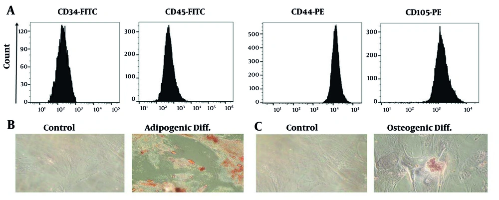
In our study, we characterized the cell surface indicators of WJ-MSCs through flow cytometry, confirming the absence of hematopoietic stem cells and monocyte macrophages, while detecting the presence of CD44 and CD105 — markers commonly associated with MSCs. In contrast, Urvi et al. (
37) utilized a different panel of markers, highlighting CD105 and CD90, while noting the lack of CD45 and CD34. This difference in marker selection (CD44 in our study versus CD90 in theirs) underscores variability in experimental design and research objectives. Nonetheless, both studies agree on the expression of CD105 as a critical MSC marker. The absence of expression of CD34 and CD45 aligns with the expected MSC phenotype, as these markers are typically linked to hematopoietic stem cells. Our findings also demonstrated the differentiation potential of WJ-MSCs into osteocytes and adipocytes, supported by Alizarin Red and Oil Red O staining. While the comparative study did not detail specific differentiation outcomes, the expression of CD90 and CD105 suggests the multipotent capabilities of MSCs, which include osteogenic and adipogenic differentiation potential.
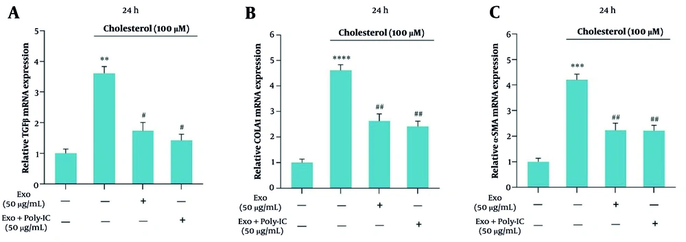
Our research revealed that cholesterol treatment significantly elevated the mRNA expression levels of TGF-β, α-SMA, and collagen1 genes in LX2 cells, indicating the development and progression of liver fibrosis. This result aligns with the conclusions of Rashidi et al. (
38), which also reported increased expression of these fibrosis-related genes following cholesterol exposure, highlighting the critical role of cholesterol in activating HSCs. Notably, treatment with unstimulated exosomes resulted in a substantial reduction in the expression of these genes, suggesting their potential therapeutic role in mitigating fibrosis, which aligns with the comparative study showing that exosomes derived from WJ-MSCs significantly decreased TGF-β, α-SMA, and collagen1α expression. Furthermore, our study revealed that poly I:C-stimulated exosomes exhibited an even greater reduction in TGF-β and α-SMA levels, indicating their enhanced therapeutic potential compared to unstimulated exosomes. These findings suggest that stimulated exosomes may offer a promising strategy to amplify the protective effects against cholesterol-induced liver fibrosis. Overall, the consistency between our results and those of prior studies emphasizes the significant influence of cholesterol on fibrosis development and underscores the therapeutic promise of exosomes in the management of liver fibrosis.
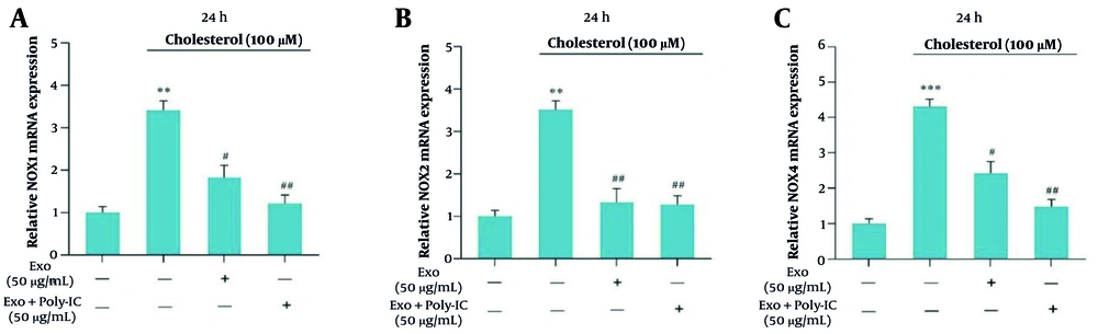
In our study, we examined the therapeutic effects of exosomes on the mRNA expression of NOX1, NOX2, and NOX4 genes in LX2 cells exposed to 100 μM cholesterol for a 24-hour period. We discovered that unstimulated exosomes significantly decreased NOX gene expression, while poly I:C-stimulated exosomes led to an even more pronounced reduction, particularly for NOX1 and NOX4, in fibrotic cells. This aligns with the findings of Afarin et al. (
17), which indicated that TGF-β1 treatment increased NOX1, NOX2, and NOX4 expression in LX2 cells. Subsequent treatment of TGF-β1-activated HSCs with exosomes derived from WJ-MSCs at concentrations of 40 and 50 μg/mL resulted in a significant reduction in NOX gene expression after 24 hours. Both studies highlight the healing capabilities of exosomes in mitigating NOX gene expression, which is vital for the progression of fibrosis. The rise in NOX expression due to TGF-β1 treatment parallels our findings related to cholesterol, suggesting that both factors activate similar fibrogenic pathways.
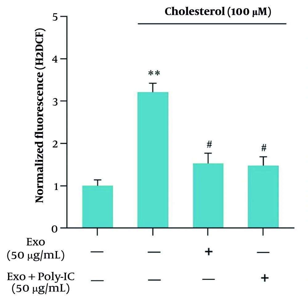
Our study investigated the influence of exosome treatment on the expression of NOX genes in LX2 cells stimulated with cholesterol. The results demonstrated that both unstimulated and poly I:C-stimulated exosomes significantly reduced the expression of NOX1 and NOX4 genes. These results align with the Asadizade et al. (
39) study, which reported increased NOX expression following TGF-β1 treatment and a subsequent decrease upon treatment with WJ-MSC-derived exosomes. Additionally, our research showed that cholesterol treatment resulted in a significant increase in ROS production in LX2 cells, whereas exosome treatment led to a notable reduction in ROS levels. These results align with the findings of Asadizade et al., which highlighted the antioxidant effects of curcumin. Together, these studies underscore the therapeutic potential of exosomes and curcumin in mitigating oxidative stress and liver fibrosis progression (
39). Overall, both investigations highlight the importance of exosomes and curcumin as promising therapeutic strategies for managing oxidative stress and treating liver fibrosis.
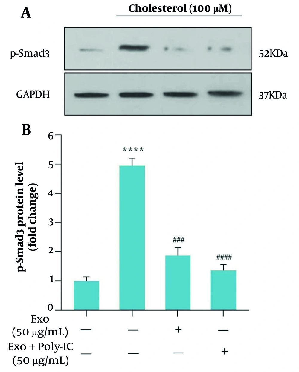
In our study, exosomes derived from WJ-MSCs significantly inhibited the activation of HSCs via the TGF-β/Smad3 pathway. Notably, treatment with these exosomes resulted in a marked reduction in Smad3 phosphorylation, a key mediator in this pathway, as demonstrated by Western blot analysis. This finding suggests a promising therapeutic potential for WJ-MSC-derived exosomes in mitigating liver fibrosis, particularly in the context of cholesterol-induced damage. Additionally, the study by Urvi et al. similarly found that curcumin inhibits Smad3 phosphorylation in TGF-β-activated HSCs, reinforcing the notion of its antifibrotic properties. While both WJ-MSC-derived exosomes and curcumin target the same signaling pathway, they differ in their mechanisms of action; curcumin acts as a small molecule inhibitor, while WJ-MSC-derived exosomes operate as biological entities (
37). This distinction highlights their unique therapeutic strategies, as curcumin demonstrates concentration-dependent effects, whereas WJ-MSC-derived exosomes exhibit efficacy at lower concentrations over shorter incubation times. Overall, both approaches underscore the critical role of the TGF-β/Smad3 pathway in liver fibrosis and present valuable options for therapeutic intervention.
Rashidi et al. further corroborate the inhibitory effect of exosomes derived from WJ-MSCs on Smad3 phosphorylation in HSCs treated with TGF-β (
38). In contrast to our study, which focused on cholesterol-induced damage, Rashidi et al. employed a direct TGF-β challenge. Their results showed that exosomes significantly reduced Smad3 phosphorylation across various concentrations, highlighting the versatility and robustness of exosomal intervention (
38). This study complements our findings by providing additional evidence that exosomes can counteract TGF-β-induced cellular responses, reinforcing the potential of exosomal therapies in diverse pathological contexts.
Collectively, these studies and our results present a consistent narrative where both natural compounds, such as curcumin, and biological products, like WJ-MSC exosomes, can effectively disrupt the TGF-β/Smad3 cascade, which is pivotal for HSC activation and the advancement of liver fibrosis. This comparison underscores the diverse strategies available for modulating this pathway and suggests the potential for integrative approaches that combine the advantages of small molecules and bioengineered products in the treatment of hepatic fibrosis. However, it is important to note that our study was conducted in vitro, and there were no specific eligibility criteria or control interventions, which should be considered in future studies. Additionally, while the study provides promising results, further research is required to evaluate the efficacy of exosome-based therapies in in vivo models and clinical settings, where factors such as bioavailability, delivery mechanisms, and long-term effects may influence therapeutic outcomes.
5.1. Conclusions
Our study demonstrates that exosomes derived from WJ-MSCs, particularly when stimulated with poly I:C, have significant therapeutic potential in modulating liver fibrosis pathways. Cholesterol accumulation in NASH patients leads to HSC activation, excessive ECM synthesis, and oxidative stress, primarily driven by TGF-β and NOX enzymes. Our results indicate that WJ-MSC-derived exosomes can inhibit Smad3 phosphorylation and the TGF-β/Smad pathway, thereby reducing fibrosis-related gene expression and oxidative stress. The enhanced efficacy of stimulated exosomes compared to unstimulated ones highlights the importance of exosome activation in therapy optimization. Overall, this research underscores the multifaceted nature of liver fibrosis and the promising function of WJ-MSC-derived exosomes in its management, paving the way for future therapeutic applications.