
1. Introduction
Calcium amorphous tumor (CAT) is a rare non-malignant neoplasm that is diagnosed based on pathological findings. The pathogenesis and natural history of this condition are still unknown. Since its initial report in 1997 (1), fewer than 100 cases have been documented up to 2023 (2). Given the limited number of reported cases, numerous critical inquiries regarding its management and treatment remain unanswered (CAT is deferent from MAC- Mitral annular calcification [MAC] is a chronic degenerative process that implies calcification on the mitral support structure).
In most CAT cases, the mass is identified due to its complications such as cerebral stroke (3, 4), acute myocardial infarction (5), and dyspnea (5).Since the nature of the cardiac mass and its distinction from malignant tumors with internal calcification (such as calcified myxoma and osteosarcoma) are not accurately achievable through echocardiography or CT scan (2),it is recommended to remove the mass by surgery. In fact, CAT is typically diagnosed post-surgery. So, introducing new cases of this condition can help enrich available information and develop comprehensive management and treatment guidelines. This report presented one of the largest CAT masses reported so far, and most previous reports have described relatively smaller masses.
2. Case Presentation
A 72-year-old man was referred with complaints of dyspnea, dizziness and, fainting over the past month. The patient reported a history of hypertension and asthma. Furthermore, an ECG examination revealed that the patient had flutter rhythm/normal axis/T tall in V1-6. The findings of echocardiography revealed the following:
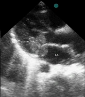
Ejection fraction (EF = 55%), severe left atrial enlargement (LAE), Doming of anterior mitral valve leaflet (AMVL) with restricted motion the posterior mitral valve leaflet (PMVL), severe mitral valve stenosis (Mitral valve Area (MVA) = 1.17 cm2), moderate to severe mitral regurgitation, a large homogenous encapsulated mass in posterior left ventricular (LV) wall (56 × 34 mm), and mild to moderate TR (tricuspid regurgitation) (TRG = 45 MMHG, PAP = 55 MMHG).
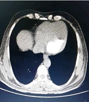
The patient underwent angiography to evaluate coronary arteries, which rendered normal findings. A calcified mass was observed in the chest CT scan (Figures 1 and 2). The patient had no history of renal disorder, calcium/phosphorus abnormalities, or diabetes. Given the patient’s presentation of dyspnea and the presence of a mass observed in echocardiography, the clinical decision was made to perform heart surgery and mitral valve replacement.
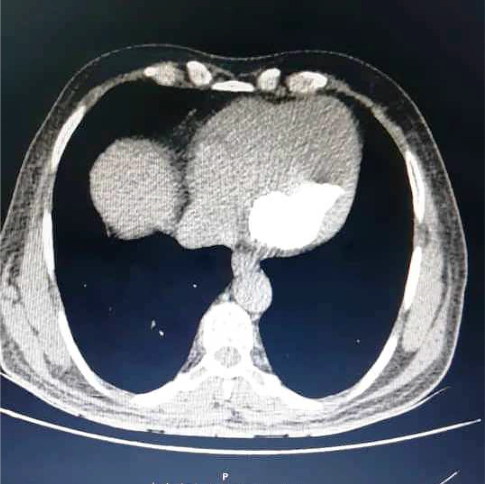
Figure 1.
The CT scan image of an intracardiac intensely calcified mass
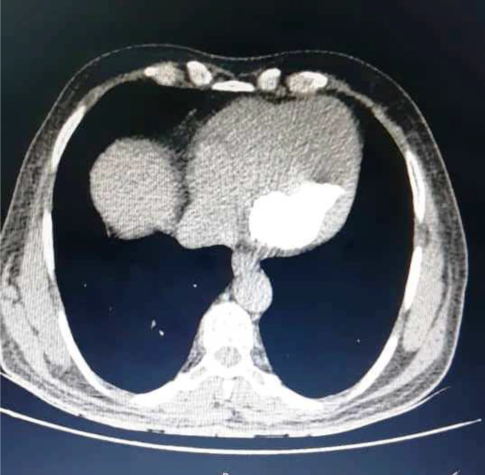
Figure 2.
The CT scan image of the mass
After middle sternotomy and cannulation of the aorta and both Vena cava veins, CPB (cardio pulmonary bypass) was established. After aortic clamping, cardiac arrest was induced via the antegrade cardioplegia injection of the Del Nido solution. After opening the left atrium, evidence of rheumatic involvement of the mitral valve and MAC was observed. In the posterior wall of the left ventricle and posterior to the mitral valve, a firm mass with a calcified wall was palpated within the muscles intramurally. Gradually, a mass with a size of approximately 40 × 60 cm was released, followed by the leak of its pasty white content. The mass capsule and its content were completely removed, and the residual empty space was debrided and cleaned. Then using a thin strip from Teflon felt, the empty space was closed, and the posterior mitral annulus was reconstructed. Valve leaflets were resected, and using the interrupted suturing method, a biological valve (Magna Ease) was placed in the mitral position. Other surgical procedures were performed according to standard protocols. The pathological examination of the mass confirmed the diagnosis of a calcified amorphous tumor. The patient underwent treatment with an anticoagulant (warfarin) and was then discharged. In serial echocardiography examinations during a six-month follow-up, no evidence of mass recurrence was observed.
3. Discussion
Calcium amorphous tumor is a non-neoplastic and non-malignant cardiac mass that can present with different complications depending on its origin cardiac cavity. Based on a systemic review conducted by De Hemptinne et al. (2), the most common manifestations of CAT were reported to be dyspnea (45%), followed by syncope (21%), and pulmonary or systemic embolization (31%). In addition, 17% of patients were identified by chance. With a growing body of literature from diverse geographic regions, other complications, such as acute myocardial infarction (5), stroke (3), perforation of non-endocardial mitral leaflets (4), or rapid growth over six months (6), have been reported.
The average age of patients has been recorded at 54 years, with a wide range of spanning from 16 - 85 years. This condition has recently been reported in a 4-month-old neonate (H). In our specific case, the patient was a 72-year-old man. This disease is slightly more prevalent in women (64% of all cases) than in men (2).
Calcium amorphous tumor can originate from any of the cardiac cavities, but it is more common in the left ventricle, with frequencies of 36%, 21%, and 17% in the mitral valve ring, right atrium, and right ventricle, respectively (2). The etiology of CAT is still uncertain; however, 21% of patients suffer from end-stage renal disease (ESRD); 14% associated with diabetes, and 21% present with coronary artery disease. Our patient did not have any of the above-mentioned risk factors. In addition, phosphocalcic metabolic dysfunction (1, 7) or hypercoagulable states (8, 9) have been proposed as possible causes of CAT. However, our patient did not present any of these two risk factors.
The average size of the tumor in CAT has been variable from small sizes of 1.7 mm to larger sizes of 90 × 20 mm. In a study on 84 patients, the average size of the tumors was reported to be 17 × 29 mm. In our patient, the size of the CAT was 34 × 56 mm, which is one of the largest masses reported so far. In some cases, diffuse infiltration has also been reported (2).
Because cardiac masses cannot be discerned without histopathological assessment, in most cases, the mass must be removed and characterized based on the pathological examination. Also, if the mass is accompanied by complications (such as dyspnea), cardiac surgery must be considered to remove the mass as soon as possible (10). In a study on 84 patients, 93% of the cases underwent surgery, and two patients underwent pharmaceutical treatment due to diffuse calcified infiltration in the left ventricle.
Following surgical intervention in patients presenting with CAT, there is a notable post-surgery mortality rate of 5%. Additionally, residual calcification has been observed in 16% of cases and mass recurrence in 2% of patients (2, 8). These findings underscore the critical significance of follow-up echocardiography in managing CAT patients.
5.1. Conclusions
Calcium amorphous tumor often manifests as a calcified mass leading to variable complications. Since a definite diagnosis is not possible without histopathological examination, surgical removal is strongly recommended as the preferred therapeutic method in order to rule out malignancies and prevent further complications. Due to the possibility of recurrence or partial resection, follow-up echocardiography is recommended. As there are limited reports of CAT, there are still many unanswered questions about this condition. By presenting this case and similar reports, we aim to contribute to the understanding of CAT’s natural history and pave the way for the development of comprehensive management guidelines.