


1. Background
| Molecular Alteration | Target |
|---|---|
| Upregulation | |
| miR-19 | β-catenin/TCF4 |
| miR-21 | β-catenin and Sox2 |
| miR-22-3-p | β-catenin |
| miR-22-5-p | |
| miR-24 | β-catenin/TCF4 |
| miR-27a | β-catenin/TCF4 and SFRP1 |
| miR-92b | β-catenin/TCF4 and NLK |
| miR-106a-5p | APC |
| miR-135b | GSK-3 β |
| miR-296-3p | β-catenin |
| miR-603 | β-catenin, WIF1, and CTNNBIP1 |
| miR-1249 | APC2 |
| miR-4476 | APC |
| Downregulation | |
| miR-34a | GSK-3β |
| miR-101 | GSK-3β |
| miR-124a | IQGAP1 and β-catenin |
| miR-126-3p | β-catenin and Sox2 |
| miR-137 | EZH2 and β-catenin |
| miR-138 | AKT and MMP2 |
| miR-138-2-3p | β-catenin |
| miR-139-5p | Flt1 and β-catenin |
| miR-142-5p | Wnt3a and β-catenin |
| miR-144-3p | β-catenin |
| miR-146b-5p | β-catenin |
| miR-150-5p | β-catenin |
| miR-181c | β-catenin |
| miR-188 | β-catenin |
| miR-206 | Frizzled 7 |
| miR-211 | β-catenin |
| miR-320a | β-catenin, cyclin D1, and MMPs (2, 7) |
| miR-370-3p | β-catenin |
| miR-370 | β-catenin |
| miR-449b-5p | Wnt2b |
| miR-505-3p | AKT |
| miR-577 | LRP6 and β-catenin |
| miR-708 | β-catenin |
| miR-769-3p | ZEB2 |
| miR-1825 | CDK-14 |
Abbreviation: miRNA, microRNA.
2. Objectives
3. Methods
3.1. Literature and Database Mining
3.2. Protein Network Analysis
| Hob Proteins | Method | Rank |
|---|---|---|
| FN1 | MCC/MNC/Degree | 2, 4, 4 |
| JUN | MCC/MNC/Degree | 3, 2, 1 |
| RHOA | MCC/MNC/Degree | 5, 3, 3 |
| PTEN | MNC/Degree | 1, 2 |
| SIRT1 | MNC/Degree | 5, 5 |
| CD44 | MCC | 1 |
| IGF1 | MCC | 4 |
| AGK | DMNC | 1 |
| BCLAF1 | DMNC | 2 |
| TRAK1 | DMNC | 3 |
| SGCE | DMNC | 4 |
| AEBP2 | DMNC | 4 |
| Hob Proteins | Method | Rank |
|---|---|---|
| EGFR | MNC/Degree | 1, 1 |
| KRAS | MNC/Degree | 2, 2 |
| STAT3 | MNC/Degree | 3, 3 |
| SIRT1 | MNC/Degree | 4, 5 |
| GRIA2 | MNC/Degree | 5, 4 |
| SNAP25 | MCC | 1 |
| SYP | MCC | 2 |
| SLC17A7 | MCC | 3 |
| CPLX2 | MCC | 4 |
| SLC17A6 | MCC | 5 |
| PANK1 | DMNC | 1 |
| NCALD | DMNC | 1 |
| SV2B | DMNC | 3 |
| CCNJL | DMNC | 4 |
| GNS | DMNC | 5 |
Abbreviations: MNC, maximum neighborhood component; EGFR, epidermal growth factor receptor; MCC, maximal clique centrality.
3.3. Functional and Pathway Enrichment Analysis
3.4. Analysis of the Network's Clusters
3.5. Promoter Motif Analysis of Hub Genes
4. Results
4.1. Protein-Protein Interaction Networks and Hub Analysis of Dysregulated MicroRNA-Targeted Genes
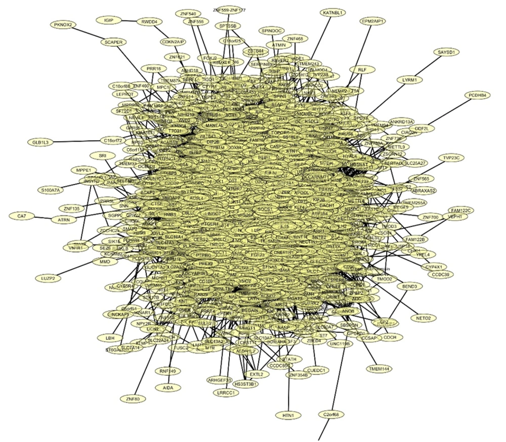
Network of proteins targeted by the downregulated microRNA (miRNA) presented by cytoscape software
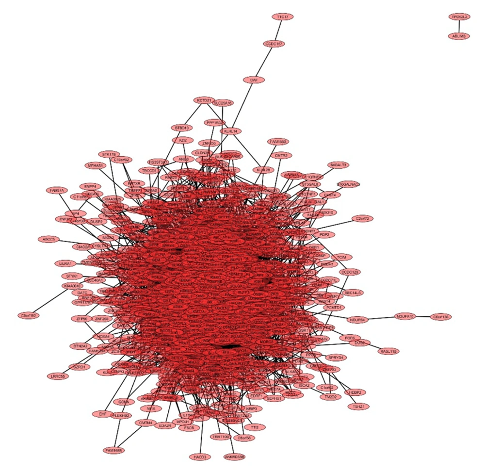
Network of proteins targeted by the upregulated microRNA (miRNA) presented by cytoscape software
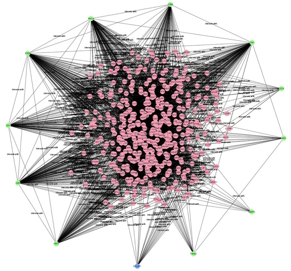
Subnetwork of hob proteins targeted by downregulated microRNA (miRNA) shown in Table 3.
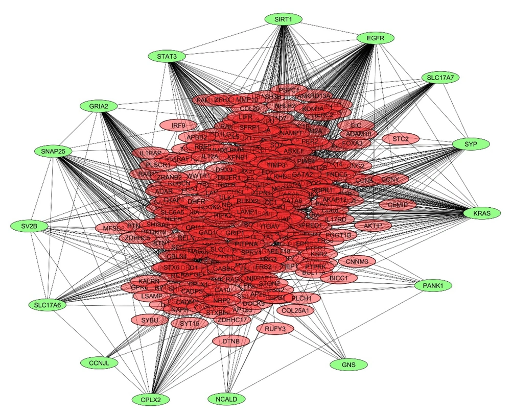
Subnetwork of hob proteins targeted by upregulated microRNAs (miRNAs) shown in Table 4.
| Clusters | Ranks | Nodes | Edges | Functions |
|---|---|---|---|---|
| 1 | 1 | 28 | 249 | |
| Metabolism of inositol phosphate | ||||
| EGFR tyrosine kinase inhibitor resistance | ||||
| Endocrine resistance | ||||
| MAPK pathway | ||||
| ErbB pathway | ||||
| 2 | 2 | 26 | 216 | |
| Bacterial attack of epithelial cells | ||||
| Renal cell carcinoma | ||||
| EGFR tyrosine kinase inhibitor resistance | ||||
| Aldosterone-regulated sodium reabsorption | ||||
| T cell receptor pathway | ||||
| 3 | 3 | 26 | 199 | |
| EGFR tyrosine kinase inhibitor resistance | ||||
| Colorectal cancer | ||||
| FoxO pathway | ||||
| AGE-RAGE pathway in diabetic complications | ||||
| Apoptosis - multiple species | ||||
| 4 | 4 | 24 | 195 | |
| Adherens junction | ||||
| TGF-beta pathway | ||||
| AGE-RAGE pathway in diabetic complications | ||||
| Colorectal cancer | ||||
| Bacterial invasion of epithelial cells | ||||
| 5 | 5 | 24 | 204 | |
| EGFR tyrosine kinase inhibitor resistance | ||||
| Bacterial invasion of epithelial cells | ||||
| Renal cell carcinoma | ||||
| Aldosterone-regulated sodium reabsorption | ||||
| Central carbon metabolism in cancer |
Abbreviation: EGFR, epidermal growth factor receptor
4.2. Functional and Pathway Enrichment Analysis
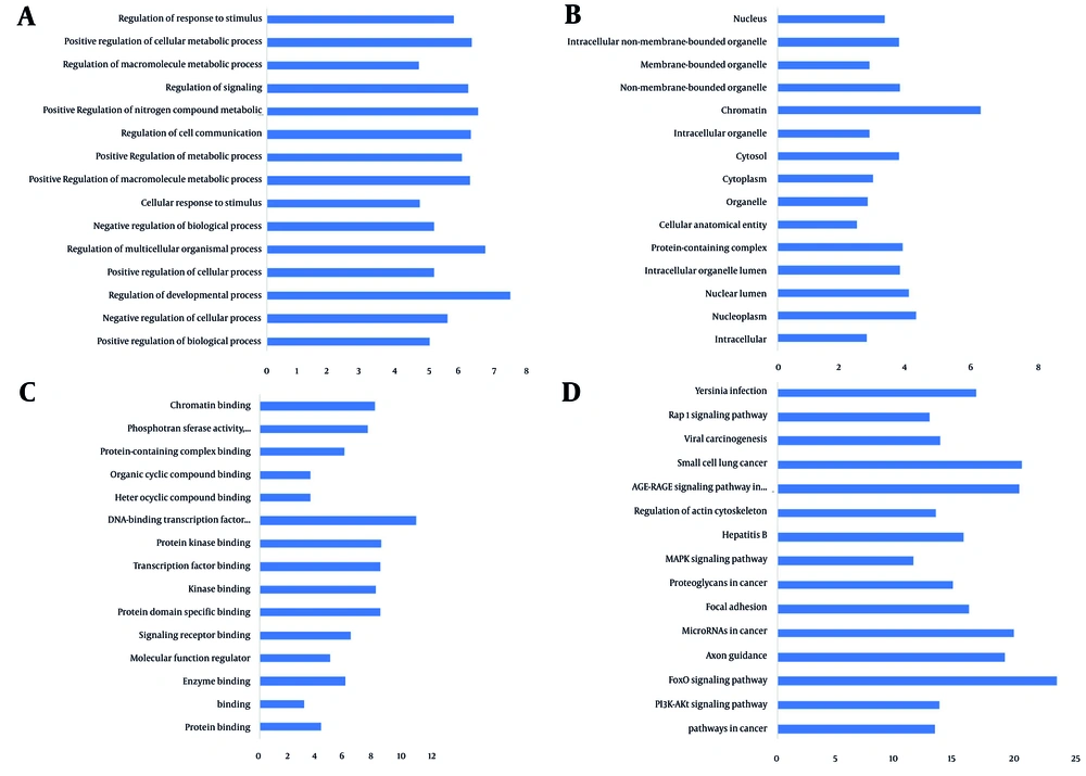
A, Biological process; B, Gene Ontology (GO): Cellular component (CC); C, GO: Molecular function (MF); D, KEGG pathway enrichment for subnetwork genes modulated by downregulated microRNAs (miRNAs).
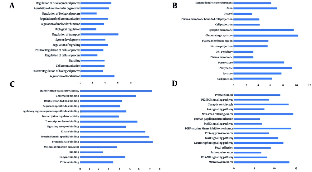
A, Biological process; B, Gene Ontology (GO): Cellular component (CC); C, GO: Molecular function (MF); D, KEGG pathway enrichment for subnetwork genes modulated by upregulated microRNAs (miRNAs).
4.3. Analysis of the Network by Clusters
| Clusters | Ranks | Nodes | Edges | KEGG Pathway Enrichment of Nodes |
|---|---|---|---|---|
| 1 | 1 | 20 | 125 | |
| Nicotine addiction | ||||
| Synaptic vesicle cycle | ||||
| Glutamatergic synapse | ||||
| Retrograde endocannabinoid signaling | ||||
| GABAergic synapse | ||||
| 2 | 2 | 20 | 124 | |
| Nicotine addiction | ||||
| Synaptic vesicle cycle | ||||
| Glutamatergic synapse | ||||
| Retrograde endocannabinoid signaling | ||||
| GABAergic synapse | ||||
| 3 | 3 | 18 | 102 | |
| Nicotine addiction | ||||
| Synaptic vesicle cycle | ||||
| Glutamatergic synapse | ||||
| Retrograde endocannabinoid signaling | ||||
| 4 | 4 | 18 | 114 | |
| Nicotine addiction | ||||
| Synaptic vesicle cycle | ||||
| GABAergic synapse | ||||
| Retrograde endocannabinoid signaling | ||||
| 5 | 5 | 18 | 110 | |
| Nicotine addiction | ||||
| Synaptic vesicle cycle | ||||
| Glutamatergic synapse | ||||
| Retrograde endocannabinoid signaling |
4.4. Promoter Motif Analysis of Hub Genes
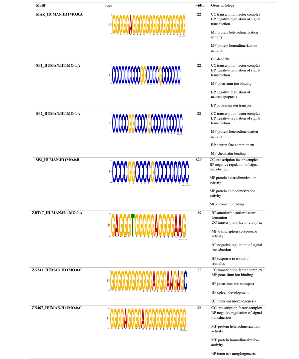
Promoter analysis of downregulated microRNA (miRNA)-targeted hub proteins
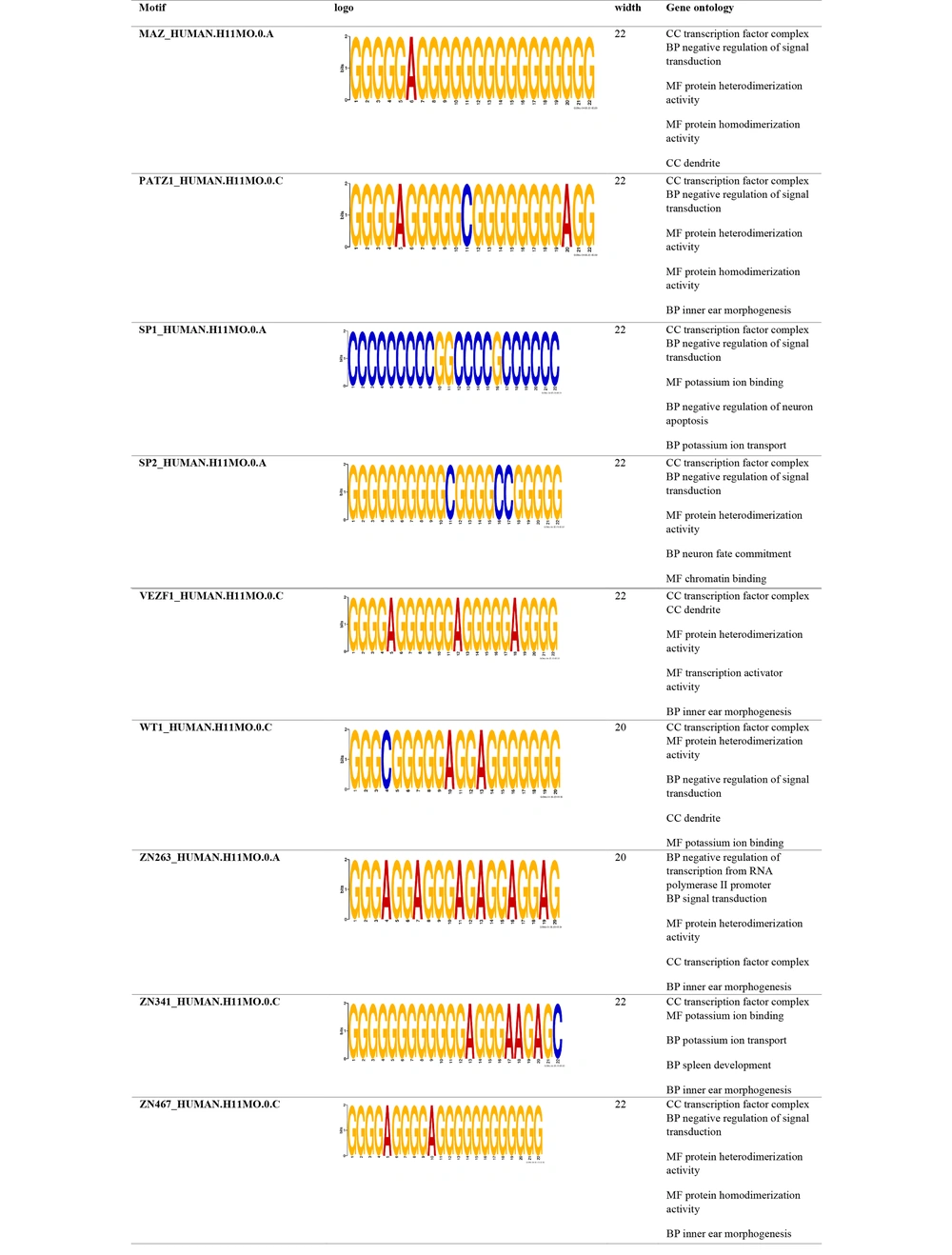
Promoter analysis of upregulated microRNA (miRNA)-targeted hub proteins




