
1. Background
The presence of metastasis in axillary lymph nodes reflects a pivotal stage in breast cancer progression and indicates a higher risk of systemic spread (1). Breast cancer accounted for the highest incidence of cancer diagnoses among women in 2020, representing 24.5% of all female malignancies and 15.5% of cancer-related deaths globally (2). Accurate assessment of these nodes informs staging, prognosis, and treatment decisions. Advances in preoperative evaluation have enabled improved prediction of nodal metastasis, supporting more personalized and targeted therapeutic approaches (3).
Sentinel lymph node biopsy (SLNB) has become the standard approach for axillary staging, replacing axillary lymph node dissection (ALND) due to its lower associated morbidity. However, SLNB remains invasive and carries risks, including lymphedema, seroma, paresthesia, chronic pain, and limited arm mobility (4). These complications can significantly impair quality of life and should be carefully considered in diagnostic planning. Nodal metastasis correlates with poorer prognosis and higher recurrence risk. European guidelines recommend routine physical examination and axillary ultrasound (US) for preoperative assessment. When suspicious nodes are identified, ultrasound-guided tissue sampling is performed. Combined US and tissue sampling yield a sensitivity of 50.0% (95% CI: 43.0 - 57.0) and specificity of 98.3% (5). Although effective, these methods still lack optimal accuracy and remain partially invasive. Magnetic resonance imaging (MRI) has gained attention as a valuable tool for identifying metastatic involvement of axillary lymph nodes, offering potential benefits in preoperative surgical planning (6). Magnetic resonance imaging offers a broader and more detailed view of the axilla than other imaging tools. However, its diagnostic accuracy may vary depending on tumor subtype and stage, with the magnitude of this variation still uncertain (7). Understanding the relationship between imaging findings and pathological characteristics could refine diagnostic algorithms and improve patient outcomes.
One of MRI’s main advantages is its ability to visualize deep axillary levels and facilitate bilateral comparison of node morphology, size, and number (8). Nevertheless, several studies have failed to show a clear diagnostic benefit of incorporating dedicated axillary sequences into MRI protocols. For instance, Ha et al. found similar diagnostic accuracy between standard breast MRI and axillary-dedicated sequences, reporting sensitivities of 64.7% and 66.2%, specificities of 94.0% and 93.3%, and negative predictive values (NPV) of 94.3% and 94.4%, respectively (9). The utility of dedicated axillary sequences in MRI remains controversial. These sequences may reduce motion artifacts by modifying the phase encoding direction and can be acquired using either standard breast coils or separate surface coils positioned over the axilla (10).
Considering the critical role of axillary staging in guiding treatment decisions and avoiding both undertreatment and overtreatment, which is one of so important part of complete resection and also decision on type of following chemotherapy and radiotherapy, and also due to possible complication of every kind of manipulation in the axillary region. This study assessed and compared the diagnostic accuracy of ultrasound and mammography with MRI for evaluating axillary nodal metastasis in breast cancer. Key diagnostic metrics — sensitivity, specificity, positive predictive value (PPV), and NPV — were calculated for each imaging modality.
2. Objectives
The objective was to determine whether MRI enhances diagnostic precision in nodal staging prior to surgery and to inform improved imaging strategies for clinical decision-making.
3. Methods
3.1. Study Design
The research team conducted this retrospective observational study between 2016 and 2021 at the Cancer Research Center of Shahid Beheshti University of Medical Sciences, following approval from the institutional ethics committee.
3.2. Patient Selection and Eligibility
The study enrolled women diagnosed with a first episode of invasive breast cancer confirmed through histopathological analysis. All included patients underwent preoperative imaging, including mammography, ultrasound, and/or MRI. Exclusion criteria were male with breast cancer, absence of preoperative axillary imaging, and incomplete clinical data.
3.3. Imaging Assessment and Patient Stratification
Preoperative imaging protocols were used to stratify participants into two diagnostic groups. The MRI group underwent combined sonography, mammography, and MRI. The sonography and mammography (SM) group received sonography and mammography without an MRI.
3.4. Surgical Management
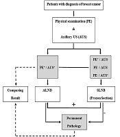
Preoperative imaging results guided surgical decision-making. Patients with no radiological evidence of axillary involvement underwent SLNB. Those with suspected nodal metastases or whom their SLNB was positive in the surgery frozen section result proceeded to ALND.
3.5. Clinical Data Collection
Clinical and pathological data were extracted from the institutional database spanning 2016 to 2021. Collected variables included patient demographics, tumor characteristics, imaging findings, and histopathological results related to axillary lymph node status.
3.6. Statistical Methodology
SPSS software (version 26.0; IBM Corp., Armonk, NY, USA) was utilized to perform statistical analyses, including descriptive procedures to outline patient characteristics and calculate frequencies for imaging methods and surgical approaches. Diagnostic accuracy was assessed through sensitivity, specificity, PPV, and NPV, using postoperative pathology as the reference standard. Agreement between imaging interpretations and pathological findings was quantified using Kappa statistics. A P-value below 0.05 was considered indicative of statistical significance.
4. Results
The study population consisted of 150 individuals diagnosed with breast cancer who fulfilled all inclusion criteria and were selected from the institutional database. Of the cohort, 115 patients received an MRI as part of their preoperative evaluation, while 34 were evaluated using the SM protocol. Based on preoperative imaging, 70% (n = 105) of patients underwent ALND, while 30% (n = 45) received SLNB. Pathological analysis confirmed lymph node involvement in 83 patients. Of these, 60 patients, classified as true positives, were diagnosed via ALND. The remaining 23 patients were considered false negatives, as preoperative imaging failed to detect nodal involvement, and SLNB later confirmed the diagnosis.
Among the 67 individuals whose pathology confirmed the absence of nodal metastasis, 22 were correctly identified as negative by imaging and subsequently underwent SLNB, representing true negative cases. However, 45 patients were incorrectly categorized as positive on imaging and received ALND, representing false positives.
Table 1 summarizes diagnostic performance in the MRI group: True positives accounted for 40.5% (47 patients), true negatives for 15.5% (18 patients), false positives for 28.4% (33 patients), and false negatives for 15.5% (18 patients).
Table 1.
Distribution of Absolute and Relative Frequency of Diagnoses of Lymph Nodes Magnetic Resonance Imaging Compared to Pathology Results
| MRI Report of Lymph Nodes | Pathology Report | Total | |
|---|---|---|---|
| Positive (%) | Negative (%) | ||
| Positive report | 47 (40.5) | 33 (28.4) | 80 |
| Negative report | 18 (15.5) | 18 (15.5) | 36 |
| Total | 64 | 52 | 116 |
Abbreviation: MRI, magnetic resonance imaging.
Table 2 presents outcomes for the SM group: True positives represented 38.2% (13 patients), true negatives 11.8% (4 patients), false positives 35.3% (12 patients), and false negatives 14.7% (5 patients).
Table 2.
Distribution of Absolute and Relative Frequency of Diagnoses of Lymph Nodes in Ultrasound and Mammography Compared to Pathology Results
| SM Report of Lymph Nodes | Pathology Report | Total | |
|---|---|---|---|
| Positive (%) | Negative (%) | ||
| Positive report | 13 (38.2) | 12 (35.3) | 25 |
| Negative report | 5 (14.7) | 4 (11.8) | 9 |
| Total | 18 | 16 | 34 |
Abbreviations: SM, sonography and mammography.
According to Table 3, MRI demonstrated 72.3% sensitivity and 35.3% specificity, with PPV and NPV measured at 58.8% and 50%, respectively. When compared with surgical pathology, MRI correctly detected axillary node involvement in 72.3% of cases. In contrast, the SM group showed slightly lower diagnostic precision, with sensitivity at 72.2%, specificity at 25%, and PPV and NPV values of 52% and 44.4%, respectively.
Table 3.
The Sensitivity, Specificity and Positive and Negative Predictive Value of Magnetic Resonance Imaging and Mammography Along with Ultrasound in Diagnosing the Involvement of Axillary Lymph Nodes in Breast Cancer Patients
| Accuracy Measurement Indicators | MRI | SM |
|---|---|---|
| Sensitivity | 72.3 | 72.2 |
| Specificity | 35.3 | 25 |
| Positive predictive value | 58.8 | 52 |
| Negative predictive value | 50 | 44.4 |
Abbreviations: MRI, magnetic resonance imaging ; SM, sonography and mammography.
Kappa analysis indicated that the diagnostic agreement between MRI and SM in detecting nodal metastasis was not statistically meaningful. While sensitivity values were similar, both methods showed limited specificity, indicating challenges in excluding axillary involvement. Moderate predictive values further suggest that both techniques have inherent diagnostic limitations. Overall, the findings indicate that both MRI and SM are viable options for preoperative axillary evaluation. However, neither method demonstrated clear superiority. These results highlight the importance of integrating imaging data with clinical judgment and considering additional diagnostic tools to enhance decision-making in breast cancer management.
5. Discussion
The comparative diagnostic value of MRI versus conventional imaging in axillary staging remains a key concern in breast cancer management. Magnetic resonance imaging demonstrated 72.3% sensitivity and 35.3% specificity in this study, corresponding PPV and NPV values of 58.8% and 50%. The SM group demonstrated similar sensitivity (72.2%) but lower specificity (25%), with PPV of 52% and NPV of 44.4%. These metrics underscore the limited discriminatory capacity of both methods and merit careful contextualization within existing evidence.
Our study's relatively limited diagnostic accuracy of MRI aligns with prior findings; for instance, Zhou et al. in conducted a meta analyze by searching databases for open access published studies relevant to the use of MRI for the detection of axillary lymph node metastasis in breast cancer patients. Their results reported a sensitivity of 0.77 and a specificity of 0.90 for MRI in identifying nodal metastases (6). The variation in specificity may reflect differences in imaging protocols and interpretative criteria. Notably, our study did not employ dedicated axillary coils, which may have influenced accuracy. Axillary assessment in the context of neoadjuvant chemotherapy (NACT) presents additional challenges. Reported nodal pathological complete response (pCR) rates post-NACT range from 23% to 41% (7-11). Given MRI’s relatively low NPV in our cohort, reliance on MRI alone in this subgroup may be inadequate, particularly in strategies aiming to de-escalate axillary surgery.
Multimodal imaging approaches have shown promise in overcoming the limitations of single techniques. Qi et al. (11) reported that the combined use of mammography, shear wave elastography (SWE), and MRI achieved 94.6% sensitivity, 86.5% specificity, and a PPV and NPV of 91.4%. These findings suggest synergistic potential in combining modalities to improve diagnostic precision. Interpretation of axillary imaging is often hampered by factors unrelated to imaging quality alone. Zaiton et al. (12) demonstrated that despite negative findings across physical exams, mammography, ultrasound, and MRI, approximately 14% of patients still had positive sentinel nodes on final pathology. This highlights the inherent limitations of current imaging technologies in detecting microscopic disease.
Following NAC, the lymphatic drainage of the breast can be disrupted and subsequently the underarms can have an uneven pattern, which increases the negative rate (13).
The MRI performance is also affected by technical and physiological variables. While MRI excels in dynamic tissue contrast for primary breast lesions, its axillary application is restricted. Post-NACT changes, respiratory motion artifacts, and physiologic variations in nodal morphology reduce interpretability (14-16). Additionally, enhancement characteristics used in breast tumor evaluation are less specific in axillary nodes due to similar enhancement in normal lymphatic tissue (17, 18). On the other way Gadolinium contrast agents used in the diagnosis of breast cancer lymph nodes, although generally safe, can cause side effects ranging from mild and temporary to more serious and potentially long-lasting. These primarily affect the kidneys, skin, muscles, and other organs and can include nephrogenic systemic fibrosis, gadolinium deposition, and allergic reactions (19).
Cost and accessibility further complicate routine MRI use. Although MRI offers detailed anatomical and vascular information, it remains resource-intensive. Qi et al. (11) concluded that selective use of MRI in complex or ambiguous cases may be the most cost-effective strategy. Emerging strategies may help address current diagnostic gaps. Placement of markers in abnormal nodes before NACT and clipping biopsy-proven metastatic nodes may improve surgical targeting and reduce false negatives (14). Integration of advanced imaging, such as diffusion-weighted imaging and dedicated axillary coils, also holds promise but requires further validation. In summary, while MRI contributes valuable information in axillary evaluation, its diagnostic advantage over conventional imaging remains limited. A multimodal imaging framework — potentially including SWE and advanced MRI protocols — may offer greater reliability in staging. Advancing imaging strategies for specific clinical contexts — especially in patients undergoing neoadjuvant therapy — remains a key direction for future investigation.
5.1. Conclusions
This study evaluated the diagnostic accuracy of MRI compared to conventional imaging methods — specifically sonography and mammography — for identifying axillary nodal metastasis in patients with breast cancer. Results indicated that both methods provide moderate diagnostic value, with no significant difference in overall performance. Magnetic resonance imaging’s limited specificity and predictive values, consistent with prior studies, underscore the ongoing challenges in precise axillary staging, particularly in neoadjuvant therapy and microscopic disease. Improving axillary assessment will likely require optimized imaging protocols and the integration of multiple modalities. Future research should prioritize individualized imaging strategies based on tumor characteristics and treatment pathways. A personalized, multimodal imaging strategy may improve the precision of preoperative staging in breast cancer while also offering greater efficiency in resource utilization.
5.2. Limitations
Integration of advanced imaging, such as diffusion-weighted imaging and dedicated axillary coils, may improve the predictive value that should be designed and evaluated in multi center and larger number studies. Small number of patients was one of our limitation that can be corrected in future studies. Placement of markers in abnormal nodes before NACT and clipping biopsy-proven metastatic nodes may improve surgical targeting and reduce false negatives that we did not reviewed. This study was retrospective but prospective studies can include more number of patients and use controlled MRI protocols to evaluate axillary lymph node metastasis