
1. Introduction
Nicotine addiction is one of the most prevalent addictions worldwide, particularly in the United States in 2021. The use of nicotine in various forms, including smokeless tobacco products such as SNUS, a blend of tobacco and lime, paan, gutkha, and, more recently, nicotine patches, has increased significantly worldwide, particularly among young populations. Smokeless tobacco is available either as chewing tobacco that is not mixed with other ingredients or as a blend of tobacco with additional substances, such as betel nut, areca nut with tobacco, and SNUS (1, 2).
There are more than 350 million smokeless tobacco users worldwide. The prevalence of tobacco use among patients with cancer in Iran is estimated at an overall consumption rate of 43%. This rate varies significantly across individual studies, ranging from 10% to 88%. Subgroup analyses further highlight disparities in tobacco consumption rates across different demographic groups, geographic regions, and cancer types (3). Among these products, SNUS has attracted considerable attention because of its widespread use in certain regions, particularly in South Asia and Europe (4).
Tobacco consumption in Iran remains alarmingly high, indicating that the current level of tobacco control policy implementation is ineffective and insufficient. This situation underscores the need to adopt, implement, and enforce comprehensive packages of evidence-based tobacco control policies (5). The high levels of nicotine and carcinogens in smokeless tobacco, compared with those in cigarette smoke, increase the potential risk of cancer (6).
The incidence of oral cancer in Southeast Asia and India is estimated to be approximately 50%, accounting for approximately 35000 new cases per year. Oral cancer is the sixth most common cancer worldwide and is considered one of the leading causes of mortality in developing countries (7), with approximately 275000 new cases reported each year (6). Because smokeless tobacco is one of the most important risk factors for the development of precancerous lesions, such as submucous fibrosis and leukoplakia, as well as head and neck cancers and other potentially malignant oral disorders, numerous studies have demonstrated a strong association between smokeless tobacco use and the incidence of oral cancer (8).
Because the mouth is the first site of contact with smokeless tobacco, the oral mucosa is exposed to the toxic effects of nicotine and other harmful chemicals present in these products. Changes in the oral mucosa can vary depending on the duration and frequency of use, as well as the specific type of product consumed (9). The localized nature of these lesions, particularly in the areas where the product is applied, suggests that, in addition to chemical exposure, physical irritation from these products also plays a key role in their development. Of particular concern is that these lesions are often asymptomatic in the early stages, which makes early detection and the implementation of preventive measures challenging (10).
All commercial tobacco products, including smokeless tobacco, cigarette smoke, and cigar smoke, contain tobacco-specific nitrosamines, which are formed during tobacco processing. Nitrosamines result from the reaction of tobacco alkaloids with nitrite, and they may also contribute to the formation of androgens, particularly through the nitrosation of nor-nicotine. Additionally, the pH and moisture content of different smokeless tobacco products vary considerably, which may influence the concentrations of carcinogens present. Higher pH levels have been associated with increased bioavailability of nicotine and carcinogens. Interactions among these substances may enhance mutagenic effects on oral mucosal cells and increase the likelihood of malignant transformation (9).
The global prevalence and distribution of smokeless tobacco use are likely underestimated (4). Although smokeless tobacco use is high in certain regions, including southern and eastern Iran, accurate statistics on its prevalence are lacking. The reported figures are primarily based on the frequency of SNUS use among students at the University of Zahedan (11, 12).
In light of the increasing global and Middle Eastern use of smokeless tobacco, the absence of pathognomonic symptoms, the frequent incidental detection of oral lesions during routine dental examinations, and the limited number of studies investigating its objective effects, many studies have examined the harmful effects of smokeless tobacco on the oral cavity. Therefore, the aim of the present study was to investigate alterations in the oral mucosa of smokeless tobacco users through visual and clinical assessment.
Here, we report 45 cases of smokeless tobacco use over 1 year at a dental school. This article presents the only study conducted in Yazd, central Iran, which is recognized as the therapeutic hub for the country’s southern region.
2. Case Presentation
2.1. Ethical Considerations
This study was approved by the Committee of Ethics in Human Research at Shahid Sadoughi University of Medical Sciences, Yazd, under ethics code IR.SSU.DENTISTRY.REC.1404.004. All participants provided informed consent for the collection, photography, and publication of images of their oral cavities, as well as for the publication of the study. The study was conducted in accordance with the Declaration of Helsinki.
2.2. Inclusion Criteria
Examinations were conducted in 4000 patients referred to the Department of Oral and Maxillofacial Medicine at the Dental School of Yazd, central Iran, over a 1-year period from February 2024 to April 2025. All medical and dental records were completed by a trained examiner. Data on various parameters, including medical and dental history, dietary habits, and tobacco consumption, were collected. Patients were selected for further investigation based on the following inclusion criteria: age 18 to 80 years; absence of systemic disease or medical conditions; no daily medication use or systemic antibiotic use for at least 1 year; not pregnant; no periodontitis; and daily use of smokeless tobacco (SNUS) for at least 6 months.
2.3. Oral Examinations and Sample Collection
Patients who met the inclusion criteria (n = 45) underwent a comprehensive oral cavity examination, including dental history assessment and oral cancer screening. Data on the history of SNUS consumption were recorded, and a single examiner was responsible for the complete evaluation of the oral mucosa and patient follow-up. Patients were also asked about their smoking habits, including cigarette, hookah, and hubble-bubble use.
In addition to comprehensive extraoral and intraoral examinations, any oral lesions were documented in the patients’ medical records. Of the 4000 patients examined, 45 reported a history of smokeless tobacco use (SNUS), including 31 males and 14 females. Among these patients, 17, including 11 males and 6 females aged 24 to 80 years, presented with oral mucosal lesions. Five cases had missing data because of a lack of cooperation with the treatment team.
This finding highlights a significant prevalence of oral changes among SNUS users and emphasizes the need for more detailed studies in this area. Examinations were performed using a dental mirror and appropriate unit lighting and included assessment of hard and soft tissue status, dental and periodontal conditions, and oral cancer screening (12, 13). Two periodontists performed the examinations.
Among the 4000 patients evaluated, 45 reported a history of smokeless tobacco use. Of these patients, 17 had oral mucosal lesions; 6 patients presented with lesions as their chief complaint, whereas lesions in 9 patients were discovered incidentally. Malignancy was suspected in 10 patients, of whom 5 consented to biopsy. All 5 biopsy results indicated squamous cell carcinoma (SCC). In 2 additional cases, vital staining was performed to guide biopsy; however, neither patient consented to the procedure, and only follow-up sessions were recommended. One patient showed improvement during follow-up after discontinuing SNUS use and adhering to health recommendations.
The SNUS users ranged in age from 24 to 80 years. All patients were free of systemic diseases or underlying health conditions, had not taken daily medication or systemic antibiotics for at least 1 year (14), were not pregnant (15), had no known periodontitis, used SNUS or other forms of smokeless tobacco daily, and did not use other nicotine products. The history of SNUS use, intraoral examination findings, histopathological analyses, and vital staining of lesions are described. Patterns of use, duration, affected areas, suspicion of malignancy, and biopsy results are presented in Table 1.
Table 1.
Pattern of Lesions Observed in Smokeless Tobacco Users
| Case No. | Lesion | Gender | Age | Involved Area | Duration of Use | Suspicion of Malignancy | Biopsy |
|---|---|---|---|---|---|---|---|
| 1 | SCC | Male | 51 | Anterior vestibule of the mandible | 20 years | + | ✓ |
| 2 | SCC | Male | 50 | Buccal vestibule of the mandible | 12 years | + | ✓ |
| 3 | SCC | Male | 49 | Buccal vestibule of the mandible | 15 years | + | ✓ |
| 4 | SCC | Female | 65 | Buccal vestibule of the mandible | 25 years | + | × |
| 5 | Erythroplakia | Male | 37 | Right buccal mucosa | 2 years | + | Vital staining |
| 6 | Leukoplakia | Male | 24 | Labial vestibule of the maxilla | 6 months | + | ✓ |
| 7 | SCC | Female | 72 | Labial vestibule of the mandible | 30 years | + | ✓ |
| 8 | Erythroleukoplakia | Male | 35 | Labial vestibule of the left maxilla | 4 years | __ | × |
| 9 | Leukoplakia | Male | 49 | Buccal vestibule of the left mandible | 10 years | __ | × |
| 10 | Leukoplakia | Male | 38 | Buccal vestibule of the left mandible | 2 years | __ | × |
| 11 | Leukoplakia | Male | 25 | Buccal vestibule of the left mandible | 4 months | __ | × |
| 12 | Leukoplakia | Male | 35 | Labial vestibule of the maxilla | 1 year | __ | × |
| 13 | Leukoplakia | Female | 68 | Right maxillary buccal vestibule | 15 years | + | × |
| 14 | Leukoplakia | Female | 60 | Left buccal mucosa | 12 years | + | × |
| 15 | SCC | Female | 58 | Left maxillary buccal vestibule | 10 years | + | ✓ |
| 16 | Leukoplakia | Female | 80 | Buccal vestibule of the left mandible | 20 years | + | × |
| 17 | Leukoplakia | Male | 24 | Vestibule of the maxilla | 6 months | - | × |
| 18 and others | No lesions | Females and males | 18 - 80 | ----- | Variable | - | × |
2.4. Case 1
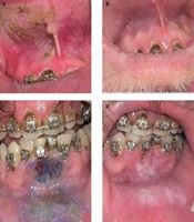
A 24-year-old man presented with a complaint and requested dental scaling. His dental history revealed that he brushed once daily using a regular toothbrush and an interdental brush but did not floss or use mouthwash. Intraoral examination revealed leukoplakia on the mucosa of the anterior maxillary vestibule. During the health history interview, the patient reported using SNUS once daily for the past 6 months, with each use lasting 30 to 60 minutes. Additionally, he had been undergoing orthodontic treatment for 1 year.
2.5. Intraoral Examination
A homogeneous leukoplakia with well-defined borders and a cracked-clay appearance was observed beneath the upper lip, deep in the maxillary vestibule, parallel to teeth 1, 2, and 3 on the right side. Yellowish-brown pigmentation was evident in some areas of the lesion, with involvement of the maxillary anterior frenum (Figure 1A). The alternative SNUS placement site was the anterior vestibule of the mandible (Figure 1B). Oral hygiene was moderate, and cervical tooth discoloration was noted. Because an ulcer was present in the attached gingiva of the mandibular incisors, a vital staining technique using toluidine blue was performed (Figure 1C).

Figure 1.
A, Yellowish-brown plaque in the maxillary anterior vestibule in a young man; B, improvement of the lesion after quitting SNUS; C, leukoplakia in the mandibular anterior vestibule with an erosive area; D, detection of high-risk areas with the vital staining technique.
2.6. Case 2
A 34-year-old man presented with complaints of tooth decay. He reported brushing his teeth once daily but did not use dental floss or mouthwash. Intraoral examination revealed a reddish-white plaque on the mucosa of the anterior mandibular vestibule. During the health history interview, he reported placing SNUS twice daily for more than 30 minutes in the lower lip vestibule over the past 4 years.
2.7. Intraoral Examination
A red and white leukoplakic lesion with a reticular pattern was observed on the lower lip near the right and left mandibular fourth teeth, extending into the mandibular vestibule. The patient’s oral hygiene was moderate, with visible dental calculus and tooth discoloration (Figure 2).

Figure 2.
Red and white leukoplakia on the lower lip with a reticular pattern and irreversible gingival recession.
2.8. Case 3
A 51-year-old man presented with oral and maxillofacial pain. Intraoral examination revealed a large ulcer with indurated edges. His history indicated that he had used SNUS spray for 20 years, 2 to 3 times per day, with each use lasting approximately 30 minutes. He placed the SNUS in the depth of the mandibular anterior vestibule. The patient reported that he did not use a toothbrush or dental floss.
2.9. Intraoral Examination
A large ulcer with indurated edges was observed on the mandibular alveolar mucosa, extending to the edentulous ridge, and was painful on palpation. The teeth in the affected area exhibited grade 3 mobility. The patient’s oral hygiene was poor, with evident plaque and calculus (Figure 3A).

Figure 3.
A, Exophytic mass in the mandibular vestibule; B, histopathological view at 10× magnification showing hyperkeratinized stratified squamous epithelium with long and wide rete ridges. Infiltration of inflammatory cells in the underlying connective tissue was evident.
A biopsy of the ulcer was performed, including a margin of healthy tissue. Local infiltration anesthesia was administered using 2% lidocaine with 1:100000 epinephrine. Soft-tissue incisions measuring approximately 5 mm in diameter and 3 mm in thickness were made using a No. 15 scalpel, and the specimens were placed in 5 mL of 37% formalin solution at a 1:15 tissue-to-formaldehyde ratio. The samples were subsequently stained with hematoxylin-eosin and examined under a microscope at 4× and 10× magnification. After the biopsy, the patient received oral care instructions and guidance on nicotine cessation.
Microscopic examination revealed dysplastic stratified squamous epithelium with epithelial islands invading the underlying connective tissue. The epithelial islands exhibited pleomorphism and abundant keratin pearl formation. A biopsy of another area of the lesion showed hyperkeratinized stratified squamous epithelium with long, broad rete ridges, along with infiltration of inflammatory cells in the underlying connective tissue (Figure 3).
3. Results
Among the 4000 patients evaluated, 45 reported a history of smokeless tobacco use. Of these patients, 17 presented with oral mucosal lesions. Six patients reported oral lesions as their chief complaint, whereas lesions in 9 patients were discovered incidentally. Malignancy was suspected in 10 patients, of whom 5 consented to biopsy (Table 1).
We evaluated the association between sociodemographic factors and oral lesions (Table 2). In the final part of the Results, the presence of oral lesions was examined in a combined analysis (Table 3).
Table 2.
Association of Sociodemographic Factors
| Study Variables | Absent Lesion | Present Lesion |
|---|---|---|
| Age, y | ||
| 18 - 30 | 8 | 3 |
| 30 - 45 | 4 | 5 |
| 45 - 60 | 6 | 5 |
| 60 - 75 | 8 | 3 |
| 75 - 90 | 2 | 1 |
| Gender | ||
| Male | 19 | 31 |
| Female | 9 | 14 |
| Location | ||
| Yazd | 4 | 2 |
| Southeastern Iran | 15 | 10 |
| South of Iran | 9 | 5 |
| Presence of systemic disease | ||
| None | 11 | 9 |
| Diabetes | 12 | 13 |
| Hypertension | 16 | 7 |
| Cancer | 3 | 1 |
| History of cancer in family | ||
| Yes | 4 | 6 |
| No | 24 | 11 |
Table 3.
Various Combinations of Oral Lesions
| Line | Sign | N |
|---|---|---|
| 1 | Plaque + ulceration | 1 |
| 2 | Leukoplakia | 9 |
| 3 | Erythroplakia | 1 |
| 4 | Exophytic lesion | 5 |
| 5 | Ulceration | 1 |
| 6 | No lesion | 28 |
| Total | - | 45 |
4. Discussion
The use of SNUS is often influenced by cultural and social factors or by misconceptions regarding its effects (16, 17). Several studies have demonstrated that smokeless tobacco, including SNUS, is associated with an increased risk of precancerous lesions, head and neck cancers, and particularly oral SCC (11, 12, 18). Frequent and localized use of SNUS can cause a range of mucosal lesions. In a study of smokeless tobacco use among male university students in Zahedan, Iran, a significant relationship was observed between a history of smokeless tobacco use and factors such as the university or college attended, place of residence, and the mother’s level of education (12).
A significant association was also observed between knowledge about smokeless tobacco and its prevalence of use. These lesions can range from benign changes to severe oral malignancies, depending on factors such as the duration and intensity of use, the type of SNUS, oral hygiene status, and the presence of aggravating factors such as smoking or alcohol consumption (8, 19). In a study conducted in Iran, Rezaeian et al. assessed the levels of arsenic, lead, nickel, and cadmium in SNUS brands available on the Iranian market. They reported that 81% of the samples contained the highest detectable levels of arsenic, a well-established carcinogen (11, 20).
Lesions caused by SNUS primarily appear as white or red plaques, including homogeneous and nonhomogeneous leukoplakia, in areas of the oral mucosa that are in constant contact with SNUS. Similar oral findings have been reported by Muller et al. (8, 20). This finding was also observed in the present study (Figure 1). Vital staining is a diagnostic technique that aids in the clinical detection of dysplastic lesions. Toluidine blue, a metachromatic nuclear stain, highlights areas of dysplasia or neoplasia by binding to the DNA of cells with high mitotic activity. It is a low-cost, noninvasive, and rapid method for screening malignant and precancerous lesions; however, its sensitivity and specificity are lower than those of biopsy (21-23).
Chronic, long-term exposure to SNUS can cause repeated mucosal irritation and ulceration, which may indicate a neoplastic process (8). In the third case presented in this article, the patient exhibited a lesion suggestive of SCC (Figure 3). Biopsy and histopathological examination revealed epithelial islands invading the underlying connective tissue, cellular pleomorphism, and keratin pearl formation, all of which are characteristic features of oral SCC (Figures 3B and C) (21, 24-26).
Among the 45 patients using SNUS, 28 showed no evidence of snuff pouch lesions, whereas 17 presented with mucosal lesions. Four patients showed improvement following advice to discontinue SNUS use and regular periodic examinations, whereas 6 patients were referred to the Department of Oral and Maxillofacial Surgery for definitive treatment with a diagnosis of oral cancer (SCC). There were 5 cases with missing data. Previous research has shown that oral lesions in healthy individuals may resolve within 6 weeks to 6 months after discontinuing smokeless tobacco. If lesions persist, a biopsy is strongly recommended (27).
Although the prevalence of smokeless tobacco use (SNUS) in the general population of Iran is relatively low, this study examined oral manifestations in a subset of patients who self-reported SNUS use and were referred to the School of Dentistry. Although this group was limited in size, it provides valuable insight into the localized effects of smokeless tobacco in clinical settings.
4.1. Limitations
One limitation of the present study was the small sample size. In addition, the study was conducted at a single center. Because most SNUS users reside in the southern and eastern regions of Iran, larger multicenter studies encompassing these areas are recommended. Furthermore, future research should consider categorizing participants according to the duration of SNUS use and examining its association with oral lesion occurrence.
4.2. Conclusions
As a common form of smokeless tobacco, SNUS may have harmful effects on oral mucosal health. The findings of the present study provide clinical evidence of a strong association between SNUS use and the development of precancerous and malignant lesions. Although the inclusion of a control group would have strengthened comparisons and enabled more definitive conclusions, future research should incorporate broader screening protocols to allow such analyses. Accordingly, the observed association between SNUS use in Iran and the occurrence of precancerous and malignant lesions should be interpreted with caution.