1. Background
Breast cancer is a biologically heterogeneous disease, characterized by diverse molecular subtypes that influence prognosis and therapeutic response (1). These subtypes are typically defined by the expression of key histopathological biomarkers, including estrogen receptor (ER), progesterone receptor (PR), human epidermal growth factor receptor 2 (HER2), and the proliferation marker Ki-67 (2). Accurate molecular classification is critical for selecting appropriate treatment strategies and improving patient outcomes (3).
Estrogen receptor- and PR-positive tumors, especially luminal A types with low Ki-67 indices, are generally associated with a favorable prognosis and responsiveness to endocrine therapy (4). In contrast, luminal B subtypes — often HER2-positive or with high Ki-67 — are more aggressive and may require combined endocrine and targeted therapies (5). Human epidermal growth factor receptor 2 -positive tumors, although aggressive, benefit from targeted agents such as trastuzumab (6). Triple-negative breast cancer (TNBC), defined by the absence of ER, PR, and HER2, is particularly challenging due to its aggressive behavior and lack of targeted therapies, making chemotherapy the mainstay of treatment (7).
While these molecular biomarkers have become indispensable in clinical decision-making, several limitations persist. Tumor heterogeneity and variability in immunohistochemical assessment can lead to diagnostic inaccuracies, potentially resulting in suboptimal treatment (8). Additionally, resistance to endocrine or HER2-targeted therapy and treatment-related toxicities further complicate disease management. In particular, the absence of actionable targets in TNBC underscores the urgent need for additional prognostic and predictive tools (9).
Imaging modalities such as mammography and ultrasonography remain first-line diagnostic tools (10), while dynamic contrast-enhanced MRI (DCE-MRI) offers superior sensitivity in detecting invasive disease and evaluating vascularity (11). However, each modality has limitations in terms of specificity and reproducibility (12). Diffusion-weighted imaging (DWI), a non-contrast MRI technique, has gained attention as it provides insights into tissue microstructure by measuring water diffusivity through the apparent diffusion coefficient (ADC) (13). This method is particularly valuable for patients who cannot receive gadolinium-based contrast agents and may enhance tumor characterization beyond morphology (13).
Several studies have investigated the role of ADC in differentiating benign from malignant breast lesions and in predicting tumor aggressiveness. Lower ADC values have been consistently associated with higher cellular density, higher histologic grade, and elevated Ki-67 Index (14, 15). Furthermore, recent evidence suggests that ADC may correlate with hormone receptor status, although findings have been mixed and remain inconclusive (16, 17). The association between ADC values and lymph node metastasis has also emerged as a potential marker of tumor invasiveness, but remains underexplored in prospective settings (18, 19).
Despite growing interest in ADC as a biomarker, challenges remain in standardizing acquisition protocols and interpreting values across different tumor types and imaging platforms (20). Recent advances in computational imaging, such as curvelet-based feature extraction combined with artificial neural networks, have shown promising potential for automated classification of breast lesions and improved diagnostic accuracy (21). Thus, there is a pressing need for prospective studies evaluating ADC in relation to established prognostic markers using consistent imaging methods.
2. Objectives
This study aimed to evaluate the potential of ADC as a non-invasive imaging biomarker for breast cancer characterization by investigating its correlation with histopathological and molecular prognostic factors, including ER/PR status, HER2 expression, Ki-67 Index, and lymph node metastasis. By integrating imaging data with molecular profiles, this work seeks to support the utility of ADC in improving early risk stratification and informing personalized treatment strategies.
3. Methods
3.1. Patient Selection
This prospective study included 35 consecutive patients (mean age 46 ± 10.2 years) with histologically confirmed malignant breast lesions who were recruited through the private radiology office in Tehran, Iran, between December 2017 and August 2018.
Eligible patients were identified through their histopathology reports, which confirmed breast malignancy and met the inclusion criteria. After confirmation, all patients underwent breast MRI at Athari Imaging Center, Tehran, using a 1.5 T scanner with a dedicated breast coil.
Inclusion criteria comprised biopsy-proven malignancy, no prior breast surgery or radiation therapy, absence of MRI contraindications (severe claustrophobia, metallic implants, or pacemakers), and ability to provide informed consent.
Exclusion criteria included prior neoadjuvant chemotherapy, lesions smaller than 5 mm, or motion artifacts on DWI sequences.
Histopathology was used as the reference standard because it remains the gold standard for confirming breast cancer diagnosis and assessing prognostic biomarkers.
All patients successfully completed both MRI and histopathological evaluation, and no missing or indeterminate data were encountered.
The study was conducted in accordance with the Declaration of Helsinki and approved by the Institutional Review Board of Shahid Beheshti University of Medical Sciences (IR.SBMU.RETECH.REC.1396.1312). All participants provided written informed consent prior to inclusion.
3.2. MRI Acquisition Protocol
All examinations were performed using a 1.5 Tesla scanner (Signa HDxt, GE Healthcare) with a dedicated bilateral breast coil. Patients were imaged in the prone position. The imaging protocol included axial T2-weighted spin-echo with fat suppression, pre-contrast axial and sagittal T1-weighted imaging, and post-contrast axial fat-suppressed T1-weighted imaging. Dynamic contrast-enhanced MRI (DCE-MRI) was acquired using seven dynamic series (one pre-contrast and six post-contrast) following intravenous administration of gadopentate dimeglumine (0.1 - 0.2 mmol/kg) with a 20 mL saline flush. T1-weighted axial fat-suppressed 3D gradient-echo parameters included TR/TE 5.4/2.6 ms, 384 × 384 matrix, 2 mm slice thickness, 30 cm FOV, iPAT 2, and 80 s temporal resolution. Post-processing utilized specialized subtraction software to eliminate pre-contrast background signals.
3.3. Diffusion-Weighted Imaging Protocol
Diffusion-weighted imaging was performed in the transverse plane using a spin-echo single-shot echo planar imaging sequence with the following parameters: TR/TE 11,000/73 ms, b-values 0/200/800 s/mm², 256 × 192 matrix, 280 × 280 mm FOV, 5 mm slice thickness, 90° flip angle, iPAT 2, and 183 s acquisition time. The b-value of 800 s/mm² was selected to minimize perfusion effects from tumor angiogenesis. Apparent diffusion coefficient maps were generated using the equation:
Where S1 and S2 represent signal intensities at b-values b1 (0 s/mm²) and b2 (200 or 800 s/mm²) respectively.
3.4. Image Analysis
A radiologist with 10 years of breast MRI experience performed all measurements. Manual region-of-interest (ROI) placement (15 - 55 mm²) avoided cystic or necrotic areas with T2 shine-through effects. For correlation with DCE-MRI, ROIs were placed in the enhancing solid tumor component. Three ROIs demonstrating the lowest ADC values were selected, and their average was used as the final ADC value.
3.5. Histopathological Assessment
Tumor grading followed Elston and Ellis' modified Scarff-Bloom-Richardson system, evaluating tubule formation, nuclear pleomorphism, and mitotic count (grade 1: 3 - 5; grade 2: 6 - 7; grade 3: 8 - 9) (22). Receptor status interpretation employed the Allred scoring system for ER/PR (positive ≥ 20%) (23), ASCO/CAP guidelines for HER2 (0 - 1+: Negative; 3+: Positive; 2+: equivocal requiring FISH confirmation) (24), and standard thresholds for Ki-67 (positive ≥ 18%) (25) (Table 1).
Table 1.
Histopathological Grading and Biomarker Classification
| Parameter and Classification | Criteria |
|---|---|
| Tumor grade (Elston-Ellis) | |
| Grade 1 | Score 3 - 5 |
| Grade 2 | Score 6 - 7 |
| Grade 3 | Score 8 - 9 |
| ER/PR status (%) | |
| Positive | Expression ≥ 20 |
| Negative | Expression < 20 |
| HER2 status | |
| 0 - 1+ | Negative |
| 2+ | Equivocal (FISH required) |
| 3+ | Positive |
| Ki-67 Index (%) | |
| Positive | ≥ 18 |
| Negative | < 18 |
Abbreviation: HER2, human epidermal growth factor receptor 2.
3.6. Statistical Analysis
Apparent diffusion coefficient values were compared across histological subtypes, lymph node status, tumor size, and biomarker expressions (ER, PR, HER2, Ki-67) using paired-sample t-tests. Tumor sizes, measured via DCE-MRI subtraction, were categorized as ≥ 2 cm or < 2 cm. Statistical analyses were performed using IBM SPSS Statistics for Windows (Version 25.0, IBM Corp., 2018), with a significance threshold of P < 0.05.
A receiver operating characteristic (ROC) curve was generated to determine optimal ADC thresholds for differentiating biomarkers with significant associations. Sensitivity, specificity, and the area under the curve (AUC) were calculated for each identified ADC cutoff.
3.7. Sample Size Justification
This study included 35 patients, reflecting the total number of eligible and consenting individuals who met the inclusion criteria during the recruitment period. As a prospective single-center imaging study, the sample size was determined based on feasibility, constrained by the availability of patients within the designated timeframe and the strict exclusion criteria (e.g., lesion size, prior treatment, motion artifacts). Despite the relatively limited cohort, statistically significant correlations were observed for key prognostic indicators, supporting the adequacy of the sample for exploratory, and hypothesis-generating analysis.
4. Results
A total of 35 patients with pathologically confirmed malignant breast cancer underwent MRI, and ADC values were measured.
4.1. Histopathological Findings
Among the 35 patients, 19 (54.2%) had invasive ductal carcinoma (IDC), 10 (28.6%) had invasive ductal carcinoma not otherwise specified (IDC-NOS), and 6 (17.2%) had ductal carcinoma in situ (DCIS). Lymph node metastasis was present in 13 (37.2%) patients and absent in 22 (62.8%) patients. Tumor grading was NG 1 in 9 (25.7%) lesions, NG 2 in 19 (54.3%) lesions, and NG 3 in 7 (20%) lesions. HER2 status was negative in 21 (60%) lesions, equivocal in 4 (11.4%) lesions, and positive in 10 (28.6%) lesions.
Hormone receptor analysis showed that 10 (28.6%) tumors were ER-negative, and all PR-negative tumors also showed ER negativity. A total of 25 (71.4%) tumors were ER-positive, and all PR-positive tumors also showed ER positivity. Ki-67 expression was negative in 17 (48.6%) lesions and positive in 18 (51.4%) lesions.
4.2. Patient and Tumor Characteristics
Sixteen (45.7%) patients were younger than 46 years, and 19 (54.3%) were 46 years or older. Tumor size was < 2 cm in 8(22.8%) cases and ≥ 2 cm in 27 (77.2%) cases.
4.3. Apparent Diffusion Coefficient Analysis
The mean ADC values for IDC-NOS, special subtypes of IDC, and DCIS were 0.912 × 10⁻³ mm²/s, 0.988 × 10⁻³ mm²/s, and 1.158 × 10⁻³ mm²/s, respectively (P = 0.212). There were no statistically significant differences in mean ADC values based on histologic grade (P = 0.182), tumor size (P = 0.207), or HER2 status (P = 0.780).
The mean ADC value was significantly lower in tumors with lymph node metastasis compared to those without (0.912 × 10⁻³ mm²/s vs. 1.046 × 10⁻³ mm²/s; P = 0.016) (Figure 1).
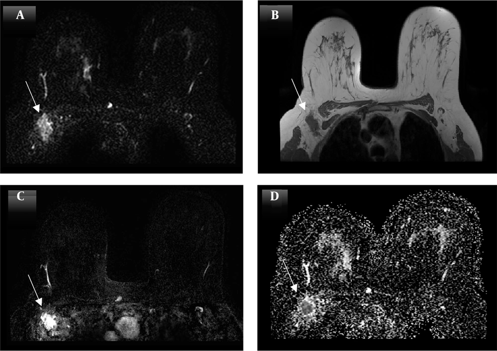
Figure 1.
Axial MRI images of a patient diagnosed with right breast invasive ductal carcinoma (NG 2) with lymph node metastasis, showing positivity for estrogen receptors (ERs), progesterone receptors (PgRs), and Ki67 but negativity for human epidermal growth factor receptor 2 (HER2). A, Subtraction images reveal a heterogeneously enhancing right breast lesion (arrows); B, axial non-fat-saturated T1-weighted MR image displays the lesion as a low-signal area; C, diffusion-weighted imaging (DWI) demonstrates a hyperintense lesion; and D, axial apparent diffusion coefficient (ADC) map confirms restricted diffusion (arrows) with an ADC value of 0.835 × 10⁻³ mm²/s.
High Ki-67 expression was associated with lower ADC values compared to Ki-67-negative tumors (0.927 × 10⁻³ mm²/s vs. 1.061 × 10⁻³ mm²/s; P = 0.042) (Figure 2). The mean ADC value was higher in ER/PR-negative tumors compared to ER/PR-positive tumors (1.023 × 10⁻³ mm²/s vs. 0.950 × 10⁻³ mm²/s; P = 0.031) (Table 2).
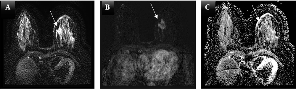
Figure 2.
Axial MRI images of a patient diagnosed with low-grade invasive ductal carcinoma in the left breast, without lymph node metastasis, showing positivity for estrogen receptors (ERs), progesterone receptors (PgRs), and the Ki67 Index but negativity for human epidermal growth factor receptor 2 (HER2). A, Axial contrast-enhanced T1-weighted subtraction MR image demonstrates an enhancing mass with irregular margins (arrows); B, diffusion-weighted imaging (DWI) reveals a hyperintense lesion; and C, apparent diffusion coefficient (ADC) mapping shows restricted diffusion, with an ADC value of 1.129 × 10⁻³ mm²/s.
Table 2.
Apparent Diffusion Coefficient Values and Prognostic Factors in 35 Patients with Breast Cancer
| Prognostic Factor Subgroup | No. of Case (N = 35) | ADC (× 10-3 mm2/s) | P-Value |
|---|---|---|---|
| Lymph node metastasis | 0.016 | ||
| Metastasis | n = 13 | 0.91246 | |
| No metastasis | n = 22 | 1.04655 | |
| Histological type | 0.212 | ||
| IDC nos | n = 10 | 0.91260 | |
| IDC | n = 19 | 0.98882 | |
| DCIS | n = 6 | 1.15833 | |
| Histological grade | 0.182 | ||
| Grade 1 | n = 9 | 1.05167 | |
| Grade 2 | n = 19 | 0.99114 | |
| Grade 3 | n = 7 | 0.97279 | |
| size | 0.207 | ||
| < 2 | n = 8 | 1.00915 | |
| ≥ 2 | n = 27 | 0.95488 | |
| ER, PR | 0.031 | ||
| Positive | n = 25 | 0.95010 | |
| Negative | n = 10 | 1.02308 | |
| Ki67 | 0.042 | ||
| Positive | n = 18 | 0.92782 | |
| Negative | n = 17 | 1.06183 | |
| HER2 | 0.780 | ||
| Positive | n = 10 | 1.01875 | |
| Equivocal | n = 4 | 1.01936 | |
| Negative | n = 21 | 0.97990 |
Abbreviations: HER2, human epidermal growth factor receptor 2; IDC, invasive ductal carcinoma; NOS, not otherwise specified; DCIS, ductal carcinoma in situ; ADC, apparent diffusion coefficient.
4.4. Receiver Operating Characteristic Curve Analysis
Receiver operating characteristic (ROC) curve analysis was performed to determine ADC thresholds for differentiating biomarkers. The area under the curve (AUC), ADC cut-off values, sensitivity, and specificity are summarized in Table 3 and Figure 3.
Table 3.
Area Under the Curve, Sensitivity, and Specificity for the Apparent Diffusion Coefficient Cut-off
| Prognostic Factor | Area Under Curve | ADC Cut-off (× 10-3 mm2/s) | Sensitivity (%) | Specificity (%) |
|---|---|---|---|---|
| Metastasis | 0.879 | 0.981 | 83.3 | 82.4 |
| ER | 0.864 | 1.030 | 84.6 | 90.9 |
| PR | 0.864 | 1.030 | 84.6 | 90.9 |
| Ki67 | 0.837 | 0.951 | 94.4 | 76.5 |
Abbreviations: AUC, area under the curve; ADC, apparent diffusion coefficient; ER, estrogen receptors.
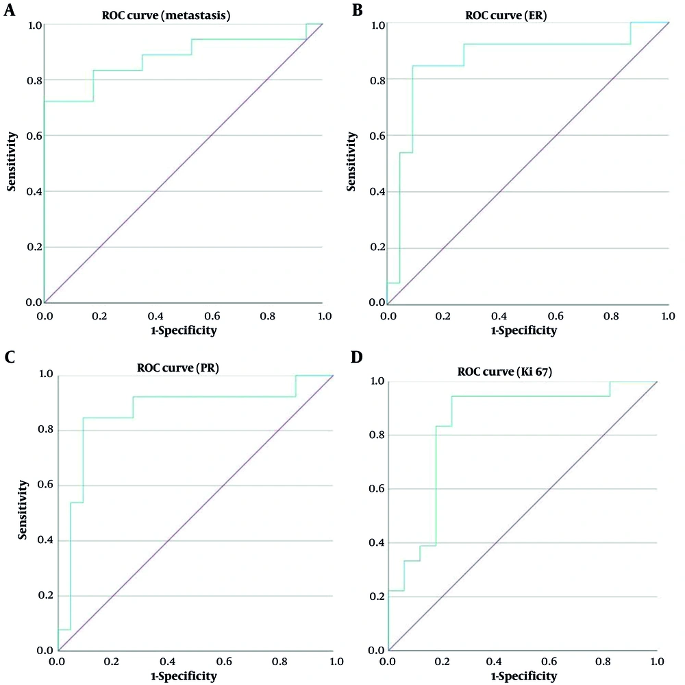
Figure 3.
Receiver operating characteristic (ROC) curves evaluating various prognostic parameters: A, ROC curve for differentiating lymph node metastasis from non-metastatic cases (AUC = 0.879); B, ROC curve for distinguishing estrogen receptor-positive (ER⁺) from ER-negative (ER⁻) lesions (AUC = 0.864); C, ROC curve for differentiating progesterone receptor-positive (PR⁺) from PR-negative (PR⁻) lesions (AUC = 0.864); and D, ROC curve for discriminating between Ki67-positive and Ki67-negative lesions (AUC = 0.837).
• Lymph node metastasis: AUC = 0.879, ADC cut-off = 0.981 × 10⁻³ mm²/s, sensitivity = 83.3%, specificity = 82.4%.
• ER/PR expression: AUC = 0.864, ADC cut-off = 1.030 × 10⁻³ mm²/s, sensitivity = 84.6%, specificity = 90.9%.
• Ki-67 index: AUC = 0.837, ADC cut-off = 0.951 × 10⁻³ mm²/s, sensitivity = 94.4%, specificity = 76.5%.
5. Discussion
Our study provided compelling evidence supporting the role of ADC values in breast cancer characterization. The significant correlations observed between ADC values and both hormone receptor status (ER/PR: P = 0.031) and proliferation markers (Ki-67: P = 0.042) underscore the potential of diffusion-weighted imaging as a complementary tool for molecular profiling (18, 26). The elevated ADC values in ER/PR-negative tumors compared to their positive counterparts suggest distinct microstructural properties that warrant further investigation, particularly given the known association between hormone receptor negativity and aggressive tumor biology (27).
The inverse relationship between ADC values and Ki-67 expression represents one of our most clinically relevant findings. The significant correlation (P = 0.042) between higher Ki-67 levels and lower ADC values strengthens the potential role of DWI in non-invasive assessment of tumor proliferation activity (15). This observation aligns with the known biological basis of diffusion restriction in highly cellular tumors and supports ADC's utility as an imaging biomarker of tumor aggressiveness (14). Furthermore, our observation is consistent with recent findings reporting that proliferative markers such as Ki-67 and SOX-10 are significantly associated with higher tumor grade and more aggressive molecular subtypes of breast cancer (28), reinforcing the biological link between cellular proliferation and diffusion restriction.
Moreover, the ROC curve analysis demonstrated high diagnostic performance for several prognostic indicators. Specifically, ADC thresholds effectively differentiated ER/PR-positive from ER/PR-negative tumors (AUC = 0.864, sensitivity = 84.6%, specificity = 90.9%) and metastatic from non-metastatic lymph nodes (AUC = 0.879, sensitivity = 83.3%, specificity = 82.4%). These values highlight the potential of ADC as a reliable non-invasive biomarker for early tumor stratification and suggest a meaningful role in clinical decision-making (29).
Importantly, we also observed a significant inverse association between ADC values and lymph node metastasis (P = 0.016), with lower ADC values found in patients with nodal involvement. This finding reinforces the clinical utility of ADC in identifying more aggressive tumor phenotypes and provides additional evidence supporting its value in predicting metastatic potential. This relationship may also reflect underlying molecular mechanisms, such as the overexpression of long non-coding RNAs like MIR4435-2HG, which has been linked to TP53 mutation, hormone receptor activity, and poor prognosis in breast cancer (30). Given that lymph node status is a pivotal determinant of prognosis and treatment planning in breast cancer, integrating ADC measurements could enhance the preoperative assessment and guide surgical and therapeutic strategies more precisely.
From a clinical perspective, these findings suggest that ADC mapping may serve as a valuable adjunct in the diagnostic workup of breast cancer, particularly in cases where histopathological information is incomplete or delayed. By incorporating ADC into preoperative MRI protocols, clinicians could more confidently assess tumor aggressiveness, hormone receptor status, and likelihood of nodal involvement (20). This non-invasive insight may help tailor the extent of surgical intervention, prioritize biopsy targets, or prompt earlier initiation of systemic therapy — ultimately contributing to more individualized and effective patient care.
Notably, the high specificity observed for ER/PR (90.9%) and Ki-67 (76.5%) reinforces the diagnostic precision of ADC and may help reduce false-positive rates in imaging-based assessments. Although no significant differences in ADC values were observed between HER2-positive and HER2-negative tumors (P = 0.780), the overall findings affirm ADC’s value in capturing key biological characteristics of breast tumors (31).
These results suggest that ADC may serve not only as a supportive parameter in imaging but also as a predictive imaging biomarker that aids in clinical triage and personalized treatment planning. The integration of ADC with conventional imaging and molecular profiling has the potential to enhance non-invasive tumor characterization and streamline therapeutic decisions in breast cancer management.
5.1. Limitations and Future Directions
Several methodological considerations must be acknowledged. First, the variability in ADC measurements due to technical factors (including b-value selection and ROI placement) and tissue heterogeneity (such as fibrosis and necrosis) presents challenges for clinical implementation (31). Second, the overlap in ADC values between benign and malignant lesions may limit diagnostic specificity (14). Third, our sample size (n = 35) restricts the generalizability of our findings and highlights the need for larger, multicenter validation studies.
Future research should focus on several key areas, including the standardization of DWI acquisition protocols across institutions, prospective validation of our findings in larger cohorts, and investigation of ADC's potential for monitoring treatment response. Additionally, AI-driven approaches for automated ROI selection could further enhance the reproducibility and efficiency of ADC measurements in breast cancer studies (32). The development of consensus guidelines for ADC quantification, incorporating advanced AI tools, will be particularly crucial for clinical translation and widespread adoption in multicenter studies.
5.2. Conclusions
This study demonstrates that ADC values not only correlate significantly with ER/PR status and Ki-67 expression but also show high diagnostic accuracy in distinguishing key prognostic factors. Notably, the significant association between lower ADC values and lymph node metastasis further highlights ADC’s role in assessing tumor aggressiveness and metastatic potential. These findings highlight ADC’s potential as a reliable, non-invasive imaging biomarker in breast cancer evaluation. With further standardization and larger-scale validation, ADC mapping could play an important role in improving early risk stratification and guiding personalized treatment strategies.