1. Introduction
Brachytherapy is an important treatment modality for managing locally advanced rectal cancer, particularly when dose escalation is required or as a boost to residual tumor after external beam radiation therapy (EBRT) (1, 2). Accurate cumulative dose assessment is essential for optimizing tumor control while minimizing complications in organs at risk (OARs), such as the bladder, rectum, and small intestine (3, 4). Conventional dose accumulation across multiple brachytherapy fractions is often performed using simple dose-volume histogram (DVH) summation, based on the assumption that organ geometry remains constant throughout treatment (5). This simplification can lead to inaccuracies because pelvic anatomy, particularly the rectal tumor and adjacent structures, undergoes considerable deformation and displacement due to factors such as rectal filling, gas motion, or applicator-induced anatomical changes (6, 7).
Deformable image registration (DIR) is a promising method for improving cumulative dose estimation by mapping dose distributions onto a common anatomical frame while accounting for inter-fraction organ deformation (8, 9). Several studies have demonstrated the utility of DIR in gynecological and rectal cancer radiotherapy (10). Rigaud et al. (6) and Nesvacil et al. (11) highlighted substantial dosimetric uncertainties introduced by ignoring anatomical changes during fractionated brachytherapy, underscoring the need for image-based dose accumulation. However, despite its potential, DIR introduces its own uncertainties, particularly in deformable tissues with low image contrast. Therefore, DIR accuracy must be validated, often using metrics such as the Dice similarity coefficient (DSC), before it can be considered for clinical implementation.
This study evaluates the dosimetric impact of DIR-based dose accumulation in fractionated brachytherapy for rectal cancer, with a novel emphasis on integrating EBRT and brachytherapy doses to account for inter-fraction motion and anatomical variability. By comparing conventional DVH summation with DIR-based cumulative dosing across multiple fractions, we aimed to assess the feasibility, accuracy, and clinical utility of this approach. This integrated evaluation provides a foundation for adaptive treatment-planning strategies that may enhance tumor control while minimizing late toxicities, thereby addressing a critical gap in the current literature. We hypothesized that DIR-based dose accumulation would reduce cumulative OAR doses compared with conventional DVH summation while maintaining or improving target coverage.
2. Methods
2.1. Study Design, Setting, and Participants
This retrospective observational cohort study was conducted at a single institution. Consecutive patients with locally advanced rectal cancer who were treated with EBRT followed by a fractionated HDR endorectal brachytherapy boost were identified from the institutional database. Eligibility criteria were as follows: (1) an EBRT dose of 50.4 Gy in 28 fractions followed by 4 HDR brachytherapy fractions of 6 - 7 Gy per fraction; (2) CT-based planning for each brachytherapy fraction; and (3) availability of Digital Imaging and Communications in Medicine (DICOM) CT, structure, and dose datasets for all fractions and the EBRT plan. Exclusion criteria were prior pelvic radiotherapy, incomplete datasets, or severe imaging artifacts that prevented reliable DIR.
The sample size (n = 12) included all eligible patients treated during the study period with complete datasets. No formal a priori sample-size calculation was performed. This work was designed as an exploratory methodological analysis to quantify the impact of DIR on cumulative dose estimation; therefore, a formal power calculation was not performed. All primary dosimetric endpoints were available for the analyzed cohort, and no imputation was required.
To mitigate potential sources of bias, we included consecutive eligible patients, applied standardized preparation instructions (including rectal emptying and bladder filling before each CT scan), used consistent contouring and planning personnel, and performed both qualitative clinical review and quantitative DSC-based quality assurance (QA) of the DIR results. As an inherent limitation of retrospective studies, residual selection and information bias cannot be completely excluded despite these mitigation measures. Baseline patient and disease characteristics are summarized in Table 1, and detailed patient-level baseline and treatment information, including the exact tumor distance from the anal verge, tumor size/volume parameters, interstitial needle details when applicable, and the interval between completion of EBRT and initiation of HDR brachytherapy, are provided in Supplementary Table S1.
Table 1.
Baseline Characteristics of the Study Cohort (n = 12) a
| Characteristic | Overall (n = 12) |
|---|---|
| Age (range), y | 58.3 ± 9.1 (42 - 73) |
| Sex | |
| Male | 8 (66.7) |
| Female | 4 (33.3) |
| ECOG performance status | |
| 0 | 7 (58.3) |
| 1 | 5 (41.7) |
| Clinical T stage | |
| cT2 | 2 (16.7) |
| cT3 | 8 (66.7) |
| cT4 | 2 (16.7) |
| Clinical N stage | |
| cN0 | 5 (41.7) |
| cN1 | 5 (41.7) |
| cN2 | 2 (16.7) |
| Tumor location, distance from anal verge | |
| Low (≤ 5 cm) | 5 (41.7) |
| Mid (5 - 10 cm) | 6 (50.0) |
| High (> 10 cm) | 1 (8.3) |
| Tumor length (range), cm | 4.8 ± 1.2 (3.0 - 7.0) |
| HDR brachytherapy technique | |
| Intracavitary only | 7 (58.3) |
| Intracavitary + interstitial | 5 (41.7) |
| Number of interstitial needles, if used | 4 (3 - 5) |
| EBRT regimen | 50.4 Gy in 28 fractions (100%) |
| HDR boost regimen | 4 fractions of 6 - 7 Gy (100%) |
| Planning CT slice thickness (mm) | 3 (100%) |
| Applicator re-insertion each fraction | Yes (100%) |
Abbreviations: CT, computed tomography; EBRT, external beam radiation therapy; ECOG, Eastern Cooperative Oncology Group; HDR, high-dose-rate.
a Values are expressed as mean ± SD or No. (%).
2.2. Patient Treatment and Imaging Protocol
EBRT was delivered using volumetric modulated arc therapy (VMAT) with 6 MV photons to a total dose of 50.4 Gy in 28 fractions. Brachytherapy was delivered in 4 fractions of 6 - 7 Gy each, using either an intracavitary approach alone or a combined intracavitary–interstitial technique. For the intracavitary approach, a rectal cylinder applicator was used to treat superficial or mid-wall rectal lesions. In cases of tumor extension beyond the rectal wall or irregular tumor geometry, transperineal interstitial needles were inserted in conjunction with the rectal cylinder to improve target coverage and achieve optimal dose conformity. Among patients who received interstitial needles, the median number of needles was 4 (range, 3 - 5). Applicator type and placement were determined individually based on tumor location, endoscopic assessment, and radiological imaging.
Applicators, including both the intracavitary cylinder and interstitial needles, were reinserted before each brachytherapy fraction to account for anatomical changes and ensure accurate placement. CT-based treatment planning was performed before each fraction using a Siemens Somatom scanner (120 kV, 162 mA, 3-mm slice thickness). Before imaging, patients were instructed to empty their rectum and fill their bladder with 80 cc of sterile saline to reduce organ motion and improve consistency across treatment sessions. All methods were carried out in accordance with relevant guidelines and regulations. This study used anonymized patient data obtained from previously treated patients and was approved by the Azad University Research Committee. All participants provided written informed consent.
2.3. Treatment Planning
Treatment plans were developed using the Oncentra Brachy treatment planning system (version 4.6.2; Elekta, Stockholm, Sweden) with the TG-43 dose calculation algorithm, as recommended by the AAPM TG-43 report. A single radiation oncologist contoured the clinical target volume (CTV), defined as the tumor-involved segment of the rectum, and OARs, including the uninvolved rectal wall, bladder, small intestine, and sigmoid colon, on each CT scan to ensure consistency. The uninvolved rectal wall was contoured separately and evaluated for potential toxicity, distinct from the CTV, to enable accurate assessment of dose constraints. All plans were optimized by a single medical physicist using inverse planning to meet institutional dose constraints, with priority given to maximizing CTV coverage (D90) while respecting OAR dose limits. For equivalent dose in 2 Gy fractions (EQD2) calculations, α/β ratios were set at 10 Gy for the CTV (rectal tumor) and 3 Gy for OARs (uninvolved rectum, bladder, small intestine, and sigmoid colon), according to the linear-quadratic model.
2.4. Deformable Image Registration and Dose Accumulation Workflow
Figure 1 illustrates the overall dose accumulation workflow, including rigid alignment, deformable image registration, EQD2 conversion, and voxel-wise dose summation across fractions. The CT datasets, dose files, and structure sets from each fraction were imported into MIM Maestro v7.0.6. Simple EQD2 summation (D_simple) was calculated across fractions, assuming identical anatomy. DIR-based accumulation (D_deform) was performed using the MIM deformable dose summation workflow. The fourth CT scan was designated as the reference, and the other scans were mapped onto it using rigid registration followed by grayscale-based free-form DIR. To improve registration accuracy, applicators were overridden to 0 Hounsfield units (HU), and the bladder/bowel was overridden to 1000 HU. After registration, anatomical alignment was assessed both qualitatively by a radiation oncologist and quantitatively using DSC. No density overrides were applied apart from voxel HU modifications.
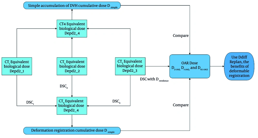
Figure 1.
Calculation and comparison process for cumulative doses obtained using deformable image registration and simple dose-volume histogram summation.
2.5. Evaluation Metrics
For each fraction and accumulation method, dose parameters for the CTV (D90) and OARs (D2cc, D1cc, and D0.1cc) were extracted. Differences between D_simple and D_deform were calculated as D_diff. Pearson correlation was used to examine the relationship between D_diff and DSC values. Paired 2-tailed t-tests were used to compare DIR-based and simple summation dose metrics, with statistical significance defined as P < 0.05. Normality of within-patient paired differences was assessed visually using histograms and Q–Q plots. Given the small sample size, findings were interpreted as exploratory.
2.6. Total Dose Accumulation Including External Beam Radiation Therapy
To address the full treatment course and enhance clinical relevance, cumulative dose calculations were extended to include the EBRT component. Total dose accumulation was performed by registering the EBRT dose distribution to the same anatomical reference frame used for brachytherapy dose accumulation, namely the CT scan of the fourth brachytherapy fraction. Rigid registration was applied between the EBRT planning CT and the reference CT using bony landmarks, followed by manual verification and adjustment as needed. The EBRT dose distribution, originally delivered as 50.4 Gy in 28 fractions (1.8 Gy per fraction), was converted to EQD2 using an α/β ratio of 10 for the tumor and 3 for OARs. The EQD2-EBRT dose was then added voxel by voxel to the DIR-accumulated brachytherapy dose map in MIM software to obtain the final cumulative dose. Final dose-volume parameters, including D90 for the CTV and D2cc, D1cc, and D0.1cc for OARs, were extracted from this combined dose distribution to evaluate total therapeutic exposure and assess the impact of DIR in the context of the complete treatment course.
3. Results
3.1. Participant Flow and Inclusion
During the study period, 18 consecutive patients with locally advanced rectal cancer who received EBRT followed by a fractionated HDR endorectal brachytherapy boost were assessed for eligibility. Six patients were excluded because of incomplete data or non-evaluable imaging, including missing DICOM dose/structure datasets for 1 or more HDR fractions (n = 3), unavailable EBRT planning dose/CT data preventing full-course accumulation (n = 2), and severe imaging artifacts or suboptimal image quality precluding reliable DIR (n = 1). The remaining 12 patients met all eligibility criteria and were included in the final analysis. Baseline characteristics are presented in Table 1, with additional patient-level details provided in Supplementary Table S1.
3.2. Deformable Image Registration vs Simple Dose-Volume Histogram Summation in Brachytherapy Fractions Only
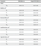
The comparison between DIR-based and simple DVH summation methods for brachytherapy alone is summarized in Table 2. DIR resulted in statistically significant reductions in cumulative doses to OARs, particularly the rectum and bladder. The reductions ranged from 1.4 to 2.8 Gy in EQD2 values. No significant differences were observed in the sigmoid colon or target volume D90, likely because of the relatively stable anatomy in these regions or limitations in DIR accuracy for small, high-dose areas. These reductions, particularly in bladder and rectal doses, may translate into lower rates of late toxicity, although clinical validation is required. Table 2 presents the mean ± standard deviation for each parameter.
Table 2.
Comparison of Cumulative Doses to Targets and Organs at Risk Between Deformable Image Registration and Simple Dose-Volume Histogram Summation (Gy, x ± s) a
| Parameter | DIR (EQD2), Gy | Simple DVH (EQD2), Gy | Dose Difference, Gy | T Value | P-Value |
|---|---|---|---|---|---|
| Target | |||||
| D90 | 29.63 ± 7.35 | 28.37 ± 4.96 | 1.26 ± 3.31 | 0.50 | 0.63 |
| Bladder, cm3 | |||||
| D2 | 24.26 ± 5.78 | 26.35 ± 6.02 | 2.09 ± 1.23 | -2.65 | 0.02 |
| D1 | 25.34 ± 4.77 | 26.51 ± 6.46 | 1.17 ± 2.26 | -3.93 | 0.002 |
| D0.1 | 33.54 ± 5.96 | 35.04 ± 7.41 | 1.50 ± 1.98 | -4.15 | 0.002 |
| Rectum (OAR), cm3 | |||||
| D2 | 19.46 ± 7.13 | 21.75 ± 6.63 | 2.29 ± 1.53 | -3.02 | 0.01 |
| D1 | 20.96 ± 8.13 | 23.03 ± 4.19 | 2.07 ± 3.34 | -2.52 | 0.03 |
| D0.1 | 25.44 ± 6.41 | 28.32 ± 5.17 | 2.88 ± 2.21 | -3.12 | 0.01 |
| Small intestine, cm3 | |||||
| D2 | 9.27 ± 6.84 | 10.71 ± 5.75 | 1.44 ± 1.52 | -2.96 | 0.01 |
| D1 | 10.13 ± 6.13 | 11.95 ± 5.76 | 1.82 ± 2.45 | -1.05 | 0.32 |
| D0.1 | 13.43 ± 8.21 | 15.14 ± 6.19 | 1.71 ± 2.79 | -1.12 | 0.29 |
| Sigmoid colon, cm3 | |||||
| D2 | 13.63 ± 5.85 | 14.65 ± 7.22 | 1.02 ± 3.16 | -0.75 | 0.47 |
| D1 | 15.63 ± 6.35 | 16.73 ± 6.13 | 1.10 ± 2.47 | -1.05 | 0.32 |
| D0.1 | 19.63 ± 6.29 | 21.74 ± 8.13 | 2.11 ± 3.07 | -0.73 | 0.48 |
Abbreviations: DIR, deformable image registration; DVH, dose-volume histogram; EQD2, equivalent dose in 2 Gy fractions; OAR, organ at risk.
a Values are expressed as mean ± SD.
3.3. Correlation Between Deformable Image Registration Accuracy and Dose Differences
Table 3 shows no statistically significant correlation between DSC values and dose differences (D_diff), indicating that geometric similarity measured by DSC alone may not fully explain dose accumulation outcomes. Notably, negative correlation coefficients, such as -0.55 for rectum D1cc, suggest that higher DSC values tended to correspond to smaller dose differences, although this trend was not statistically significant.
Table 3.
Correlations Between Dose Difference and Dice Similarity Coefficient (x ± s) a
| Organ (cm3) | Dose Difference, Gy | DSC | Correlation Coefficient | P-Value |
|---|---|---|---|---|
| Bladder | ||||
| D2 | 2.09 ± 1.23 | 0.83 ± 0.21 | -0.22 | 0.82 |
| Rectum (OAR) | ||||
| D2 | 2.29 ± 1.53 | 0.81 ± 0.11 | -0.29 | 0.78 |
| D1 | 2.07 ± 3.34 | 0.79 ± 0.20 | -0.55 | 0.51 |
| D0.1 | 2.88 ± 2.21 | 0.77 ± 0.15 | -0.56 | 0.33 |
| Small intestine | ||||
| D2 | 1.44 ± 1.52 | 0.66 ± 0.10 | -0.28 | 0.52 |
| D1 | 1.82 ± 2.45 | 0.64 ± 0.15 | -0.48 | 0.65 |
| D0.1 | 1.71 ± 2.79 | 0.61 ± 0.12 | -0.25 | 0.60 |
Abbreviations: DSC, Dice similarity coefficient; OAR, organ at risk.
a Values are expressed as mean ± SD.
3.4. Evaluation of Deformable Registration Results
All deformable registrations were automatically performed in MIM software and subsequently reviewed by an experienced radiation oncologist for clinical plausibility. Quantitative evaluation was conducted using DSC to assess anatomical agreement between the registered and reference contours. As shown inTable 4, the bladder and rectum achieved DSC values > 0.80, indicating good alignment. According to the AAPM TG-132 report, DSC values above 0.80 are generally considered acceptable for clinical applications. The target CTV, small intestine, and sigmoid colon demonstrated lower DSC values, reflecting the greater difficulty of deformable registration in regions with high anatomical variability.
Table 4.
Dice Similarity Coefficient for Each Structure After Deformable Registration a
| Organ | DSC |
|---|---|
| CTV (rectal tumor) | 0.73 ± 0.15 |
| Bladder | 0.82 ± 0.13 |
| Rectum (OAR) | 0.80 ± 0.19 |
| Small intestine | 0.65 ± 0.22 |
| Sigmoid colon | 0.58 ± 0.23 |
Abbreviations: CTV, clinical target volume; DSC, Dice similarity coefficient; OAR, organ at risk.
a Values are expressed as mean ± SD.
It is noteworthy that although the rectum as an OAR achieved high DSC values (> 0.80), the CTV (rectal tumor) showed lower agreement. This discrepancy arises because the CTV typically encompasses a smaller, asymmetric segment of the rectal wall that is more susceptible to anatomical deformation and registration errors. In contrast, the full rectum contour as an OAR is more anatomically distinct and consistently delineated, resulting in higher registration accuracy.
3.5. Target Dose Improvement After Deformable Registration
As presented in Table 1 and 3, DIR produced measurable reductions in OAR doses across patients, with the most pronounced effects observed in the bladder and rectum. In particular, the D2cc of the bladder consistently decreased with DIR-based accumulation, and this decrease was used as a surrogate marker of registration accuracy. Based on DIR-accumulated dose distributions, treatment plans were retrospectively re-analyzed on the latest CT scan, corresponding to fraction 4, for each patient. The expected improvement in CTV dose (D90) was assessed in response to reduced OAR constraints. After EQD2 conversion, the minimum observed increase in D90 was 50.2 cGy, the maximum was 464.0 cGy, and the median was 177.0 cGy. These findings support the potential of DIR not only for more accurate dose accumulation but also for adaptive re-optimization in brachytherapy planning to enhance tumor coverage while sparing surrounding healthy tissues.
3.6. Total Dose Accumulation Including External Beam Radiation Therapy
Final cumulative doses were derived by voxel-wise summation of EQD2-converted EBRT and brachytherapy distributions after rigid registration. Cumulative EQD2 dose metrics combining EBRT and brachytherapy for each patient are presented in Table 5. The integration of EBRT did not negate the advantage of DIR, which continued to show meaningful reductions in OAR doses. DIR maintained its dosimetric advantage even when EBRT doses were integrated, with statistically significant reductions in OAR doses, including rectum D2cc (-2.5 ± 1.2 Gy, P < 0.01), and a modest but significant increase in CTV D90 (+1.3 ± 1.1 Gy, P = 0.03). These results underscore the clinical relevance of DIR for comprehensive dose optimization in combined-modality regimens.
Table 5.
Final Cumulative Equivalent Dose in 2 Gy Fractions Dose (External Beam Radiation Therapy + Brachytherapy) Using Simple Versus Deformable Image Registration-Based Accumulation a
| Structure | Simple + EBRT; Mean ± SD, Gy | DIR + EBRT; Mean ± SD, Gy | Dose Difference; Mean ± SD, Gy | P-Value |
|---|---|---|---|---|
| CTV D90 | 74.8 ± 4.2 | 76.1 ± 4.0 | +1.3 ± 1.1 | 0.03 |
| Rectum (OAR) D2 | 65.2 ± 3.6 | 62.7 ± 3.3 | -2.5 ± 1.2 | < 0.01 |
| Bladder D2 | 60.9 ± 4.1 | 59.0 ± 3.8 | -1.9 ± 1.0 | 0.02 |
| Sigmoid D2 | 58.4 ± 4.3 | 56.6 ± 3.9 | -1.8 ± 1.1 | 0.04 |
Abbreviations: CTV, clinical target volume; DIR, deformable image registration; EBRT, external beam radiation therapy; EQD2, equivalent dose in 2 Gy fractions; OAR, organ at risk.
a Values are expressed as mean ± SD.
4. Discussion
This study investigated the dosimetric implications of DIR for cumulative dose evaluation in fractionated HDR brachytherapy for rectal cancer, including the integration of EBRT doses. Our results demonstrate that DIR-based dose accumulation significantly reduced doses to OARs, particularly the rectum and bladder, while enabling modest improvements in target coverage. Integrating EBRT into DIR workflows represents an important extension of previous studies focused solely on brachytherapy and addresses a key gap in combined-modality dose optimization (Table 5).
Previous studies in gynecological and rectal brachytherapy have emphasized the risks of ignoring inter-fraction anatomical changes. Rigaud et al. (6) reported that rigid dose summation overestimates rectal doses by > 10%, potentially skewing toxicity predictions. This finding is consistent with our results, in which DIR reduced rectal D2cc by 2.5 Gy (P < 0.01) in the full-course EBRT + brachytherapy evaluation (Table 5). Similarly, Nesvacil et al. (11) advocated the use of DIR to mitigate uncertainties caused by organ motion; this recommendation is supported here by statistically significant OAR sparing, such as a bladder D2cc reduction of 1.9 Gy (P = 0.02) (Table 5). Our work extends these findings by demonstrating the robustness of DIR in hybrid EBRT-brachytherapy regimens, a scenario common in rectal cancer management but underexplored in the earlier literature. In addition, for brachytherapy-only accumulation, DIR showed consistent OAR dose reductions across multiple metrics, with overall reductions of approximately 1.4 - 2.8 Gy EQD2, as summarized in the Results (Table 2).
Although DIR accuracy remains challenging, our DSC-based evaluation showed clinically acceptable registration quality for most structures. The rectum and bladder achieved DSC values > 0.80, consistent with commonly reported DIR QA ranges and the AAPM TG-132 emphasis on quantitative evaluation (Table 4). However, the lower DSC for the rectal CTV (0.73) highlights the difficulty of registering small, irregular targets prone to deformation, a limitation also reported for tumor-specific geometries. This underscores the need for DIR algorithms tailored to tumor-specific geometries, particularly in organs such as the rectum, where gas, peristalsis, and applicator placement induce complex motion. Notably, our correlation analysis did not demonstrate statistically significant associations between DSC and dose differences (Table 3), suggesting that DSC alone may not fully explain dosimetric discrepancies in high-gradient regions and that complementary QA metrics may be beneficial.
The median 1.77 Gy EQD2 improvement in CTV D90 after DIR-based re-optimization is consistent with adaptive brachytherapy principles. By relaxing OAR constraints, DIR may facilitate safe dose escalation, a strategy that is critical for improving local control in rectal cancer. However, the observed variability in CTV D90 improvement (range, 50 - 464 cGy) suggests that patient-specific characteristics, such as tumor size, anatomical distortion, or DIR quality, may influence the degree of benefit achieved through DIR-guided adaptation. Future studies should stratify outcomes by tumor characteristics and DIR QA metrics, such as the DSC values in Table 4, to refine patient selection.
4.1. Limitations and Future Directions
This study has several limitations. First, reliance on TG-43 dose calculations ignores tissue heterogeneity, potentially underestimating doses to air-filled regions, such as the rectal lumen. Transitioning to model-based algorithms, such as Acuros BV, could improve accuracy. Second, although MIM’s DIR algorithm demonstrated clinical utility, independent biomechanical validation is needed to assess its precision in high-deformation regions. Third, rigid EBRT-to-brachytherapy registration may not fully capture soft-tissue motion; deformable EBRT dose mapping could further enhance cumulative dose accuracy. In addition, this work is primarily dosimetric and does not directly link DIR-based cumulative dose metrics with clinical endpoints, such as toxicity or local control, which should be addressed in prospective studies.
4.2. Clinical Implications
The 2.5 Gy reduction in rectal D2cc has direct relevance for toxicity mitigation because even small dose increments may correlate with the risk of late proctitis. Similarly, the 1.3 Gy increase in CTV D90 (P = 0.03) demonstrates the potential of DIR to enhance tumor control without exceeding OAR tolerances in the combined EBRT + brachytherapy setting (Table 5). These findings support further evaluation of DIR integration into adaptive brachytherapy protocols, particularly for patients with high baseline toxicity risks. Baseline cohort characteristics supporting the clinical context are provided in Table 1, with patient-level details in Supplementary Table S1.
4.3. Bias and Generalizability
As a retrospective single-center study, our findings may be influenced by selection and measurement biases. We minimized observer-related variability by using a single radiation oncologist for contouring and a single physicist for plan optimization, and all DIR results were clinically reviewed in addition to quantitative DSC evaluation. However, excluding patients with incomplete datasets or non-evaluable imaging may bias the cohort toward higher-quality imaging and more stable anatomy, which could overestimate DIR performance and the degree of OAR sparing achievable in routine practice. Additionally, the magnitude of dosimetric differences should be interpreted as exploratory given the small sample size and software-specific implementation, and it may vary across institutions, DIR algorithms, and dose engines. External validation in larger, multi-institutional cohorts, ideally including independent DIR verification and model-based dose calculation, will be important before broader generalization. This potential selection bias is likely directional, potentially inflating apparent DIR performance, although its magnitude remains uncertain given the small cohort size.
4.4. Conclusion
This study highlights the clinical and dosimetric potential of DIR in cumulative dose assessment for patients with rectal cancer treated with combined EBRT and HDR brachytherapy. By explicitly modeling inter-fraction anatomical variations, DIR may improve dose estimation accuracy for both the CTV and OARs. Key findings include significant reductions in rectal and bladder doses (eg, ΔRectum D2cc = -2.5 Gy, P < 0.01; ΔBladder D2cc = -1.9 Gy, P = 0.02) and a modest but clinically relevant improvement in CTV coverage (ΔD90 = +1.3 Gy, P = 0.03), demonstrating the dual utility of DIR for toxicity mitigation and target optimization in a dosimetric analysis.
The integration of EBRT into DIR workflows represents an important advancement toward comprehensive and adaptive radiotherapy planning. Given the retrospective single-center design, limited cohort size, and software-/parameter-specific implementation, these findings should be interpreted as exploratory and hypothesis-generating. Rather than establishing routine adoption, our results support further evaluation of DIR within adaptive brachytherapy workflows, especially for patients at higher risk of toxicity or with anatomically complex disease. Future work should focus on prospective validation, incorporation of model-based dose calculations, and exploration of patient-specific adaptive strategies to fully realize the potential of DIR in rectal cancer treatment, including studies that link DIR-based cumulative dose metrics to clinical outcomes, such as toxicity and local control.