1. Background
Breast cancer (BC) remains the most commonly diagnosed cancer and the leading cause of cancer-related death among women worldwide (1). One of the key challenges in BC management is its marked heterogeneity across genomic, epigenomic, transcriptomic, and proteomic levels (2, 3). This biological complexity results in substantial variation in tumor growth, treatment response, and patient outcomes (4). Prognosis is influenced by several factors, including patient characteristics such as age; tumor features such as size and lymph node involvement (5, 6); and molecular biomarkers such as estrogen receptor (ER), progesterone receptor (PR), and human epidermal growth factor receptor 2 (HER2) status (7). However, these traditional markers often provide an incomplete assessment, and their accuracy in predicting survival or treatment response remains limited.
Recent advances in breast cancer treatment, including chemotherapy, radiotherapy, and surgery, have significantly improved survival and quality of life. Breast-conserving surgery (BCS) followed by adjuvant radiotherapy achieves locoregional recurrence rates of 2% - 3% for ER-positive and HER2-positive subtypes and 5% for triple-negative breast cancer (TNBC), comparable to outcomes after mastectomy alone (8, 9). Chemotherapy, particularly for TNBC and HER2-positive subtypes, not only improves pathologic complete response rates but can also convert some mastectomy candidates to BCS candidates (10, 11). Standard chemotherapy regimens often combine taxanes with anthracyclines, whereas platinum agents may offer additional benefit in TNBC, although their long-term impact remains under investigation (12-15). Radiotherapy remains essential after BCS, with hypofractionated whole-breast regimens preferred for early-stage disease and partial or accelerated protocols increasingly used for low-risk patients because of their favorable toxicity profiles (16-18). Postmastectomy radiotherapy is widely recommended for patients with 4 or more positive lymph nodes, and evidence also supports regional nodal irradiation in node-positive cases; however, the value of internal mammary node irradiation remains debated because of its limited benefit and potential for added toxicity (19-21).
Despite these therapeutic advances, predicting which patients will benefit most from specific treatments or which may relapse remains a major challenge. Tumor heterogeneity continues to limit the predictive value of traditional biomarkers such as ER, PR, and HER2, and newer biomarkers, such as circulating tumor DNA and immune gene signatures, remain limited by insufficient validation for widespread clinical use (22). Integrating multi-omics data, including genomics, proteomics, transcriptomics, and imaging, could improve risk stratification; however, this approach is challenging because of missing data, small sample sizes, and inconsistencies across datasets, all of which limit machine learning model performance (23). Predicting response to radiotherapy or chemotherapy is particularly complex because validated radiosensitivity signatures are lacking and chemoresistance mechanisms are highly variable (24, 25). Thus, although outcomes have improved, the need for precise, personalized prediction tools in BC care remains unmet.
Recent advances in artificial intelligence-based breast cancer screening have demonstrated the ability to automatically detect suspicious features and uncover patterns in imaging data, achieving diagnostic performance comparable to, or even exceeding, that of radiologists (26-28). Among these methods, unsupervised learning approaches such as ARM have emerged as powerful tools for identifying relationships that are not explicitly labeled or predefined. For example, temporal ARM has been used to identify demographic and treatment patterns associated with survival in SEER-Medicare datasets (29). These studies indicate that unsupervised learning approaches such as ARM can provide clinically meaningful insights from large, heterogeneous datasets that might be missed by traditional statistical models.
2. Objectives
Building on this foundation, the present study applied ARM as an unsupervised machine learning approach to investigate survival outcomes in patients with breast cancer treated with combined modalities, including chemotherapy, radiotherapy, and surgery. By analyzing co-occurrence patterns among patient demographics, tumor characteristics, treatment regimens, and outcomes, we aimed to identify novel prognostic associations. These findings may support more personalized and effective treatment strategies for breast cancer management, particularly in the context of integrated multimodal therapies.
3. Methods
3.1. Data Collection
This cross-sectional study analyzed clinical data from 216 patients with non-metastatic breast cancer referred to the Radiotherapy Center of Towhid Hospital in Sanandaj, Iran, between 2016 and 2023. Patients with a confirmed diagnosis and complete clinical and treatment records were included. The study was approved by the Ethics Committee of Kurdistan University of Medical Sciences (IR.MUK.REC.1399/064) and conducted in accordance with the Declaration of Helsinki. Data were extracted from medical records using a structured checklist. The variables included the following:
Demographics: Age
2. Tumor characteristics: Primary tumor size, histologic/pathology type, pathology grade, and hormone receptor status (ER, PR, and HER2). Nodal status (N stage) was not available in a consistently extractable format across the medical records and was therefore not included in the analyses.
3. Treatment details: First-line treatment modality (surgery, chemotherapy, or radiotherapy), whether each modality was performed, type of surgery (lumpectomy vs mastectomy), and prescribed radiotherapy dose/fractionation, as recorded in radiotherapy treatment charts. Radiotherapy prescriptions at our center followed institutional practice during the study period. Whole-breast or chest-wall irradiation was most commonly delivered using conventional fractionation of 45 - 50.4 Gy in 1.8 - 2.0 Gy per fraction, with a 10 - 16 Gy tumor-bed boost for high-risk margins when indicated. Hypofractionated schedules, such as 40 - 42.5 Gy in 15 - 16 fractions, were used selectively for patients judged eligible based on clinical risk and physician preference; however, hypofractionation flags were not systematically recorded in older records. Detailed radiotherapy target volumes, such as explicit chest-wall vs regional nodal fields, were not consistently coded in the electronic dataset and therefore were not used as independent variables in the present analysis.
4. Outcome: Survival status, defined as alive or dead at the end of 2023.
3.2. Data Preprocessing
All preprocessing was performed in Python using pandas and standard scientific libraries. The aim was to produce a reliable, consistently encoded transaction-style dataset suitable for ARM while minimizing biased signals introduced by inconsistently recorded variables.
3.2.1. Cleaning, Standardization, and Exclusion of Unreliable Variables
Records with missing essential data, including survival status, treatment indicators, or receptor status, were removed (n = 18, 8.3%). Categorical inconsistencies, such as “Alive” vs “alive,” were standardized. Variables with substantial missing data due to inconsistent documentation were excluded to avoid spurious associations. These variables included nodal stage (100% missing, free-text only), chemotherapy regimens (89% missing, inconsistent drug/cycle recording), radiotherapy target volumes (100% missing, not coded), fractionation schedules (76% missing, not systematically documented), endocrine therapy (96% missing, externally managed), and trastuzumab receipt (94% missing, referrals unconfirmed). These variables were excluded rather than imputed because missingness was non-random. This approach prioritized data integrity while acknowledging residual confounding as a limitation.
3.2.2. Variable Transformation and Encoding
To enable rule mining, all continuous variables were converted into clinically meaningful categorical bins, as summarized in Table 1. Prescribed total radiotherapy dose was used as a pragmatic proxy for radiotherapy exposure because fractionation schedules were not systematically recorded.
Table 1.
Summary of Variable Transformations and Encodings
| Variables | Original Type | Transformation |
|---|---|---|
| Age (y) | Continuous | Categorized as ≤ 40, 41 - 50, 51 - 60, and > 60 years |
| Tumor size (cm) | Continuous | Categorized as 0.5 - 2 (small), 2 - 5 (medium), and > 5 (large) |
| Prescribed dose (Gy) | Continuous | Coded as no radiotherapy, < 45 (low), 45 - 50.4 (standard), 50.5 - 60 (moderate-high), and > 60 (high) |
| ER, PR, and HER2 | Binary | Standardized as positive or negative |
| Survival outcome | Binary | Standardized as alive or dead |
| Surgery, radiotherapy, and chemotherapy | Categorical | Coded as yes or no to indicate whether treatment was received |
3.2.3. Transaction Formatting for Association Rule Mining
Each patient record was converted into a transaction consisting of item-like entries, such as “Age > 60,” “HER2 Negative,” “Received Chemotherapy,” and “Alive.” This transaction-style format was required to extract meaningful association rules using ARM algorithms. These preprocessing steps ensured that the dataset was clean, consistent, and ready for mining meaningful patterns related to tumor characteristics, treatment decisions, and survival outcomes in patients with breast cancer.
3.3. Association Rule Mining
Association rule mining enables the identification of clinically relevant patterns that may influence survival and treatment outcomes, thereby supporting clinical decision-making. The Apriori algorithm, implemented in Python, was used to derive these associations. The algorithm works in 3 main steps: 1) identifying frequent single items that meet the support threshold, 2) combining them into larger frequent itemsets, and 3) deriving rules that satisfy the confidence and lift criteria. The Apriori algorithm was used to identify frequent combinations of clinical features and treatments associated with survival. Rules are expressed as antecedent → consequent, such as {Tumor Size > 5 cm, Received Chemotherapy} → {Alive}. Rule quality was assessed using the following standard metrics:
1. Support: Frequency of the itemset in the data.
2. Confidence: Conditional probability of the consequent given the antecedent.
3. Lift: The extent to which the consequent is more likely given the antecedent. A lift > 1 indicates a positive association.
To ensure clinical relevance and statistical robustness, we applied thresholds of support ≥ 5% and confidence ≥ 60% and retained only rules with lift > 1. The top 10 rules by support were selected for interpretation. Figure 1 illustrates the overall analytical workflow. To assess rule robustness given the modest sample size, we performed sensitivity analyses using varying minimum support thresholds (3%, 5%, 7%, and 10%) while maintaining confidence ≥ 60% and lift > 1. Rules that persisted at higher support thresholds were considered more robust. In addition, for each rule presented in the Results section, we calculated and reported the absolute number of patients supporting the rule, defined as the raw count of transactions containing both the antecedent and the consequent. This transparency allows readers to assess rule stability based on subgroup sizes.
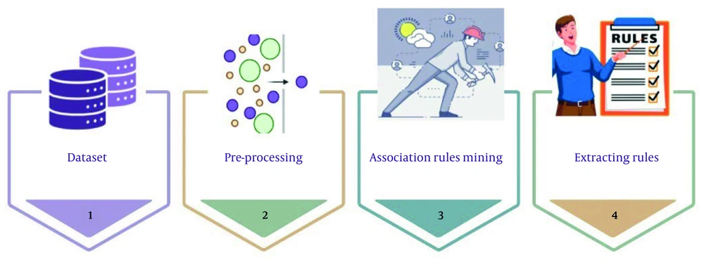
Figure 1.
Association rule mining process for exploring meaningful relationships among variables in the dataset
3.4. Statistical Analysis
To complement rule mining, we assessed monotonic relationships between all clinical variables and five-year survival status. Given the ordinal and non-normally distributed nature of the data, Spearman rank correlation was used. A correlation heatmap was generated in Python using seaborn to visualize the strength and direction of these associations, supporting the identification of feature clusters and potential multicollinearity.
4. Results
This study initially analyzed a cohort of 216 patients with non-metastatic breast cancer treated at Towhid Hospital in Sanandaj, Iran, between 2016 and 2023. After preprocessing and excluding records with incomplete or missing essential variables, 198 patients with complete records remained for ARM analysis. This exclusion may introduce selection bias if missingness was non-random. Table 2 summarizes key characteristics of the final cohort, including demographic variables such as age; tumor features such as size, grade, and pathology; treatment modalities such as surgery, chemotherapy, and radiotherapy; receptor status including HER2, ER, and PR; and survival outcomes. Survival rates across subgroups are also reported to highlight potential outcome disparities and guide further pattern discovery.
Table 2.
Characteristics of the Breast Cancer Dataset (N = 198) a
| Characteristics | Frequency | Percentage | Additional Metrics |
|---|---|---|---|
| Survival status | - | ||
| Alive | 132 | 66.7 | |
| Dead | 66 | 33.3 | |
| Age range | |||
| < 40 | 32 | 16.2 | Mean age: 48.5 years (SD: 12.3) |
| 40 - 49 | 79 | 39.9 | |
| 50 - 69 | 84 | 42.4 | |
| ≥ 70 | 4 | 2.0 | |
| Tumor size (categorized) | |||
| Small (< 2 cm) | 53 | 26.8 | Mean tumor size: 3.8 cm (SD: 2.2) |
| Medium (2 - 5 cm) | 112 | 56.6 | |
| Large (> 5 cm) | 33 | 16.7 | |
| Pathology grade | |||
| 1 | 21 | 10.6 | Survival rate (grade 1): 71.4% (15/21) |
| 2 | 119 | 60.1 | Survival rate (grade 2): 68.1% (81/119) |
| 3 | 58 | 29.3 | Survival rate (grade 3): 60.3% (35/58) |
| Pathology type | |||
| Invasive ductal carcinoma | 158 | 79.8 | Survival rate: 65.8% (104/158) |
| Infiltrating ductal carcinoma | 23 | 11.6 | Survival rate: 69.6% (16/23) |
| In situ and invasive ductal carcinoma | 6 | 3.0 | Survival rate: 66.7% (4/6) |
| Invasive lobular carcinoma | 11 | 5.6 | Survival rate: 72.7% (8/11) |
| Surgery | |||
| Yes | 180 | 90.9 | Survival rate (yes): 67.2% (121/180) |
| No | 18 | 9.1 | Survival rate (no): 61.1% (11/18) |
| Surgery type | |||
| Mastectomy | 117 | 59.1 | Survival rate: 65.8% (77/117) |
| Lumpectomy | 63 | 31.8 | Survival rate: 69.8% (44/63) |
| No surgery | 18 | 31.8 | Survival rate: 69.8% (44/63) |
| Received radiotherapy | |||
| Yes | 181 | 91.4 | Survival rate (yes): 66.3% (120/181) |
| No | 17 | 8.6 | Survival rate (no): 70.6% (12/17) |
| Dose range (Gy) | |||
| Low (< 40) | 19 | 9.6 | Survival rate: 63.2% (12/19) |
| Standard (45 - 50.5) | 155 | 78.3 | Survival rate: 67.1% (104/155) |
| Moderate-high (> 50.4) | 6 | 3.0 | Survival rate: 66.7% (4/6) |
| No radiotherapy | 17 | 8.6 | Survival rate: 70.6% (12/17) |
| Received chemotherapy | |||
| Yes | 106 | 53.5 | Survival rate (yes): 64.2% (68/106) |
| No | 92 | 46.5 | Survival rate (no): 69.6% (64/92) |
| HER2 status | |||
| Positive | 67 | 33.8 | Survival rate (positive): 61.2% (41/67) |
| Negative | 131 | 66.2 | Survival rate (negative): 69.5% (91/131) |
| ER status | |||
| Positive | 158 | 79.8 | Survival rate (positive): 67.1% (106/158) |
| Negative | 40 | 20.2 | Survival rate (negative): 65.0% (26/40) |
| PR status | |||
| Positive | 153 | 77.3 | Survival rate (positive): 66.7% (102/153) |
| Negative | 45 | 22.7 | Survival rate (negative): 66.7% (30/45) |
a Abbreviation: SD, Standard deviation.
4.1. Association Rules Predicting Five-Year Mortality
Using the Apriori algorithm implemented in Python with a minimum support of 5%, minimum confidence of 60%, and lift > 1, 10,000 rules were generated from the full breast cancer dataset. Of these, 7306 rules met all 3 thresholds: support ≥ 0.05, confidence ≥ 0.60, and lift > 1. The 10 rules with the highest support are presented below and grouped according to their consequents (“Survival = Dead” vs “Survival = Alive”) to highlight the most frequent predictors of five-year mortality and survival, respectively. Before presenting the rules, we emphasize that ARM identifies co-occurrence, not causation. The associations below reflect patterns in the data that may be influenced by unmeasured confounders, selection bias, or local practices. These patterns do not imply causation and should not be used for clinical decision-making without prospective validation.
Tables 3 and 4 present the top 10 rules by support, including the absolute number of patients supporting each rule. Given the modest sample size, we consider rules supported by fewer than 20 patients to require particularly cautious interpretation because they may be unstable or overfitted. Sensitivity analyses across support thresholds (3%, 5%, 7%, and 10%) showed that rules predicting mortality among HER2-positive patients persisted at higher thresholds (7% - 10%), indicating relative robustness. However, rules involving highly specific combinations, particularly the grade 3, HER2-negative, PR-positive survival profile, did not persist above 5% support, confirming their dependence on smaller subgroups.
Table 3.
Top Ten Association Rules (Support ≥ 5%, Confidence ≥ 60%, Lift > 1) Predicting Five-Year Mortality (Survival = Dead)
| Antecedent (X) | Support | Confidence | Lift | N Supporting a |
|---|---|---|---|---|
| Chemotherapy = Yes, Pathology Type = Invasive Ductal Carcinoma, Dose Range = Standard, HER2 = Positive | 0.061 | 0.706 | 2.118 | 12 |
| Radiotherapy = Yes, Chemotherapy = Yes, Pathology Type = Invasive Ductal Carcinoma, Dose Range = Standard, HER2 = Positive | 0.061 | 0.706 | 2.118 | 12 |
| Radiotherapy = Yes, Chemotherapy = Yes, Pathology Type = Invasive Ductal Carcinoma, HER2 = Positive | 0.066 | 0.684 | 2.053 | 13 |
| Chemotherapy = Yes, ER = Positive, Pathology Type = Invasive Ductal Carcinoma, Dose Range = Standard, HER2 = Positive | 0.051 | 0.667 | 2.000 | 10 |
| Radiotherapy = Yes, Chemotherapy = Yes, Pathology Type = Invasive Ductal Carcinoma, Surgery = Yes, HER2 = Positive | 0.051 | 0.667 | 2.000 | 10 |
| Radiotherapy = Yes, Chemotherapy = Yes, Pathology Type = Invasive Ductal Carcinoma, HER2 = Positive, PR = Positive | 0.051 | 0.667 | 2.000 | 10 |
| Radiotherapy = Yes, Chemotherapy = Yes, ER = Positive, Pathology Type = Invasive Ductal Carcinoma, HER2 = Positive | 0.056 | 0.647 | 1.941 | 14 |
| Chemotherapy = Yes, Dose Range = Standard, HER2 = Positive | 0.071 | 0.636 | 1.909 | 14 |
| Radiotherapy = Yes, Chemotherapy = Yes, Dose Range = Standard, HER2 = Positive | 0.071 | 0.636 | 1.909 | 10 |
| Tumor Size Categorized = Medium, Chemotherapy = Yes, Pathology Grade = 2, First Treatment = Surgery, Surgery Type = Mastectomy | 0.051 | 0.625 | 1.875 | 10 |
a N Supporting: absolute number of patients out of 198 who exhibited both the antecedent conditions and the consequent (Survival = Dead). Values are rounded to the nearest integer. Rules with N < 20 should be interpreted cautiously because of potential instability.
Table 4.
Top Ten Association Rules (Support ≥ 5%, Confidence ≥ 60%, Lift > 1) Predicting Five-Year Survival (Survival = Alive)
| Antecedent (X) | Support | Confidence | Lift | N Supporting |
|---|---|---|---|---|
| Pathology Type = Invasive Ductal Carcinoma, Pathology Grade = 3, HER2 = Negative, PR = Positive, Surgery Type = Mastectomy | 0.066 | 1.000 | 1.500 | 13 |
| Radiotherapy = Yes, Pathology Type = Invasive Ductal Carcinoma, Pathology Grade = 3, Age Range = 50 - 69, HER2 = Negative | 0.056 | 1.000 | 1.500 | 11 |
| Pathology Type = Invasive Ductal Carcinoma, Pathology Grade = 3, Age Range = 50 - 69, Dose Range = Standard, HER2 = Negative | 0.056 | 1.000 | 1.500 | 11 |
| Tumor Size Categorized = Small, Pathology Type = Invasive Ductal Carcinoma, Surgery Margin = Negative, Age Range = 50 - 69, PR = Positive | 0.071 | 1.000 | 1.500 | 14 |
| Tumor Size Categorized = Small, Surgery = Yes, Surgery Margin = Negative, Age Range = 50 - 69, PR = Positive | 0.076 | 1.000 | 1.500 | 15 |
| Tumor Size Categorized = Small, Surgery Margin = Negative, Age Range = 50 - 69, First Treatment = Surgery, PR = Positive | 0.071 | 1.000 | 1.500 | 14 |
| Tumor Size Categorized = Small, Surgery Margin = Negative, Age Range = 50 - 69, Dose Range = Standard, PR = Positive | 0.071 | 1.000 | 1.500 | 14 |
| Tumor Size Categorized = Small, Chemotherapy = Yes, Age Range = 50 - 69, Dose Range = Standard | 0.051 | 1.000 | 1.500 | 10 |
| Tumor Size Categorized = Small, Radiotherapy = Yes, Chemotherapy = Yes, Age Range = 50 - 69, Dose Range = Standard | 0.051 | 1.000 | 1.500 | 10 |
| Tumor Size Categorized = Small, Surgery = Yes, Surgery Margin = Negative, Age Range = 50 - 69 | 0.071 | 0.947 | 1.421 | 14 |
4.1.1. Rules Predicting Five-Year Mortality (Survival = Dead)
Table 3 shows the top 10 rules associated with a higher likelihood of death within 5 years. In every rule, the tumor type was invasive ductal carcinoma, and most rules involved HER2-positive status. For example, rules 1 and 2 (support = 0.061, confidence = 0.706, lift = 2.118) indicate that patients with HER2-positive invasive ductal carcinoma who received standard-dose chemotherapy, regardless of whether they also received radiotherapy, were approximately 2.1 times more likely to die within 5 years than the average patient. Beyond HER2-positive cases, rule 10 (support = 0.051, confidence = 0.625, lift = 1.875) identifies another high-risk group: patients with medium-sized tumors (2 - 5 cm), grade 2 pathology, and surgery as the first treatment, mostly mastectomy, followed by chemotherapy. In other words, even patients with moderately large and moderately aggressive tumors had worse outcomes when treated with surgery plus chemotherapy.
4.1.2. Rules Predicting Five-Year Survival (Survival = Alive)
Table 4 reveals 2 clear low-risk profiles for five-year survival. The first profile included patients aged 50 - 69 years with small (< 2 cm), PR-positive tumors and negative surgical margins. Seven rules, including rules 4 - 9, describe variations of this profile, all with perfect confidence (100% survival) and a lift of 1.5, indicating that these patients were 1.5 times more likely to survive than the average patient. This favorable outcome was independent of whether they received chemotherapy or radiotherapy. The second profile consisted of patients aged 50 - 69 years with grade 3, HER2-negative, PR-positive invasive ductal carcinoma, as shown in rules 1 - 3. Despite high-grade histology, these patients also achieved 100% five-year survival (lift = 1.5) when treated with mastectomy or standard-dose radiotherapy. This finding suggests that hormone receptor positivity and HER2 negativity can mitigate the prognostic impact of high tumor grade in this age group. Rule 10 (support = 0.071, confidence = 0.947, lift = 1.421) further reinforces the excellent prognosis associated with small, margin-negative tumors in patients aged 50 - 69 years, even without considering receptor status.
4.2. Correlation Analysis
To complement rule mining, we assessed monotonic relationships between clinical variables and five-year survival using Spearman rank correlation (Figure 2). Categorized tumor size showed the strongest negative correlation with survival (ρ = -0.14), confirming that larger tumors were associated with poorer outcomes. Receipt of chemotherapy showed a modest positive correlation (ρ = 0.10), suggesting a survival benefit. Surgery was negatively correlated with survival (ρ = -0.15), likely reflecting that sicker patients underwent more aggressive surgery rather than indicating a detrimental effect of surgery itself. Strong intercorrelations were observed between ER and PR (ρ = 0.75), surgery and surgery type (ρ = -0.57), and radiotherapy and dose range (ρ = 0.48), indicating redundancy among these variables. These findings informed feature selection for ARM, in which we prioritized nonredundant and clinically informative variables, such as tumor size and receptor status, to ensure interpretable rules.
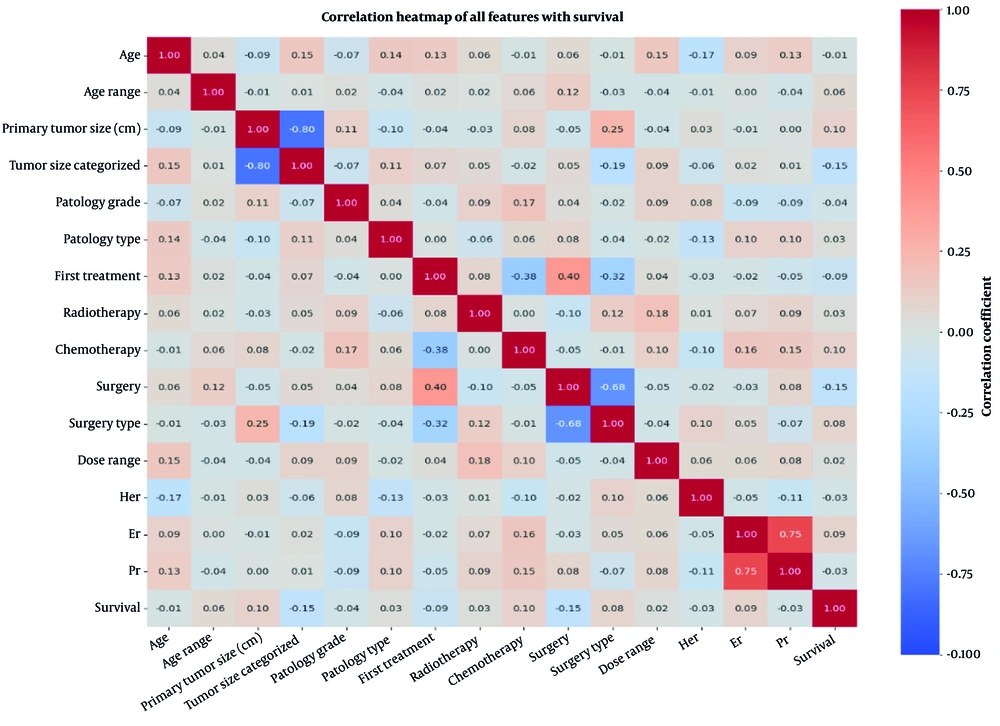
Figure 2.
Heatmap of pearson correlation coefficients for clinical features and five-year survival in breast cancer patients
5. Discussion
In this study, we applied ARM to a cohort of 198 patients with non-metastatic breast cancer treated with multimodal therapy to identify clinically interpretable patterns associated with five-year survival. Our analysis revealed distinct prognostic profiles: HER2-positive invasive ductal carcinoma treated with standard-dose radiotherapy and/or chemotherapy emerged as a high-risk pattern, whereas patients aged 50 - 69 years with small, PR-positive, margin-negative tumors, as well as an unexpectedly favorable subset with grade 3, HER2-negative, PR-positive disease, demonstrated excellent outcomes. Although these findings are associative, they generate hypotheses about how tumor biology and patient characteristics interact with treatment modalities to influence survival.
Our findings are broadly consistent with previous literature emphasizing the prognostic importance of tumor burden, receptor status, and multimodal therapy (30). The high-risk profile associated with HER2-positive disease aligns with large-scale analyses showing that HER2 positivity confers increased recurrence risk (31) and can be associated with resistance to standard chemotherapy regimens in the absence of targeted therapy (12). Our finding that this risk persisted despite receipt of standard radiotherapy and chemotherapy underscores the critical importance of anti-HER2 agents, such as trastuzumab, which were not systematically recorded in our dataset. This omission likely explains the elevated mortality observed in this subgroup and highlights a potential gap in guideline-concordant care that warrants further investigation.
The favorable prognosis observed in patients with small, PR-positive, margin-negative tumors is well supported by the literature. More notably, our identification of a low-risk profile among patients aged 50 - 69 years with grade 3, HER2-negative, PR-positive tumors adds nuance to the understanding of high-grade disease. Although grade 3 histology is typically associated with aggressive behavior, our results suggest that, in the context of hormone receptor positivity, HER2 negativity, and appropriate locoregional therapy, outcomes may be excellent. This finding echoes subtype-adjusted analyses showing that hormone receptor-positive, HER2-negative cancers have favorable prognoses even when other high-risk features are present (31). The correlation analyses reinforced the centrality of tumor size as a prognostic factor and revealed expected collinearities, such as ER/PR and surgery/surgery type, which informed our feature selection for ARM and ensured rule interpretability.
Figure 3 illustrates the hypothesis-generating risk patterns identified by ARM. HER2-positive patients had higher mortality despite standard treatment, underscoring the need for early targeted therapies such as trastuzumab. Patients with medium-sized, grade 2 tumors treated with mastectomy and chemotherapy remained at risk; closer follow-up or genomic profiling might be helpful in this subgroup. Two low-risk groups performed exceptionally well: women aged 50 - 69 years with small, PR-positive, margin-negative tumors and those with grade 3, HER2-negative, PR-positive disease. For the first group, shorter radiation or endocrine therapy alone could reduce toxicity. For the second group, multiagent chemotherapy might be unnecessary. High-risk patients may require more intensive monitoring, whereas low-risk patients could shift to less frequent follow-up focused on quality of life. These hypotheses require prospective validation before clinical implementation.
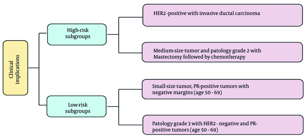
Figure 3.
Clinical implications of risk-stratified subgroups in early-stage breast cancer
5.1. Strengths and Novel Contributions
This study offers several strengths and novel contributions to breast cancer prognosis. Unlike many ARM studies that focus only on demographics or baseline tumor features (29), this work integrates treatment variables with tumor biology within an ARM framework and generates directly interpretable rules that complement traditional survival analyses. This hypothesis-generating approach can reveal patterns that prompt further investigation, such as the unexpected finding that a subset of patients with grade 3 disease had excellent outcomes. Furthermore, by focusing on a uniformly treated real-world cohort from a single center, we minimized practice variation that can confound larger registry-based analyses.
5.2. Limitations
Despite these strengths, several important limitations should be considered. First and most importantly, ARM identifies co-occurrence rather than causation. Our rules represent statistical associations that may be driven by unmeasured confounders, such as nodal stage, comorbidity, performance status, and socioeconomic factors, as well as selection biases or local treatment practices. All findings are therefore hypothesis-generating, and clinical implications, such as treatment de-escalation or intensified surveillance, are presented only as hypotheses to be tested in future interventional studies, not as recommendations. The single-center retrospective design and exclusion of 18 patients with incomplete records may introduce selection bias, thereby limiting external validity. Multicenter validation is essential before any clinical translation.
We lacked consistently extractable data on nodal staging, detailed chemotherapy regimens, systematic fractionation or target-volume coding, and reliable records of endocrine or HER2-targeted therapy receipt; these omissions limited our ability to account for key prognostic and treatment modifiers. Total prescribed dose categories served as a pragmatic proxy for radiotherapy exposure, although different fractionation schedules with equivalent total doses have different biological effects. The dataset exhibited moderate class imbalance, with 66.7% alive vs 33.3% dead, which may have biased rule discovery toward the majority class, as ARM inherently favors frequent itemsets. We did not perform balanced sampling or correction techniques because our primary goal was hypothesis generation using the full real-world cohort, and any correction method would introduce its own biases given our modest sample size. Therefore, minority-class patterns, particularly the grade 3, HER2-negative, PR-positive survival profile, should be interpreted with particular caution. Finally, some subgroup rules are based on small numbers and therefore carry wide uncertainty despite high apparent confidence; readers should avoid overinterpretation.
5.3. Future Directions
Several steps are needed to increase robustness and generalizability: 1) validating ARM-derived rules in larger external cohorts with complete nodal staging and treatment data; 2) applying survival-specific methods, such as survival trees and time-dependent ARM, that can accommodate censored data; 3) addressing class imbalance through balanced sampling or synthetic minority oversampling in larger datasets; 4) integrating genomic signatures, such as Oncotype DX, to enhance biological plausibility; and 5) prospectively evaluating whether rule-based risk stratification improves clinical decision-making. We are actively collecting a multicenter dataset to address these goals. If validated, ARM rules could be incorporated into decision-support prototypes that present interpretable risk patterns alongside traditional survival models.
5.4. Conclusions
In summary, applying ARM to routine clinical data revealed clear multivariable patterns that are consistent with established prognostic factors, such as tumor size, receptor status, and treatment approach, and that point to possible high- and low-risk patient groups. Some of these patterns confirmed well-known relationships, whereas others, such as excellent short-term survival in certain older patients with grade 3, HER2-negative, PR-positive tumors, were less expected and merit further study. These findings are hypothesis-generating and should be validated in larger, prospective, multicenter studies. With the addition of more detailed clinical, genomic, and radiomic data, ARM-derived rules could be integrated into decision-support tools to help guide personalized treatment strategies and potentially improve patient outcomes.