
1. Background
Ovarian cancer is one of the three most common malignant tumors in the female reproductive system.Early diagnosis is difficult and the onset of the disease is much unpredictable (1). The most common type of ovarian cancer is epithelial cell tumors. Epithelial ovarian cancer constitutes 90% of ovarian malignancies and 25% of women's genital malignancies. It is usually fatal due to late diagnosis. Epithelial ovarian cancer includes various types i.e. serous (the most common, 50% of all ovarian cancers), mucinous (15% - 20%), endometriosis (10% - 25%), clear cell (10.5%), undifferentiated (5%), and Brenner (5%) (2). Since the symptoms of this disease are not usually easy to diagnose and mimic other complications, the prompt diagnosis becomes difficult. In fact, more than 60% of patients with ovarian cancer are diagnosed when the cancer has progressed to a great extent so that their prognosis is poor (3).
In approximately 70% of all cases of ovarian cancer, the disease is not diagnosed before reaching an advanced stage (4). The 5-year survival rate associated with ovarian cancer is less than 30% (5). The early diagnosis of ovarian malignancies is an important issue to increase the survival rate of patients. The most available tools to recognize the low and high-risk patients are the tumor markers such as CA 125 and ROMA (6).
The tumor marker CA l25 has been used for 30 years for monitoring the afflicted patients, and assessing the recurrence rate of cancer (7). CA 125 has a widespread usage in clinical aspects; however, much more investigation is required to ensure its ability as a suitable biomarker of a malignant tumor or early diagnosis of ovarian cancer (8). Currently, CA-125 is frequently used to detect ovarian cancer before the onset of clinical signs, but CA-125 can increase in association with some physiological conditions such as pre-menopausal women and benign diseases in women suspicious of cancer (9). The other negative points about CA-125 biomarker properties are its low sensitivity for early-stage detection, and low specificity related to ovarian cancer. High level of Ca125 in the other cancers such as endometrial, cervix, and lung cancers is reported (10).
ROMA index value is developed to indicate the impact of current detection methods, to intensify the power of early diagnosis, and to measure the risk of ovarian cancer (11). CA-125 and human epididymis protein4 (HE4) levels and the menopausal status are the main fundamentals of ROMA, a useful method to integrate applied research and statistical analyses. This value has shown remarkable ability to distinguish epithelial ovarian carcinoma (EOC) from benign ovarian tumors (12). The sensitivity and specificity of ROMA were obtained 88.7% and 74.7%, respectively, when applied in cohorts of pre and post-menopausal women (13). As it is described in reference 13, ROMA is calculated as follow:
Pre-menopausal: predictive index (PI) = -12.0 + 2.38 LN [HE4] + 0.0626LN [CA125]
Post-menopausal: predictive index (PI) = -8.09 + 1.04 [HE4] + 0.732LN [CA125]
Predictive probability (PP) = exp (PI)/[1 + exp (PI)]
CEA is a glycoprotein of fetal tissues and some carcinomas. In patients with colorectal cancer, elevated CEA level depends on the stage of the disease that is confirmed (14). The important role of CEA in colorectal cancer is corresponding to the relationship between above 20 ng/mL concentrations of CEA and metastatic stage of disease (15). This marker is used to monitoring of patients after surgery for colorectal cancer, where a rise in CEA is indicative of disease progression (16).
In spite of some limitations, ROMA is a valuable index to predict of malignancies. High amount of ROMA in patients with early-stage serous ovarian cancer relative to benign tumors is reported and discussed (17). The aim of this study is determining appropriate ROMA, CA-125, and CEA levels to evaluate the efficacy of this biomarker panel in correlation with early detection of low grade serous ovarian cancer.
2. Methods
In this experimental study, 10 female patient with low grade serous ovarian cancer and 10 women without ovarian cancer as control group who were referred to the hospitals of Guilan University of Medical Sciences in Rasht since 2014 to 2015 were sampled.
The patients with a history of any type of cancers or chemotherapy were excluded. Ten patients diagnosed with ovarian masses through sonography suspicious with serous ovarian cancer through clinical and laboratory data were chosen for low-grade serous ovarian cancer group. Finally, the differentiation between benign ovarian masses and malignant was based on the pathologist’s report. The control group included women without cancer based on the mentioned diagnostic methods. Five mL blood was collected from each sample a day before surgery. Blood samples were immediately centrifuged at 3,000 rpm for 10 minutes at 4°C; the supernatant serum was collected and kept at -70°C up to the time when CEA, CA-125, and ROMA were tested. Sampling intervals and freezing took about 1 hour. CA-125 and CEA values were assessed, using chemiluminescent enzyme immunoassay and ELISA, respectively. Results are reported as mean.
The means of the data were compared with the two groups and t test was used for validation of the findings. Ethics committee of Shahid Beheshti University of Medical Sciences approved the Haniyeh Bashizadeh Fakhar’s Ph.D. dissertation by IR.SBMU.RETECH.REC.1396.709.
3. Results
The obtained results are based on comparing 10 control and 10 cancerous serum samples; the changes were calculated by mean and standard deviation (SD). From the 10 patients with low-grade ovarian tumor, all common histological types were serous adenocarcinoma. The results of serum tumor markers in the two groups are shown in Table 1. The mean and SD of the age of healthy and cancerous groups were 34 ± 12.8 and 53.3 ± 10.33 years, respectively. Out of 10 reference individuals of this study, 2 patients (20 %) had cystic ovarian, 5 patients (50 %) had a mass body, and 3 patients (3%) indeed surgery because of crevice cancer. Moreover, in group, 1 (10 %) had cystic ovarian and 9 (90 %) had pelvic mass.
Table 1.
The Amounts of Ca125, CEA, and ROMA for the Samples in the Two Groups and the Diagnostic Aspects are Presented
| Group/R | First Recognition | Pathology | Age | Ca125, U/mL | CEA, ng/mL | ROMA |
|---|---|---|---|---|---|---|
| Healthy | ||||||
| 1 | Ovarian cyst | Benign | 44 | 9 | 2 | 5 |
| 2 | Ovarian cyst | Benign | 13 | 8 | 2 | 10 |
| 3 | Pelvic mass | Benign | 30 | 21 | 1 | 21 |
| 4 | Pelvic mass | Benign | 33 | 7 | 2 | 9 |
| 5 | Pelvic mass | Benign | 15 | 8 | 2 | 11 |
| 6 | Pelvic mass | Benign | 36 | 9 | 3 | 9 |
| 7 | Pelvic mass | No remarkable pathology | 35 | 35 | 2 | 16 |
| 8 | Cervical cancer | Cervical cancer Scc | 48 | 20 | 2 | 25 |
| 9 | Cervical cancer | Cervical cancer Scc | 48 | 20 | 2 | 25 |
| 10 | Cervical cancer | Cervical cancer Scc | 52 | 22 | 2 | 21 |
| Cancerous | ||||||
| 1 | Pelvic mass | Serous cancer | 53 | 74 | 65 | 45 |
| 2 | Pelvic mass | Serous cancer | 54 | 64.6 | 57 | 42 |
| 3 | Pelvic mass | Serous cancer | 63 | 825 | 3 | 94 |
| 4 | Pelvic mass | Serous cancer | 40 | 122 | 3 | 18 |
| 5 | Pelvic mass | Serous cancer | 63 | 982 | 3 | 92 |
| 6 | Pelvic mass | Serous cancer | 50 | 160 | 3 | 40 |
| 7 | Pelvic mass | Serous cancer | 70 | 810 | 2 | 92 |
| 8 | Pelvic mass | Serous cancer | 43 | 124 | 2 | 17 |
| 9 | Pelvic mass | Serous cancer | 48 | 155 | 2 | 35 |
| 10 | Ovarian cyst | Serous cancer | 47 | 127 | 3 | 32 |
In terms of pathology in reference group, out of 10 people, 6 (60%) had benign and 4 (40%) had non-cancerous changes. In patients with ovarian cancer, each one (100%) had serious pathology. In this study, out of 10 healthy people, 4 (40%) had a family history and in patients with cancer, 5 (50%) had reported clashes with ovarian cancer.
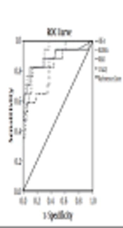
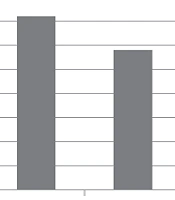
The analyzed results of the three biomarkers are tabulated in Table 2. Average amounts of CA125 in patients with low grade serous ovarian cancer and the reference group were 413.1 ± 384.0 and 15.9 ± 9.2 U/mL, respectively. Difference between the two mean values was statistically significant. Then, CEA values for the cancerous and reference groups were 2.6 ± 0.5 and 2.0 ± 0.5 ng/mL, respectively. The statistical analysis indicates a significant difference between the two groups. The two samples (row 1 - 2 in Table 1), whose CEA levels were extremely elevated relative to the other patients were excluded. Since amount of CEA level of patients is more than control samples, this exclusion does not affect the finding. ROMA amounts for the two mentioned groups are 52.5 ± 34.2 and 15.2 ± 7.2, respectively. As CA125 and CEA, the amounts of ROMA in patients and references are statistically different.
4. Discussion
This prospective study evaluated the power of the ROMA, CEA, and Ca125 in distinguishing the nature of low-grade serous ovarian cancer.
Many researchers have reported the lack of efficient biomarkers relative to early detection of cancer as a huge problem in hindering the blood-based diagnostic tools (17). Ovarian cancer has a poor prognosis, usually diagnosed when patient's status is worsening (18). So far, no screening approach is available or validated to detect ovarian cancer at an early stage, and the only factor affecting survival is the extent of surgical tumor debunking and correct surgical staging during primary surgery (19).
Zhang et al. reported that three biomarkers including (a) apolipoprotein A1 (down-regulated in cancer); (b) a truncated form of transthyretin (down-regulated); and (c) a cleavage fragment of inter-alpha-trypsin inhibitor heavy chain H4 (up-regulated) were correlated to ovarian cancer. The combination of these biomarkers and CA125 elevated the sensitivity of tests about 9% relative to CA125 alone (4).
The results of this study support previous findings, suggesting that the ROMA index is an efficient marker as CA125 or CEA in the differentiation of ovarian cancer (20).
According to Ikeda’s study, ROMA was a significant predictor of peritoneal dissemination being more powerful than CT (21). In their study, ROMA was assessed as a marker of peritoneal dissemination in patients with epithelial ovarian cancer. They established cut-off values of CA125, HE4, ROMA as 197 U/mL, 161 pmol/mL, and 86%, respectively. Their results correlated to high specificity for predicting the presence of peritoneal dissemination in epithelial ovarian cancer (21).
In the recent publication of Kaijser et al. the authors analyzed the value of serum HE4 or ROMA as second-stage tests to characterize the tumors on the basis of ultrasound findings (22). From 360 patients with pelvic tumors, 54% had a high confidence, 38% had moderate confident, and 8% were completely uncertain about their diagnosis. Most of the unclassifiable tumors were benign (79%) followed by borderline ovarian cancer (14%). The sensitivity and specificity of subjective assessment were 67% and 70%, respectively. HE4 and ROMA had a poor distinguishing power. In sum, ROMA and HE4 as second-test after transvaginal ultrasonography declined the power of the test (22).
In another study conducted by Pitynski, the clinical significance of the combination of CA 125, HE4, and ROMA was evaluated for the identification of ovarian masses in patients with suspected early stage ovarian cancer on 225 women with a pelvic mass of suspected ovarian origin. Median CA-125 and HE4 levels were significantly higher in patients with OC compared with women with benign ovarian tumors .The ROMA was significantly more accurate at detecting OC but only in premenopausal patients (23).
More performed investigations on Ca125 combined with image findings in asymptomatic women have not been merged (24). The positive predictive value of Ca125 is low to be used as an initial step in the screening ovarian cancer (25). Moreover, Ca125 is not a suitable tool to be used in the diagnosis of ovarian cancer because of the lack of its specificity, particularly in premenopausal women with benign gynecological diseases, mainly related to endometriosis.It can be considered as a complementary tumor marker to assess the risk of ovarian cancer (9, 26). Previously published studies of HE4 have reported a higher specificity than Ca125 in different benign and malignant tumors. According to reports, ROMA improved sensitivity and specificity, and both tumor markers have been highlighted as complementary biomarkers (11, 27).
Serum CEA is elevated in approximately 35% of all ovarian patients with cancer, 88% in mucinous and 19% in serous tumors (28). In some studies such as Sorensen’s study, by using CEA, a larger proportion of patients with non-ovarian cancers were identified. In their study, the use of CA-125/CEA ratio would spare 67 out of 107 patients with non-ovarian cancers from a planned unnecessary operation. Their findings suggest that any patient referred to the hospital with an undiagnosed tumor in the pelvis should, in addition to malignancy risk index (RMI) be tested by using the CA-125/CEA ratio < 25 as a criterion for further examination such as computed tomography of the abdomen, colonoscopy, mammography, magnetic resonance imaging (29). Mediu et al. reported that CA125, CEA, and He4 can be used to discriminate ovarian cancer from other benign gynecologic diseases (30). In a meta-analysis by Junhong et al. diagnostic property of combination of CA125, CA199, and CEA ovarian cancer was evaluate. They suggested that the introduced panel is useful tool epithelial ovarian cancer diagnosis (31).
In the present study, serum level alteration of the three tumor markers ROMA, CEA, and CA125 in patients with low grade serous ovarian cancer was confirmed. However, ROMA and CEA are not more sensitive in differentiating malignancy before surgery in comparison to CA125 (32). It can be concluded that a combined test including ROMA, CEA, and CA125 is a more cost-effective method for patients rather than the single CA125 test (33). Here, the combined values of ROMA, CEA, and CA125 are suggested for diagnosis of low-grade serous ovarian cancer. So, it seems that positive response to indicators of the three biomarkers can be an efficient tool relative to early detection of ovarian cancer.
4.1. Conclusions
It was concluded that the combined test including three tumor markers, CEA, CA125, and ROMA (which it is a function of CA125 and HE4 for pre- and post-menopausal) can be considered as a suitable biomarker panel related to early detection of ovarian cancer. This panel can be used to distinguish malignant from benign tumors. However, we suggested evaluating this panel in larger sample size in future studies.