
Iran J Pediatr

Image Credit:Iran J Pediatr

Correlation Between the Urine Analysis in Neonates with Renal Anomaly

Author(s):
Maryam SaeediMaryam SaeediMaryam Saeedi ORCID1, 2, Kayvan MirniaKayvan MirniaKayvan Mirnia ORCID1, 2, Razieh SangsariRazieh SangsariRazieh Sangsari ORCID1, 2,*, Golnaz AliniaGolnaz Alinia2
1Department of Pediatrics, Division of Neonatology, Faculty of Medicine, Tehran University of Medical Sciences, Tehran, Iran
2Children's Medical Center, Pediatrics Center of Excellence, Tehran, Iran
*Corresponding Author: Department of Pediatrics, Division of Neonatology, Faculty of Medicine, Tehran University of Medical Sciences, Children’s Medical Center, Pediatric Center of Excellence, Tehran, Iran. Email: [email protected]

Innovative Journal of Pediatrics:Vol. 32, issue 3; e122680
Published online:Jun 06, 2022
Article type:Research Article
Received:Jan 17, 2022
Accepted:Apr 07, 2022
How to Cite:Saeedi M, Mirnia K, Sangsari R, Alinia G. Correlation Between the Urine Analysis in Neonates with Renal Anomaly. Inn J Pediatr. 2022;32(3):e122680. doi: https://doi.org/10.5812/ijp-122680

Abstract

Background:

According to the literature, urine analysis to diagnose urinary tract infection (UTI) in neonates is not the basis. It even is not recommended as a part of the evaluation, but some abnormalities in a simple urine test in neonates with UTI may be associated with urinary tract anomaly. Therefore, a simple urine test in neonates with UTI can be helpful for early diagnosis and timely treatment of urinary tract anomalies.

Methods:

This study was performed as a cross-sectional study. First, 100 neonates hospitalized for various reasons and diagnosed with urinary tract infections were chosen. Before treatment, urine samples were taken from all neonates by catheterization or suprapubic sterility for culture and urine bag for urinalysis. Finally, neonates with positive culture were treated with antibiotics and evaluated; kidney and urinary tract ultrasound and voiding cystourethrography (VCUG) is used to diagnose renal anomalies. The obtained information from urine analysis of neonates without urinary tract anomalies was compared with urinary tract anomalies neonates by SPSS 21 software. To compare the qualitative data, the chi-square and Fisher’s exact tests were used, whereas to compare the quantitative data, we used Wilcoxon test. P < 0.05 was considered statistically significant.

Results:

Out of 100 patients who were evaluated, only 30 patients had renal anomalies. No statistically significant correlation was found between gender, cause of hospitalization, positive culture, and type of organism with the renal anomaly. However, pyuria (P-value 0.003), bacteriuria (P-value 0.016), fever (P-value 0.002), nitrite positive (P-value 0.001), and leukocyte esterase (P-value 0.001) showed a statistically significant correlation with the renal anomaly.

Conclusions:

Certain indicators such as pyuria, bacteriuria, nitrite, and leukocyte esterase can be seen in a simple urine analysis as criteria for suspicious urinary tract anomalies in neonates with UTIs.

1. Background

Neonatal infections have nonspecific manifestations, and in some cases, it is difficult to diagnose the source of the infection. One of the causes of infection in a neonate is urinary tract infection (UTI). Diagnosis of UTI is given based on the urine culture (1, 2). A definite diagnosis of UTI cannot be made based on urine analysis before the urine culture results are ready. Screening tests such as positive nitrite or gram staining sometimes show false-positive results, as it has been already observed in cases with negative urine culture, especially in neonates (3, 4).
On the other hand, the leukocyte esterase dipstick test is not accurate in diagnosing pyuria in febrile patients (4), and a negative leukocyte esterase test cannot rule out pyuria; so this test cannot detect cases of infection without microscopic examination and urine culture as far as it may increase the chance of false negatives (5, 6). In previous literature, it has been shown that the presence of less than 10 white blood cells (WBCs) in a simple urine test indicates normal urine. Although, in some cases, the presence of 15 to 20 WBCs has also been associated with negative urine culture in general, there is no definite number that is highly sensitive and specific to diagnose infection in neonates, and due to the underdevelopment of immune response in them, we may not see reliable pyuria (4, 7).
Urinary tract infection in neonates includes a wide range of symptoms; for instance, growth retardation, polyuria, oliguria, jaundice, etc. (8, 9). Since urosepsis manifestations are nonspecific in neonates, urine culture is requested before the first dose of antibiotics, and urinary tract malformations are investigated during the treatment course in neonates with UTI (2). Although neonates with structural disorders of the urinary system are at higher risk of UTIs than normal infants, the relationship between pyuria and urinary tract abnormality has not yet been established. However, the association between positive urinary culture and structural abnormalities of the urinary system has already been detected (10, 11).

2. Objectives

This study investigated the relationship between simple urine tests and renal abnormality.

3. Methods

This study was performed as a cross-sectional-analytical evaluation on 100 neonates with UTIs in Tehran Children’s Medical Center, and the relationship between renal anomalies and simple urine tests was investigated.

3.1. Study Design

Neonates who were hospitalized for various reasons and were diagnosed with UTIs were selected. Urine samples were taken from all neonates by urine bag and catheterization or suprapubic in a sterile manner for urine culture. More than 1,000 colonies in the catheter urine sample or even one count in the suprapubic sample were considered positive. Counts of WBCs, which were more than ten, bacteriuria, positive nitrite, and positive leukocyte esterase in early detection of UTI were considered valuable. None of the neonates with UTI had positive blood cultures in our study. Finally, neonates with positive urine culture were treated with antibiotics, and also renal and urinary tract ultrasound and VCUG were performed to diagnose renal abnormalities and urinary reflux. SPSS21 software was used to investigate the relationship between urinary indices and urinary tract anomalies. The alpha level has been set to 0.05, and P-values less than 0.05 were considered significant.

3.2. Inclusion Criteria

Neonates with UTIs admitted to the children’s Medical Center in 2016 - 2017 were included in this study.

3.3. Exclusion Criteria

The patients who had incomplete information were excluded from this study.

3.4. Statistical Analysis

Descriptive statistics were implemented by calculating the frequency, percentage, mean ± standard deviation (SD). To compare the qualitative data, the chi-square test was used for contingency with tables of cell counts more than 5, and Fisher’s exact test was used for contingency with tables of cell counts less than 5. For the comparison of the quantitative data, we used the Wilcoxon test. P < 0.05 was considered statistically significant.

3.5. Ethical Considerations

Since urine analysis, urine culture, sonography, and VCUG in neonates with UTI are necessary for the treatment of the disease; therefore, no additional action was taken on patients besides what should have been done in the process of the treatment. Patients’ information was recorded anonymously. The study was approved by the Ethics Committee of Tehran University of Medical Science with the following ethical code: IR.TUMS.CHMC.REC.1398.122.

4. Results

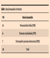
The mean age of neonates was 13.64 days. The mean gestational age was 37.17 weeks, and the mean weight was 3,310 grs. A total number of 30 patients had renal anomalies. The most common observed anomaly was urinary reflux (Table 1). The most common organism found in this study was Klebsiella (31%), which was associated with renal anomalies in 45% of cases, but none of the organisms were significantly associated with renal anomalies (P-value 0.987) (Table 2). There was a statistically significant correlation between fever with the renal anomaly (P-value 0.002). Comparison of urinalysis in the two groups with and without renal anomalies showed a statistically significant difference (Table 3). Pyuria (P-value = 0.003), bacteriuria (P-value = 0.016), nitrite positive (P-value = 0.001), and leukocyte esterase (P-value = 0.001) showed a statistically significant correlation with the renal anomaly. However, there was no statistically significant correlation between gender, cause of hospitalization, positive culture, CRP level, and type of organism with the renal anomaly.
Table 1.Renal Anomalies in Patients
No.Renal Anomalies
15Vesicourethral reflux (VUR)
8Posterior urethralvalve (PUV)
7Ureteropelvic junction obstruction (UPJO)
30Total
Table 2.The Organisms Were Significantly Associated with Renal Anomalies
OrganismRenal AnomaliesP-Values
NoYes
Enterobacter340.987
Candida108
Escherichia coli109
Staphylococcus53
Klebsiella1714
Serratia43
Enterococcus44
Total5347
Table 3.Comparison of Urine Analysis in Two Groups with and Without Renal Anomalies
IndexRenal AnomaliesP-Value
NoYes
Pyuria0.003
Yes719
No4628
Bacteriuria0.016
Few238
Many1619
Negative1420
Nitrite0.001
Positive1024
Negative4323
Leukocyte esterase0.001
Positive421
Negative4926
Reinfection0.694
Positive2722
Negative2625

5. Discussion

The findings of this study showed that some indicators in urinalysis, including pyuria, bacteriuria, nitrite, and leukocyte esterase, have statistically significant correlations with a renal anomaly. Based on the results observed in this study, these indicators are important for reevaluating antibiotics and assessing renal anomalies accurately. However, the best choice of empirical treatment for UTIs in neonates is aminoglycosides (12). In the Crain and Gershel’s study, out of 32 positive urine cultures in infants younger than eight weeks of age with UTIs, 50% had normal urine analysis. Then more than half of the UTIs in neonates would have been missed based on urine analysis (13).
In the study of Falakaflaki et al., proteinuria and hematuria in neonates were associated with ureteropelvic junction and vesicoureteral reflux (14). Mohamed et al. found that the rate of UTIs in neonates with pyuria was 5.44 times higher than in other neonates (15). Eberechukwu et al.’s study showed the presence of hematuria, glucosuria, and ketonuria in the urine of the infants was not usual, and it should be investigated in terms of the underlying factor (16).
The most common anomaly observed in our study was urinary reflux, which is similar to other studies (15, 16). The most common organism found in this study was Klebsiella (31%), which was associated with renal anomalies in 45% of cases, but none of the organisms were significantly associated with renal anomalies. In the study of Cleper et al., Klebsiella was more likely to be associated with urinary reflux (17).

5.1. Conclusions

Based on the results observed in this study, some indicators related to UTIs in neonates, including pyuria, bacteriuria, nitrite, and leukocyte esterase, which are seen in simple urinalysis, have a statistically significant correlation with renal anomaly and need to reevaluate antibiotics and assess renal anomalies accurately.
Acknowledgments

Footnotes

  • Authors' Contribution: Study concept and design: Saeedi M. Collection of data: Alinea G. Analysis and interpretation of data: Mirnia K. Drafting of the manuscript: Sangsari R. Critical revision of the manuscript for important intellectual content: Kajbaf Sangsari R. Developed the original idea: Kayvan M. Final revisions: Saeedi M.

  • Conflict of Interests: The authors declare no conflicts of interest.

  • Data Reproducibility: The data presented in this study are openly available in one of the repositories or will be available on request from the corresponding author by this journal representative during submission or after publication. Otherwise, all consequences of possible withdrawal or future retraction will be with the corresponding author.

  • Ethical Approval: The study was approved by the Ethics Committee of Tehran University of Medical Science with the following ethical code: IR.TUMS.CHMC.REC.1398.122. (link: ethics.research.ac.ir/IR.TUMS.CHMC.REC1398.122).

  • Funding/Support: The study was a medical specialty thesis supported by the Tehran University of Medical Sciences, and no extra funding was received for the study.

  • Informed Consent: There are parental informed consent forms.

References

  • 1.
    Emamghorashi F, Mahmoodi N, Tagarod Z, Heydari ST. Maternal urinary tract infection as a risk factor for neonatal urinary tract infection. Iran J Kidney Dis. 2012;6(3):178-80. [PubMed ID: 22555480].
  • 2.
    Lee JH, Choi HS, Kim JK, Won HS, Kim KS, Moon DH, et al. Nonrefluxing neonatal hydronephrosis and the risk of urinary tract infection. J Urol. 2008;179(4):1524-8. [PubMed ID: 18295269]. https://doi.org/10.1016/j.juro.2007.11.090.
  • 3.
    Kanellopoulos TA, Salakos C, Spiliopoulou I, Ellina A, Nikolakopoulou NM, Papanastasiou DA. First urinary tract infection in neonates, infants and young children: a comparative study. Pediatr Nephrol. 2006;21(8):1131-7. [PubMed ID: 16810514]. https://doi.org/10.1007/s00467-006-0158-7.
  • 4.
    Goldman M, Lahat E, Strauss S, Reisler G, Livne A, Gordin L, et al. Imaging after urinary tract infection in male neonates. Pediatrics. 2000;105(6):1232-5. [PubMed ID: 10835062]. https://doi.org/10.1542/peds.105.6.1232.
  • 5.
    Pashapour N, Nikibahksh AA, Golmohammadlou S. Urinary tract infection in term neonates with prolonged jaundice. Urol J. 2007;4(2):91-4.
  • 6.
    Ellison JS, Dy GW, Fu BC, Holt SK, Gore JL, Merguerian PA. Neonatal Circumcision and Urinary Tract Infections in Infants With Hydronephrosis. Pediatrics. 2018;142(1). [PubMed ID: 29880703]. https://doi.org/10.1542/peds.2017-3703.
  • 7.
    Garcia FJ, Nager AL. Jaundice as an early diagnostic sign of urinary tract infection in infancy. Pediatrics. 2002;109(5):846-51. [PubMed ID: 11986445]. https://doi.org/10.1542/peds.109.5.846.
  • 8.
    Ozcan M, Sarici SU, Yurdugul Y, Akpinar M, Altun D, Ozcan B, et al. Association Between Early Idiopathic Neonatal Jaundice and Urinary Tract Infections. Clin Med Insights Pediatr. 2017;11:1179556517701120. [PubMed ID: 28469520]. [PubMed Central ID: PMC5398657]. https://doi.org/10.1177/1179556517701118.
  • 9.
    Eisenberg ML, Galusha D, Kennedy WA, Cullen MR. The Relationship between Neonatal Circumcision, Urinary Tract Infection, and Health. World J Mens Health. 2018;36(3):176-82. [PubMed ID: 29623700]. [PubMed Central ID: PMC6119846]. https://doi.org/10.5534/wjmh.180006.
  • 10.
    Baz AMK, El-Agamy OAE, Ibrahim AM. Incidence of urinary tract infection in neonates with significant indirect Hyperbilirubinemia of unknown etiology: case-control study. Ital J Pediatr. 2021;47(1):35. [PubMed ID: 33596989]. [PubMed Central ID: PMC7891145]. https://doi.org/10.1186/s13052-021-00982-0.
  • 11.
    Firoozi H, Seidan Anbi S, Mousavi J, Farshidi F, Rezai MS. [Prevalence of urinary tract infection in neonates with hyperbilirubinemia]. Journal of Mazandaran University of Medical Sciences. 2019;28(169):91-7. Persian.
  • 12.
    Sangsari R, Behjati Ardakani S, Kadivar M, Fardi R, Jafari A. Do You Prefer Aminoglycosides or Cephalosporins in Neonatal Infections? J Compr Ped. 2019;10(1). https://doi.org/10.5812/compreped.64948.
  • 13.
    Crain EF, Gershel JC. Urinary tract infections in febrile infants younger than 8 weeks of age. Pediatrics. 1990;86(3):363-7. [PubMed ID: 2388785].
  • 14.
    Falakaflaki B, Mousavinasab SN, Mazloomzadeh S. Dipstick urinalysis screening of healthy neonates. Pediatr Neonatol. 2011;52(3):161-4. [PubMed ID: 21703559]. https://doi.org/10.1016/j.pedneo.2011.03.012.
  • 15.
    Mohamed W, Algameel A, Bassyouni R, Mahmoud AT. Prevalence and predictors of urinary tract infection in full-term and preterm neonates. Gaz Egypt Paediatr Assoc. 2020;68(1). https://doi.org/10.1186/s43054-020-00022-2.
  • 16.
    Eberechukwu YIL, Peace Ibo P, Tochi Ada UO. Dipstick Urinalysis in apparently healthy Neonates in Port Harcourt. Greener Journal of Medical Sciences. 2015;5(3):48-52. https://doi.org/10.15580/gjms.2015.3.030815042.
  • 17.
    Cleper R, Krause I, Eisenstein B, Davidovits M. Prevalence of vesicoureteral reflux in neonatal urinary tract infection. Clin Pediatr (Phila). 2004;43(7):619-25. [PubMed ID: 15378148]. https://doi.org/10.1177/000992280404300706.

Copyright

Copyright © 2022, Author(s). This is an open-access article distributed under the terms of the Creative Commons Attribution-NonCommercial 4.0 International License (http://creativecommons.org/licenses/by-nc/4.0/) which permits copy and redistribute the material just in noncommercial usages, provided the original work is properly cited.

Similar Articles

30
Apr
2010

Antenatal Detection of Urinary Tract Abnormalities by Ultrasonography

Om Prakash Mishra,
Nisha Pandey,
Ram Chandra Shukla,
Nisha Rani Agarwal,
Rajniti Prasad

Mishra O, Pandey N, Shukla R, Agarwal N, Prasad R. Antenatal Detection of Urinary Tract Abnormalities by Ultrasonography. Nephro-Urol Mon. 2010;2(2):. doi:

1
Nov
2020
Nephro-Urology Monthly

Urinary Tract Infections with Non-Escherichia coli Pathogens in Children: An Observational Study

Mitra Naseri,
Gholam Reza Sarvari,
Niayesh Tafazoli,
Nooshin Tafazoli

Naseri M, Sarvari GR, Tafazoli N, Tafazoli N. Urinary Tract Infections with Non-Escherichia coli Pathogens in Children: An Observational Study. Nephro-Urol Mon. 2020;12(4):e107856. doi: https://doi.org/10.5812/numonthly.107856

15
Apr
2015

Diagnostic Challenges in Urinary Tract Infections in Children

Mostafa Sharifian,
Shakiba Sharifian

Sharifian M, Sharifian S. Diagnostic Challenges in Urinary Tract Infections in Children. Arch Pediatr Infect Dis. 2015;3(2):e21386. doi: https://doi.org/10.5812/pedinfect.21386

26
Feb
2025
Arch Clin Infect Dis

Evaluation of the Laboratory and Sonographic Findings of Vesicoureteral Reflux in Children with Acute Febrile Urinary Tract Infection

Azar Nickavar,
Elham Zarei,
Mohammad Hosein Ahmadian

Nickavar A, Zarei E, Ahmadian MH. Evaluation of the Laboratory and Sonographic Findings of Vesicoureteral Reflux in Children with Acute Febrile Urinary Tract Infection. Arch Clin Infect Dis. 2025;20(3):e144861. doi: https://doi.org/10.5812/archcid-144861

19
Dec
2009

Clinical and laboratory aspects of urinary tract infections in hospitalized children

Abolhassan Seyedzadeh,
Shams Vazirian,
Tahereh Yavari,
Liela Karimi,
Akram Soliemani

Seyedzadeh A, Vazirian S, Yavari T, Karimi L, Soliemani A. Clinical and laboratory aspects of urinary tract infections in hospitalized children. J Kermanshah Univ Med Sci. 2009;13(3):e79597. doi:

Download PDF111.53 KB

Crossmark

Crossmark

Checking

Share on
Cited by
Metrics

Ordering Reprints

Articles are published under the Creative Commons license stated on each article. No permission or royalty fee is required for uses permitted by that license. CCC handles optional bulk and customized reprint orders. Any quotation covers production and delivery services only, not copyright permission. > Request Reprints from CCC 

Search Relations

Author(s):

Related Articles