
1. Background
Juvenile idiopathic arthritis (JIA) is the most prevalent chronic rheumatologic disease in children, with an incidence of 2 to 20 cases per 100,000 children and a prevalence of 16 to 150 cases per 100,000 children, depending on the region (1). A 2025 meta-analysis reported a pooled prevalence of uveitis of 12.7% (95% CI, 10.5% - 15.1%) across different populations, with a lower rate of 6.5% observed in Asian cohorts (2). Furthermore, a 2025 study evaluating Pediatric Rheumatology International Trials Organisation classification criteria found that antinuclear antibody (ANA) positivity at a titer of ≥ 1:160 combined with an age at onset younger than 7 years was associated with a substantially higher risk of new-onset uveitis and a requirement for biologic disease-modifying antirheumatic drugs (3). In contrast, lower rates are observed among patients with polyarticular rheumatoid factor (RF)-positive or systemic subtypes. Geographic and ethnic differences also affect prevalence; for example, European cohorts have reported uveitis in up to 19% of patients with JIA, whereas studies from Japan and some Middle Eastern regions have reported rates of approximately 5% to 6% (4, 5).
The clinical presentation of JIA-associated uveitis may include chronic anterior uveitis or acute anterior uveitis. Chronic anterior uveitis may be asymptomatic in its early stages and is more common in young, ANA-positive girls with oligoarticular JIA. Without timely and adequate detection strategies, posterior synechiae may lead to severe ocular complications, including cataracts, glaucoma, and permanent vision loss (4). HLA-B27 positivity usually coincides with acute anterior uveitis, which is more common in patients with the enthesitis-related arthritis subtype. Acute anterior uveitis typically has a sudden onset and is characterized by eye redness, pain, photophobia, and, in some cases, blurred vision (5). Identified risk factors in JIA include female sex, ANA positivity, shorter disease duration, and the oligoarticular subtype. Patients diagnosed at an earlier age, typically before 6 years, are considered high risk and require more frequent eye examinations (6, 7).
Because chronic anterior uveitis may be asymptomatic in its early stages, routine ophthalmologic screening is essential. The American College of Rheumatology (ACR) and other international guidelines recommend that screening begin within 6 weeks of a JIA diagnosis. Follow-up after the initial examination depends on individual risk assessment and typically ranges from every 3 months to once yearly (8). Updated 2025 recommendations from the Multinational Interdisciplinary Working Group for Uveitis in Childhood provide a simplified screening protocol that can be implemented by community eye care practitioners (9). Treatment options for mild disease include topical corticosteroids, whereas persistent or severe cases require systemic immunosuppressants such as methotrexate. Biologic agents, particularly tumor necrosis factor alpha inhibitors such as adalimumab, have shown strong efficacy in controlling inflammation and preventing complications. For resistant cases, novel interleukin 6 inhibitors, such as tocilizumab, and Janus kinase inhibitors are being actively studied and are considered promising (8, 9). A comprehensive 2026 clinical review provides updated guidance on subtle signs, screening protocols, and treatment strategies for JIA-associated uveitis (10). Furthermore, a 2025 review summarizes the pathogenesis, diagnosis, and management of this condition (11).
2. Objectives
This study highlights the clinical need to address JIA-associated uveitis. It also supports continued collaboration between pediatric rheumatology and ophthalmology services to prevent delays in eye care, improve the recognition of ocular damage, guide targeted treatment, and reduce long-term visual impairment.
3. Methods
3.1. Study Design and Setting
This retrospective cross-sectional study was conducted in the pediatric rheumatology department of Akbar Children's Hospital, a tertiary pediatric referral center, from January 2016 to January 2019.
A total of 342 children diagnosed with JIA were assessed during the study period. These patients comprised all consecutive referrals to the center within the defined timeframe. Patients were screened for uveitis through a review of rheumatologic and ophthalmologic medical records.
3.2. Participants
The inclusion criterion was a confirmed diagnosis of JIA with concomitant uveitis. Patients with unrelated ocular comorbidities, incomplete medical documentation, or a lack of follow-up were excluded.
The number of patients excluded for each reason was as follows: unrelated ocular disease (n = 150), incomplete medical records (n = 120), and lack of follow-up (n = 44). After application of the eligibility criteria, 28 patients with JIA-associated uveitis were included in the final analysis.
3.3. Data Collection and Ophthalmologic Assessment
A retrospective review of patient files and the hospital health information system yielded demographic and clinical data, including age, sex, JIA type, ANA results, disease duration, treatment modalities, and the interval between arthritis onset and uveitis development.
All patients underwent a detailed ophthalmologic evaluation by pediatric ophthalmologists. Uveitis was classified as acute anterior, chronic anterior (unilateral or bilateral), intermediate, posterior, or panuveitis. Clinical manifestations and ocular complications, including cataracts, glaucoma, posterior synechiae, elevated intraocular pressure, band keratopathy, and blindness, were recorded.
Recurrence of uveitis was defined according to the Standardization of Uveitis Nomenclature (SUN) criteria as the reappearance of anterior chamber cells ≥ 1+ (SUN grading: 1+ = 5 - 10 cells per high-power field) or vitreous haze ≥ 0.5+ after at least 3 months of well-controlled inactive disease (anterior chamber cells < 0.5+ and no vitreous haze) while the patient was receiving stable or reduced therapy. This definition is consistent with SUN criteria and the recommendations of the Multinational Interdisciplinary Working Group for Uveitis in Childhood (12, 13).
Refractory uveitis was defined according to the 2019 American College of Rheumatology/Arthritis Foundation guideline as persistent active uveitis (anterior chamber cells ≥ 1+ on SUN grading) despite at least 3 months of methotrexate at a therapeutic dose (≥ 15 mg/m2/week, administered orally or subcutaneously) in combination with topical corticosteroids (prednisolone acetate 1%, at least 4 times daily). This definition excludes patients who were intolerant to methotrexate or had contraindications to its use (14).
Ophthalmic complications, including cataract, glaucoma, posterior synechiae, increased intraocular pressure, and band keratopathy, were recorded as either 1) present at the time of uveitis diagnosis or 2) detected during follow-up. Table 1 reports the total number of patients who developed each complication at any time point; however, the footnote indicates whether the complication was present at onset or developed subsequently. Uveitis type was classified based on the anatomic localization criteria of the International Uveitis Study Group and the temporal criteria of the SUN Working Group as follows: acute anterior uveitis (duration ≤ 3 months, often with sudden onset and symptomatic), chronic anterior uveitis (duration > 3 months, typically insidious and often asymptomatic), intermediate uveitis (inflammation primarily involving the vitreous and peripheral retina), posterior uveitis (inflammation primarily involving the retina or choroid), and panuveitis (inflammation involving all 3 anatomical sections: anterior, intermediate, and posterior). All classifications were determined by pediatric ophthalmologists based on slit-lamp examination and dilated funduscopy (12, 15).
ANA testing was performed for all patients at the time of JIA diagnosis using indirect immunofluorescence on HEp-2 cells at the central laboratory of Akbar Children's Hospital. A positive result was defined as a titer ≥ 1:80 according to the manufacturer's reference range and local laboratory standards. The same protocol was applied uniformly to all patients.
3.4. Ethics Approval and Informed Consent
This study was based on a research project (grant No. 961510) and a thesis conducted at Mashhad University of Medical Sciences. The study was approved by the Ethics Committee of Mashhad University of Medical Sciences (ethics approval code: IR.MUMS.REC.1396.510). A formal waiver of informed consent was granted by the ethics committee because of the retrospective nature of the study, as all patient data were de-identified and analyzed anonymously. Patient medical records were accessed only after ethics approval and in compliance with the Declaration of Helsinki (2013 revision). All procedures were performed in accordance with relevant guidelines and regulations.
3.5. Statistical Analysis
Statistical analysis was performed using IBM SPSS Statistics version 26. Continuous variables were expressed as mean ± standard deviation (SD), whereas categorical variables were presented as frequencies and percentages. For group comparisons where sample sizes permitted, the independent t-test was used for continuous variables, and the chi-square test or Fisher exact test was used for categorical variables when expected cell counts were < 5. A P value < 0.05 was considered statistically significant.
Because of the small overall sample size (n = 28) and the limited number of patients in certain JIA subgroups (eg, oligoarticular JIA, n = 23; polyarticular RF-negative JIA, n = 2; psoriatic arthritis, n = 3), many subgroup comparisons were not statistically feasible. Therefore, for most comparisons, only descriptive statistics, including frequencies, percentages, and mean ± SD, are reported. Where applicable, 95% CIs for proportions were calculated using the Wilson score method to quantify the precision of key estimates (16).
The frequency of ophthalmologic examinations was determined according to the 2019 American College of Rheumatology/Arthritis Foundation guideline for screening, monitoring, and treatment of JIA-associated uveitis. Patients were classified into low-, moderate-, and high-risk categories based on JIA subtype, age at onset, ANA status, and disease duration. Accordingly, the interval between routine ophthalmologic examinations ranged from 3 months for high-risk patients to 12 months for low-risk patients. The actual follow-up duration for each patient was calculated from the date of uveitis diagnosis to the last documented ophthalmologic examination. The median follow-up duration was 18 months (range, 6 - 36 months). Variability in follow-up duration and examination frequency is acknowledged as a study limitation.
4. Results
During the study period from January 2016 to January 2019, 342 children diagnosed with JIA were evaluated at a pediatric rheumatology clinic. All patients were consecutive referrals during the study period. Patients with unrelated ocular comorbidities, incomplete medical records, or lack of follow-up were excluded. After application of the eligibility criteria, 28 patients (8.2%) with JIA-associated uveitis were included in the final analysis. The participant selection process is illustrated in Figure 1.
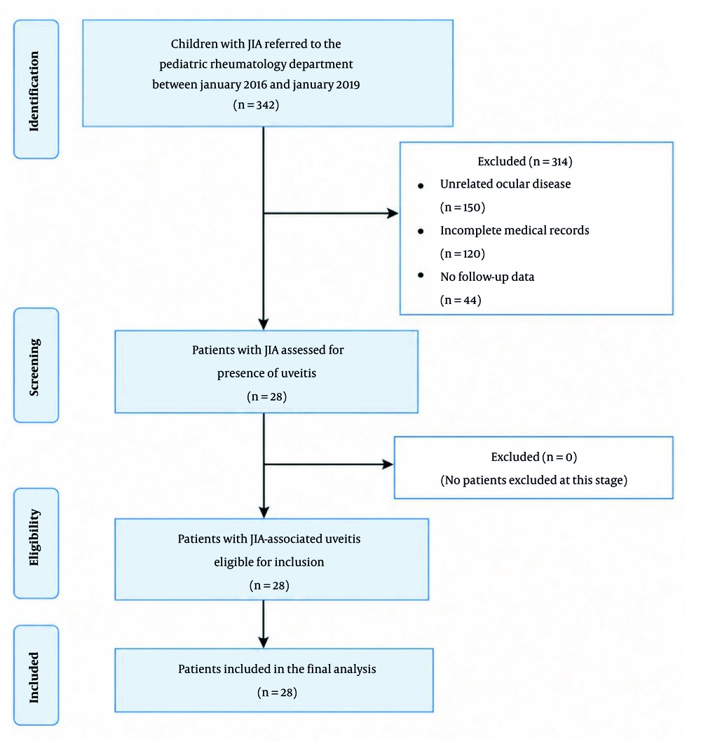
Figure 1.
Participant flow diagram according to STROBE, showing the screening, exclusion, and inclusion of children with juvenile idiopathic arthritis (JIA) in the study.
Table 2 provides an overview of the demographic and clinical characteristics of the included patients. In most children, specifically 19 cases (67.1%), uveitis developed within 2 years of arthritis onset. Notably, in 9 patients (32.1%), uveitis appeared within the first 6 months. In nearly all cases, arthritis symptoms preceded the onset of uveitis. Only 1 child (3.6%) developed uveitis before joint symptoms, with less than 6 months between the 2 conditions.
Table 1.
Type of Uveitis According to Juvenile Idiopathic Arthritis Subtype a
| Subtype of JIA | Chronic anterior uveitis | Acute anterior uveitis | Chronic intermediate uveitis | Chronic panuveitis |
|---|---|---|---|---|
| Oligoarticular JIA | 7 (25) U; 13 (46.4) B | 1 (3.6) B | 1 (3.6) B | 1 (3.6) B |
| Polyarticular RF-positive JIA | - | - | - | - |
| Polyarticular RF-negative JIA | 2 (7.1) U | - | - | - |
| Systemic JIA | - | - | - | - |
| Psoriatic arthritis | 2 (7.1) B | 1 (3.6) B | - | - |
| Enthesitis-related arthritis | - | - | - | - |
| Undifferentiated JIA | - | - | - | - |
a Values are expressed as No. (%). Abbreviations: B, bilateral; JIA, juvenile idiopathic arthritis; RF, rheumatoid factor; U, unilateral.
Table 2.
Demographic Data and Clinical Characteristics of Patients a
| Characteristics | Values |
|---|---|
| Age (mo) | 9.91 ± 3.04 |
| Age at onset (mo) | 6.08 ± 3.33 |
| Sex | |
| Female | 18 (64.3) |
| Male | 10 (35.7) |
| Parental consanguinity | 11 (46.7) |
| Family history of rheumatic disease | 9 (32.1) |
| Subtype of JIA | |
| Oligoarticular JIA | 23 (82.1) |
| Polyarticular RF-positive JIA | 0 (0) |
| Polyarticular RF-negative JIA | 2 (7.1) |
| Systemic JIA | 0 (0) |
| Psoriatic arthritis | 3 (10.7) |
| Enthesitis-related arthritis | 0 (0) |
| Undifferentiated JIA | 0 (0) |
| Duration of JIA (mo) | |
| < 6 | 1 (3.6) |
| 6 - 11 | 5 (17.9) |
| 12 - 24 | 3 (10.7) |
| > 24 | 19 (67.9) |
| Duration between arthritis and uveitis (mo) | |
| < 6 | 9 (32.1) |
| 6 - 11 | 6 (21.4) |
| 12 - 24 | 4 (14.3) |
| > 24 | 9 (32.1) |
a Values are expressed as mean ± SD or No. (%). Abbreviations: JIA, juvenile idiopathic arthritis; RF, rheumatoid factor; SD, standard deviation.
Ocular complications were common during the disease course. The most frequent complications were posterior synechiae (25%), cataracts (17.9%), and elevated intraocular pressure (7.1%). These complications were attributed either to the underlying inflammatory disease or to treatment-related adverse effects. Among the 28 study patients, 15 (53.8%) experienced at least 1 ocular complication. Posterior synechiae was the most common complication (7 patients, 25.1%), of whom 6 (21.4%) had this finding at the time of uveitis diagnosis and 1 (3.6%) developed it during follow-up. Cataract was observed in 5 patients (17.9%); all developed cataract during follow-up, and none had cataract at diagnosis. Increased intraocular pressure occurred in 2 patients (7.1%), and band keratopathy occurred in 1 patient (3.6%); both developed exclusively during follow-up. No patient in this study developed glaucoma or blindness (Figure 2). All complications recorded in this study, regardless of timing, were identified during subsequent ophthalmologic follow-up examinations; no complication was recorded solely on the basis of patient history without a confirmatory eye examination (Table 1).
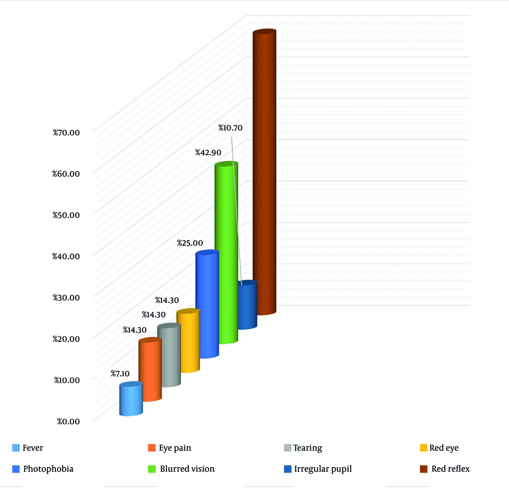
Figure 2.
Clinical symptoms and examination findings at presentation among symptomatic patients with JIA-associated uveitis. The figure shows the frequency of ocular symptoms and signs, including eye redness, pain, photophobia, blurred vision, and abnormal slit-lamp findings, documented during the initial ophthalmologic examination in patients who presented with symptomatic uveitis. Not all patients with JIA-associated uveitis were symptomatic; asymptomatic patients were identified through routine screening.
Despite treatment, 11 children (39.3%) experienced at least 1 recurrence of uveitis. In 3 cases (10.7%), uveitis was refractory to the initial therapeutic regimen and required treatment escalation. Among the 23 children with oligoarticular JIA, 9 (39.1%) experienced relapse of uveitis after treatment initiation (Table 3). Recurrence was also observed in 1 child (50%) with RF-negative polyarticular JIA and 1 child (33%) with psoriatic arthritis (Table 1).
Table 3.
Frequency of Medical Treatment Among Patients with Juvenile Idiopathic Arthritis-Associated Uveitis a
| Medical treatment | Oligoarticular JIA | Polyarticular RF-negative JIA | Psoriatic arthritis | All JIA subtypes |
|---|---|---|---|---|
| Topical corticosteroid | 22 (95.7) | 2 (100) | 3 (100) | 27 (96.4) |
| Systemic corticosteroids | 22 (95.7) | 2 (100) | 3 (100) | 27 (96.4) |
| Methotrexate | 21 (91.3) | 2 (100) | 3 (100) | 26 (92.8) |
| Infliximab | 2 (8.7) | - | - | 2 (7.1) |
| Adalimumab | 3 (13) | - | 1 (33) | 4 (14.3) |
| Cyclosporine | 2 (8.7) | - | 1 (33) | 3 (10.7) |
a Values are expressed as No. (%). The treatments listed in this table represent ever-use during the entire follow-up period, from uveitis diagnosis to the last documented visit. These data do not distinguish between initial therapy, maintenance therapy, and escalation therapy. Multiple patients received more than 1 treatment modality sequentially or in combination. Therefore, these numbers should be interpreted as descriptive management data and not as evidence of comparative treatment effectiveness or response rates.
Recurrence of uveitis was observed in 9 of 23 patients (39.1%) with oligoarticular JIA and in 2 of 5 patients (40.0%) with other JIA subtypes. This difference was not statistically significant (chi-square test: P = 0.97). However, because of the small sample size in the nonoligoarticular group (n = 5), this comparison should be interpreted with caution.
Because of the small sample size, formal statistical comparisons between JIA subtypes were not feasible for most outcomes. Therefore, only descriptive data are presented in Tables 1 - 5.
The overall frequency of uveitis among 342 patients with JIA was 8.2% (95% CI, 5.7% - 11.6%). Among the 28 patients with uveitis, the recurrence rate was 39.3% (95% CI, 23.7% - 57.3%), and the refractory disease rate was 10.7% (95% CI, 3.7% - 27.2%). Ocular complications of any type occurred in 53.8% of patients (95% CI, 35.5% - 71.2%). The most common complications were posterior synechiae (25.1%; 95% CI, 12.8% - 43.2%) and cataract (17.9%; 95% CI, 7.9% - 35.2%).
Based on available medical records, the documented reasons for switching to biologic therapy with infliximab or adalimumab included 1 or more of the following: 1) persistent active uveitis (anterior chamber cells ≥ 1+ on SUN grading) despite at least 3 months of methotrexate at a therapeutic dose (≥ 15 mg/m2/week); 2) intolerance or contraindication to methotrexate; 3) development of sight-threatening complications, such as refractory posterior synechiae or corticosteroid-induced glaucoma; or 4) recurrence of uveitis, defined as ≥ 2 flares, despite optimized conventional therapy with methotrexate and topical corticosteroids. These criteria were not applied uniformly in a prospective protocol but were extracted from clinical documentation. Therefore, the decision to escalate to biologic therapy was made at the discretion of the treating physician and may have been influenced by individual patient factors.
In Table 1, complications are classified according to their most likely etiology based on the available literature and clinical judgment. Posterior synechiae was considered disease-related because it is a direct consequence of intraocular inflammation and is not typically caused by treatment. Cataract and increased intraocular pressure were considered potentially treatment-related because they may result from either chronic inflammation or prolonged corticosteroid use, whether topical or systemic. In the absence of prospective monitoring of cumulative corticosteroid dose and intraocular pressure measurements before and after treatment initiation, these complications could not be definitively attributed to treatment or disease. Band keratopathy was considered indeterminate because it can result from chronic inflammation or corticosteroid use. Therefore, the numbers in Table 1 represent the total occurrence of each complication during follow-up without implying a specific causal pathway.
Table 4.
Frequency of Ophthalmic Complications at Onset Among Juvenile Idiopathic Arthritis Subgroups a
| Ophthalmic complications | Oligoarticular JIA | Polyarticular RF-negative JIA | Psoriatic arthritis | Total |
|---|---|---|---|---|
| Cataract | 4 (14.3) | 1 (3.6) | - | 5 (17.9) |
| Glaucoma | - | - | - | - |
| Posterior synechiae | 5 (17.9) | 1 (3.6) | 1 (3.6) | 7 (25) |
| Blindness | - | - | - | - |
| Increased intraocular pressure | 2 (7.2) | - | - | 2 (7.1) |
| Band keratopathy | 1 (3.6) | - | - | 1 (3.6) |
| Total | 12 (43) | 2 (7.2) | 1 (3.6) | 15 (53.6) |
a Values are expressed as No. (%). Posterior synechiae was considered disease-related. Cataract was considered potentially treatment-related or disease-related. Increased intraocular pressure was considered potentially treatment-related. Band keratopathy was considered to have indeterminate etiology. Because of the retrospective design, disease-related and treatment-related causes of cataract, increased intraocular pressure, and band keratopathy could not be definitively distinguished.
Table 5.
Frequency of Positive Antinuclear Antibody According to Juvenile Idiopathic Arthritis Subtype a
| Subtype of JIA | Positive ANA | Negative ANA |
|---|---|---|
| Oligoarticular JIA | 6 (26.1) | 17 (73.9) |
| Polyarticular RF-negative JIA | - | 2 (100) |
| Psoriatic arthritis | 2 (66.7) | 1 (33.3) |
a Values are expressed as No. (%). Abbreviations: ANA, antinuclear antibody; JIA, juvenile idiopathic arthritis; RF, rheumatoid factor.
5. Discussion
In this study, uveitis was diagnosed in 8% of children with JIA referred to our center, a prevalence comparable to that reported in several recent studies from neighboring countries. Previous studies have reported variable prevalence rates of uveitis in JIA, ranging from 5% to 20% depending on the population studied (1), whereas a recent meta-analysis reported a pooled uveitis prevalence of 12.7% (95% CI, 10.5% - 15.1%) across different populations (2). This variation may be attributable to differences in genetic predisposition, environmental exposures, and, in particular, the frequency and rigor of ophthalmologic screening programs.
In our setting, children undergo regular ophthalmologic evaluations in accordance with established screening intervals. Consistent with global data, oligoarticular JIA was the predominant subtype in our uveitis cohort, accounting for 82.1% of cases. Chronic anterior uveitis was the most frequent manifestation and typically involved both eyes. This pattern mirrors observations from North American and European registries, where ANA-positive oligoarticular JIA, most commonly affecting females younger than 6 years, has been closely associated with chronic anterior uveitis (3, 4).
Interestingly, the ANA positivity rate among our oligoarticular cases was 26.1% (95% CI, 11.0% - 48.7%). This rate falls within the range reported in some published cohorts but is lower than the 40% to 70% range reported in several Western studies. However, formal statistical comparisons were not performed because of differences in study populations, laboratory methods, and sample size. Multicenter prospective studies are needed to determine whether this represents a true regional variation (5). This difference could reflect regional immunogenetic variation or technical discrepancies in ANA testing protocols and thresholds.
The temporal relationship between arthritis onset and uveitis development is a key prognostic factor. In our cohort, 67.1% of children developed uveitis within 2 years of arthritis diagnosis, and in 32.1% of cases, uveitis presented within 6 months. These findings are consistent with previously reported risk markers for severe JIA-associated uveitis (6).
Ocular complications were identified in more than half of the patients (53.8%), with posterior synechiae, cataract, and increased intraocular pressure being the most prevalent. These complications are clinically important because they can lead to irreversible visual impairment if not managed promptly. The 2019 ACR/Arthritis Foundation guideline emphasizes that timely screening is critical for preventing vision-threatening complications in high-risk children (7). Notably, our cohort did not include any cases of glaucoma or blindness, which may reflect timely access to care or, alternatively, limitations in the available follow-up data.
Uveitis recurrence was noted in 39.3% of patients, and 10.7% demonstrated resistance to initial treatment, underscoring the chronic and relapsing nature of JIA-associated uveitis. These figures are comparable to those reported in longitudinal studies examining long-term disease activity and treatment outcomes (8).
With respect to treatment strategies, nearly all patients received topical and systemic corticosteroids. Methotrexate was the most frequently prescribed disease-modifying antirheumatic drug in our cohort and was used in 92.8% of patients. This reflects current clinical practice patterns rather than evidence of superior effectiveness, as methotrexate is recommended as first-line systemic therapy for JIA-associated uveitis by the 2019 ACR/Arthritis Foundation guideline (8).
Biologic therapies were reserved for patients with an inadequate response, with adalimumab being the most frequently used agent. This treatment approach aligns with current international recommendations, which advocate early initiation of biologic therapy in refractory or relapsing uveitis (9). Randomized controlled trials, including the SYCAMORE trial, have demonstrated the efficacy of adalimumab in reducing intraocular inflammation and preventing flare-ups in patients with JIA-associated uveitis (17). In our cohort, adalimumab was used in 4 patients (14.3%), and infliximab was used in 2 patients (7.1%). However, because of the retrospective design and small sample size, our study cannot assess the comparative effectiveness of different biologic agents or predict which patients are most likely to respond. The decision to initiate biologic therapy was based on clinical judgment and documented criteria (10, 11).
Although the use of infliximab and cyclosporine was limited in our study, newer therapeutic agents, including tocilizumab and baricitinib, are currently under investigation and may offer additional options in resistant cases (17). The recent ADJUST trial investigated the outcomes of stopping adalimumab in patients with controlled uveitis, providing important insights into treatment de-escalation strategies (18). A systematic review specifically focused on biologic therapy in noninfectious pediatric uveitis provides additional context (19). A 2025 comparative retrospective cohort study reported no cases of uveitis among 285 patients with JIA receiving adalimumab, compared with 30 cases among 365 patients receiving etanercept, suggesting potential differences in uveitis prevention between tumor necrosis factor inhibitors (20). A 2025 systematic review and meta-analysis found that, despite advances in biologic therapy, 16.6% of patients with JIA-associated uveitis developed severe visual impairment in adulthood. Similarly, a 2025 single-center study from Beijing Children's Hospital reported a uveitis frequency of 4.82% among 1038 Chinese patients with JIA, with ocular complications observed in 25.9% of affected individuals (21).
Overall, our findings from this single-center retrospective study are broadly consistent with global patterns reported in larger cohort studies, although direct comparisons should be made cautiously because of differences in study design, population characteristics, and potential referral bias. Prospective multicenter studies are needed to confirm the generalizability of our observations. However, the notable rates of recurrence and complications in our cohort highlight the ongoing need for systematic screening, timely initiation of systemic immunosuppression, and close collaboration between pediatric rheumatologists and ophthalmologists. As emphasized in the 2019 ACR/Arthritis Foundation guidelines, early ophthalmologic involvement remains a cornerstone of care for preventing irreversible vision loss (9).
Some distinguishing features of our study deserve further discussion. First, the low ANA positivity rate among patients with oligoarticular JIA (26.1%) was well below the rates reported in Western studies, possibly because of regional differences in immune profiles or variability in laboratory practices. Second, the rapid onset of uveitis in many cases, within 2 years in 67.1% and within 6 months in 32.1%, emphasizes the need for aggressive screening protocols immediately after arthritis diagnosis. Third, although chronic anterior uveitis was the most prevalent form, we also observed less common patterns, such as chronic panuveitis and intermediate uveitis, reflecting a broader clinical spectrum. Finally, the relatively high use of biologics, particularly adalimumab (14.3%), in our modestly sized patient population suggests a proactive shift in clinical practice that aligns with contemporary treatment paradigms.
5.1. Conclusions
Within the limitations of this single-center retrospective study, uveitis was identified in 8.2% of patients with JIA evaluated at our tertiary center, underscoring that uveitis remains a potentially serious complication in children with JIA, particularly those with the oligoarticular subtype. The high prevalence of asymptomatic chronic anterior uveitis and its related complications, such as posterior synechiae, cataracts, and increased intraocular pressure, highlights the critical need for systematic, risk-based ophthalmologic screening.
In this descriptive retrospective study, despite the use of topical corticosteroids and methotrexate, 39.3% of patients experienced at least 1 relapse, and 10.7% met the criteria for refractory disease. These findings underscore the chronic and relapsing nature of JIA-associated uveitis. Biologic therapies, including adalimumab or infliximab, were administered to 21.4% of patients, primarily those with refractory disease or intolerance to methotrexate. Prospective studies with standardized treatment protocols and predefined response criteria are needed to compare the effectiveness of different therapeutic strategies and identify predictors of treatment response. These findings are consistent with current international guidelines advocating early systemic treatment in high-risk patients.
Ultimately, preventing vision-threatening outcomes in JIA-associated uveitis depends heavily on timely diagnosis, regular monitoring, and close collaboration between pediatric rheumatologists and ophthalmologists. Longer-term studies are needed to further refine treatment strategies and reduce the burden of ocular complications in this vulnerable group.
5.2. Limitations
Several limitations of this study should be considered. Because this was a single-center retrospective analysis, the results may not be generalizable to a broader population. The relatively small sample size limited statistical power, especially for subgroup analyses. In addition, the lack of long-term follow-up data and standardized grading for uveitis severity limited the ability to fully assess disease progression and treatment outcomes. Future multicenter prospective studies with larger patient groups will be essential to validate these findings and improve clinical practice.
This study was conducted at a single tertiary pediatric rheumatology referral center. As such, our cohort likely overrepresents patients with more severe, complicated, or refractory JIA who were referred to a specialized center for advanced management. Consequently, the frequency of uveitis (8.2%) and the rates of complications (53.8%), recurrence (39.3%), and refractory disease (10.7%) may be overestimated compared with population-based cohorts. Conversely, patients with mild uveitis or those successfully managed in community settings may have been underreferred to our center. Therefore, our findings may not be generalizable to all patients with JIA and should be interpreted with caution.
As with all retrospective studies, our data were subject to information bias. Although we excluded patients with incomplete medical records, the accuracy and completeness of the recorded data depended on the original documentation by treating physicians. We cannot rule out the possibility that some cases of uveitis, particularly asymptomatic chronic anterior uveitis, were underdiagnosed or underreported in the medical records.
Not all patients had identical follow-up duration or assessment frequency. The interval between ophthalmologic examinations ranged from 3 to 12 months based on individual risk stratification according to the 2019 ACR/Arthritis Foundation guidelines. This variability may have affected the detection of uveitis recurrence and the timing of complication identification. Specifically, patients with longer intervals between examinations may have experienced delayed detection of relapses or complications.