
Lifesaving Vertebral Artery Stenting in a Child With Takayasu Arteritis

Author(s):
Afshin Borhani HaghighiAfshin Borhani Haghighi1, Golnoush Sadat Mahmoudi NezhadGolnoush Sadat Mahmoudi Nezhad2, Hamid AmoozgarHamid AmoozgarHamid Amoozgar ORCID3,*
1Neurology Department, Shiraz University of Medical Sciences, Shiraz, IR Iran
2Student Research Committee, Shiraz University of Medical Sciences, Shiraz, IR Iran
3Cardiac and Neonatology Research Center, Shiraz University of Medical Sciences, Shiraz, IR Iran
*Corresponding Author: Corresponding author: Hamid Amoozgar, Cardiac and Neonatology Research Center, Shiraz University of Medical Sciences, P. O. Box: 71937-11351, Shiraz, IR Iran. Tel: +98-9173111877, E-mail: Email: [email protected]

Innovative Journal of Pediatrics:Vol. 25, issue 2
Published online:Apr 18, 2015
Article type:Letter
Received:Feb 06, 2015
Accepted:Mar 11, 2015
How to Cite:Borhani Haghighi A, Mahmoudi Nezhad GS, Amoozgar H. Lifesaving Vertebral Artery Stenting in a Child With Takayasu Arteritis. Inn J Pediatr. 2015;25(2):-. doi: https://doi.org/10.5812/ijp.262

Dear Editor,
Takayasu arteritis (TA) is a rare granulomatous pan-arteritis that involves large vessels predominantly the thoracoabdominal aorta and its branches (1). Angiography, particularly subtraction angiography is the procedure of choice for diagnosis of patients with TA and provides an appropriate guide for endovascular procedure (2, 3).
A ten-year-old Afghan girl was referred to the pediatric cardiology ward of Namazee Hospital, Shiraz, Iran due to a history of fever, vascular claudication, and easy fatigability in both lower extremities. In physical examination, pulseless upper extremities and carotid artery bruit were detected. One year prior to this admission, the patient was labeled as a case of Henoch-Schonlein purpura due to her first presentation with rash and fever. In spite of this management, erythrocyte sedimentation rate and C reactive protein levels had not returned to the normal range. Color Doppler sonography of both upper extremities showed the significant thickening of intima and media, diffuse lumen narrowing of both subclavian arteries, subclavian and axillary arteritis. Bringing together all the aforementioned findings, the patient’s impression was changed to the TA; therefore, prednisolone and infliximab was started. The patient was followed at the outpatient clinic. Four months later, she was admitted in the hospital due to decreased level of consciousness following left sided paralysis in both upper and lower extremities and status epilepticus. In her physical exam, motor power of the left upper and lower extremities was 1/5. The brain computerized tomography showed a small size intra-cerebral hemorrhage in the frontal lobe. Brain magnetic resonance angiography showed stenosis in the posterior, middle and anterior cerebral artery. Echocardiography showed fair left ventricular function, hypertrophic left ventricle with dyskinesia of the ventricular septum. She received plasmaphresis, rituximab, prednisolone, anticonvulsants (phenobarbital and phenytoin) and antihypertensive drugs.
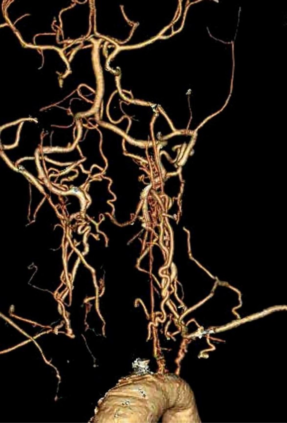
Multislice computerized tomography showed stenosis of both carotids and subclavian arteries (Figure 1).
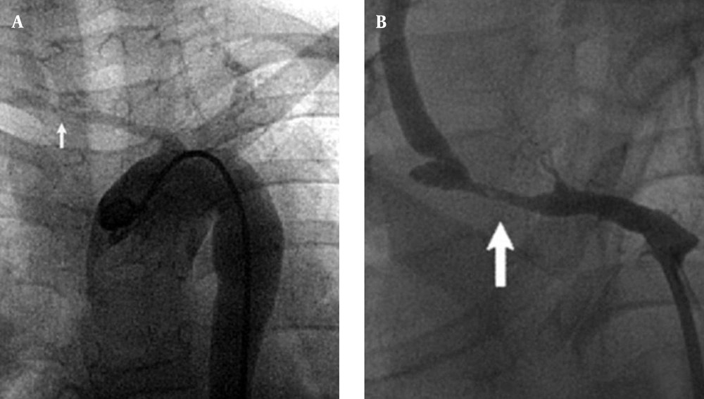
Due to repeated convulsions and poor general condition of the patient angiography for possible stenting the stenotic cranial arteries was done. The right innominate and subclavian artery was severely stenotic and totally occluded just proximal to the origin of the vertebral artery (Figure 2). Blood flow to the right cerebral hemisphere was supplied by the collateral channel, mainly from the vertebrobasilar system. The left common carotid and subclavian arteries were also stenotic.
Under conscious sedation anesthesia and systemic heparinization, a five-French guiding catheter was placed in the aortic arch via the femoral route and a hippocampus stent 6 × 20 mm stent was deployed at the stenotic lesion of the right subclavian artery but the stenosis could not be relieved by multiple inflation of balloon (Figure 3). Subsequently, another stent (hippocampus stent 6 × 20 mm) was placed in the stenotic part, proximal to the previous stent, which markedly improved the antegrade flow of the right common carotid artery (Figure 4).
The symptoms disappeared immediately after the first treatment, and the patient returned to her daily life activities. Neurologic examination showed no changes during one year follow up and Doppler sonography revealed both carotid stenosis and good flow through the stent.
In cases of TA, stenting for these supra-aortic vessels, especially for the carotid lesions, has rarely been presented (4-6). Several authors have reported using subclavian stenting in cases of adult TA (7, 8). In our case, we think that the right vertebral artery stenting was reasonable because both carotid arteries were occluded and the right vertebral artery was functioning as the main collateral pathway to the right cerebral hemisphere, which must be preserved throughout life. To our knowledge, this is the first reported case of stenting of stenotic lesions in the supra-aortic vessels in children in Iran and also there are few reports of stenting in children in the literature.
Multislice Computerize Tomography Shows Stenosis of Both Carotids And Subclavian Arteries
Figure 1.

Multislice Computerize Tomography Shows Stenosis of Both Carotids And Subclavian Arteries
A, Arch Aortogram Shows Deterioration of All the Supra-Aortic Lesions; B, Transbrachial Angiogram Reveals That the Right Subclavian Artery Was Stenotic (Arrow) Proximal to the Orifice of the Vertebral Artery. Right common carotid artery was not visualized on the aortogram.
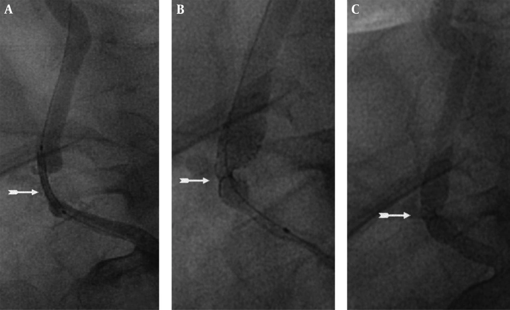
Figure 2.

A, Arch Aortogram Shows Deterioration of All the Supra-Aortic Lesions; B, Transbrachial Angiogram Reveals That the Right Subclavian Artery Was Stenotic (Arrow) Proximal to the Orifice of the Vertebral Artery. Right common carotid artery was not visualized on the aortogram.

A and B, A Hippocampus Stent 6 × 20 mm Stent Was Deployed at the Stenotic Lesion of the Right Subclavian Artery but the Stenosis Could not Be Relieved by Multiple Inflations of Balloon; C, Another Stent (Hippocampus Stent 6 × 20 mm) Was Placed in the Stenotic Part, Proximal to the Previous Stent, Which Markedly Improved the Antegrade Flow of the Right Vertebral Artery
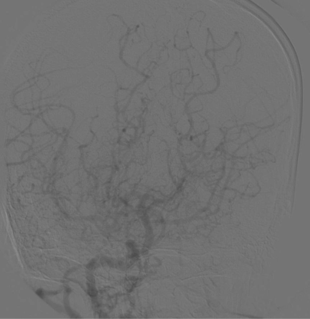
Figure 3.

A and B, A Hippocampus Stent 6 × 20 mm Stent Was Deployed at the Stenotic Lesion of the Right Subclavian Artery but the Stenosis Could not Be Relieved by Multiple Inflations of Balloon; C, Another Stent (Hippocampus Stent 6 × 20 mm) Was Placed in the Stenotic Part, Proximal to the Previous Stent, Which Markedly Improved the Antegrade Flow of the Right Vertebral Artery

Final Result Showed Significant Improvement of Antegrade Flow to Right Hemisphere
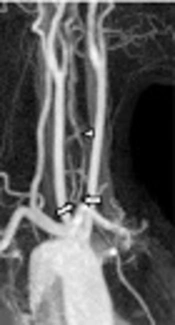
Figure 4.

Final Result Showed Significant Improvement of Antegrade Flow to Right Hemisphere
References

  • 1.
    Yamada I, Shibuya H, Matsubara O, Umehara I, Makino T, Numano F, et al. Pulmonary artery disease in Takayasu's arteritis: angiographic findings. AJR Am J Roentgenol. 1992;159(2):263-9. [PubMed ID: 1352939]. https://doi.org/10.2214/ajr.159.2.1352939.
  • 2.
    Park JH, Chung JW, Lee KW, Park YB, Han MC. CT angiography of Takayasu arteritis: comparison with conventional angiography. J Vasc Interv Radiol. 1997;8(3):393-400. [PubMed ID: 9152912].
  • 3.
    Yamada I, Numano F, Suzuki S. Takayasu arteritis: evaluation with MR imaging. Radiology. 1993;188(1):89-94. [PubMed ID: 8099751]. https://doi.org/10.1148/radiology.188.1.8099751.
  • 4.
    Hwang J, Kim SJ, Bang OY, Chung CS, Lee KH, Kim DK, et al. Ischemic stroke in Takayasu's arteritis: lesion patterns and possible mechanisms. J Clin Neurol. 2012;8(2):109-15. [PubMed ID: 22787494]. https://doi.org/10.3988/jcn.2012.8.2.109.
  • 5.
    Kim HJ, Lee CS, Kim JS, Know SU, Kim JL, Park JW, et al. Outcomes after endovascular treatment of symptomatic patients with Takayasu's arteritis. Interv Neuroradiol. 2011;17(2):252-60. [PubMed ID: 21696668].
  • 6.
    Angiletta D, Marinazzo D, Guido G, Fullone M, Pulli R, Regina G. Eight-year follow-up of endovascular repair of a brachiocephalic trunk aneurysm due to Takayasu's arteritis. J Vasc Surg. 2012;56(2):504-7. [PubMed ID: 22554423]. https://doi.org/10.1016/j.jvs.2012.02.031.
  • 7.
    Gotway MB, Araoz PA, Macedo TA, Stanson AW, Higgins CB, Ring EJ, et al. Imaging findings in Takayasu's arteritis. AJR Am J Roentgenol. 2005;184(6):1945-50. [PubMed ID: 15908559]. https://doi.org/10.2214/ajr.184.6.01841945.
  • 8.
    Brunner J, Feldman BM, Tyrrell PN, Kuemmerle-Deschner JB, Zimmerhackl LB, Gassner I, et al. Takayasu arteritis in children and adolescents. Rheumatology (Oxford). 2010;49(10):1806-14. [PubMed ID: 20562196]. https://doi.org/10.1093/rheumatology/keq167.

Copyright

Copyright © 2015, Growth & Development Research Center.This is an open-access article distributed under the terms of the Creative Commons Attribution-NonCommercial 4.0 International License (http://creativecommons.org/licenses/by-nc/4.0/) which permits copy and redistribute the material just in noncommercial usages, provided the original work is properly cited.

Similar Articles

31
Oct
2017
A Case of Duplicated Origin of the Left Vertebral Artery: CTA and MRA Findings and Literature Review

A Case of Duplicated Origin of the Left Vertebral Artery: CTA and MRA Findings and Literature Review

Seung Eun Lee,
Young-Seon Kim

Lee SE, Kim Y. A Case of Duplicated Origin of the Left Vertebral Artery: CTA and MRA Findings and Literature Review. I J Radiol. 2018;15(1):e63469. doi: https://doi.org/10.5812/iranjradiol.63469

21
Feb
2013

Aortic valve replacement and Coronary Artery Bypass Graft in a Patient with Takayasu's Arteritis and Aorcelain Aorta

Omid Assar,
Mohammadhasan Kalantarmotamedi

Assar O, Kalantarmotamedi M. Aortic valve replacement and Coronary Artery Bypass Graft in a Patient with Takayasu's Arteritis and Aorcelain Aorta. Multidiscip Cardio Annal. 2013;5(1):e8782. doi:

1
Jan
2009

Takayasu disease and cardiac manifestations, presentation of a case with Takayasu and coronary artery disease

Ali Sadeghpour Tabaee

Sadeghpour Tabaee A. Takayasu disease and cardiac manifestations, presentation of a case with Takayasu and coronary artery disease. Multidiscip Cardio Annal. 2009;2(3):e8720. doi:

25
Apr
2016

Dissection of Extracranial Internal Carotid Artery Due to Balloon Guiding Catheter Resulting in Asymptomatic Internal Carotid Artery Occlusion

Suha Akpinar,
Guliz Yilmaz

Akpinar S, Yilmaz G. Dissection of Extracranial Internal Carotid Artery Due to Balloon Guiding Catheter Resulting in Asymptomatic Internal Carotid Artery Occlusion. I J Radiol. 2016;13(3):e28209. doi: https://doi.org/10.5812/iranjradiol.28209

20
Aug
2016

Endovascular Treatment of Ruptured Vertebral Artery Dissecting Aneurysms

Byungjun Kim,
Namjoon Lee,
Keonha Kim,
Kyung-Il Jo,
Pyoung Jeon

Kim B, Lee N, Kim K, Jo K, Jeon P. Endovascular Treatment of Ruptured Vertebral Artery Dissecting Aneurysms. I J Radiol. 2017;14(2):e33070. doi: https://doi.org/10.5812/iranjradiol.33070

Download PDF1.96 MB

Crossmark

Crossmark

Checking

Share on
Cited by
Metrics

Ordering Reprints

Articles are published under the Creative Commons license stated on each article. No permission or royalty fee is required for uses permitted by that license. CCC handles optional bulk and customized reprint orders. Any quotation covers production and delivery services only, not copyright permission. > Request Reprints from CCC 

Search Relations

Author(s):

Related Articles