Animals
Thus, to follow-up the current study, 48 mature (180 ± 15 g, 6-8 weeks old) virgin female Wistar rats were randomly divided into control and experimental groups. The animals were kept in a standard condition (12-h light/12-h dark cycle, 21 ± 01 °C). All animals received the standard rat diet and water ad libitum. Following 48 days, the animals were euthanized by ketamine and xylazine (Alfasan, Woerden, The Netherland) overdose administration. All analyses and investigations were conducted based on the guideline for animals care and research of Urmia University (Ethical No: 2/PD/85). The mature, virgin and clinically healthy rats were included in the study. The animals with anatomically visible abnormalities, as well as those immature, pregnant and weak animals, were excluded in the current study. E = Total number of animals - Total number of groups was considered as a sample size formula.
Experimental design
Our previous experimental trial showed that administration of 7.5 mg/kg-1, 15 mg/kg-1 and 30 mg/kg-1 NMC in the male Wistar rats, negatively and dose-dependently affected the embryo development process. To see the effect of NMC on female reproductive potential, here in the current study, the animals in experimental groups were subdivided into 4 groups and received 7.5 mg/kg-1 (low dose), 15 mg/kg-1 medium dose) and 30 mg/kg-1 (high dose) of NMC (No: 12 rats in each group of total 4 groups, including control). The animals in control group received the same volume of solvent (saline normal), which was used for NMC. The chemicals were administrated orally.
Germinal vesicle (GV) oocytes dissection for IVM and in-vitro fertilization
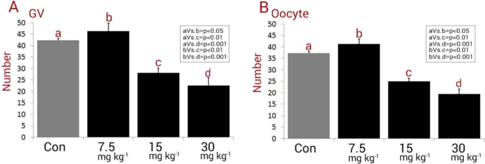
Following 48 days, in order to take GV oocytes, 25 IU (i.p.) of the pregnant mare’s serum gonadotropin (PMSG, ASKA Pharmaceutical, Tokyo, Japan) hormone was administrated. Following 48-h, the hCG (15 IU) was injected (i.p.) and thereafter followed to collect the GV oocytes after 16-18-h (
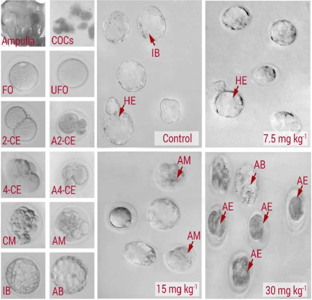
16). The animals were euthanized to collect the GV oocytes, the ovaries were dissected out and placed in Petri dishes containing TCM199 medium (Gibco, Invitrogen, USA, Cat NO: 11150059) supplemented with 5% (v/v) heat-inactivated fetal bovine serum (Merck, Germany, Cat NO: F2442). Cumulus-oocyte complexes (COCs) were released by follicular puncturing with the 30 G needles under high magnification provided by a stereomicroscope (Olympus, Japon). Only immature oocytes containing compact cumulus cells were considered. Next, by using 0.1% hyaluronidase in PB1 medium, the oocytes were freed from cumulus cells (
17).
Preparing IVM culture media and activation of oocytes after IVM
A day pre-conception, the required culturing mediums were prepared and incubated in 5% CO
2 and 37 °C. To prepare the culture media for IVM, TCM199, which was remixed with FBS 10% (Merck, Germany, Cat NO: F2442), 100 mIU of FSH and 40 mIU of LH (Merck, Germany, Cath NO: L6420-10UG) in 1 mL of medium. After mixing, the 50 µL drops were transferred into a dish and covered with mineral oil (
18). After culturing
in-vitro, artificial parthenogenetic activation was carried out with oocytes that reached MII. The MII oocytes were physically denuded with a glass capillary pipette. The oocytes were then cultured in the previously prepared medium. After culturing, the oocytes that contained a distinct pronucleus were counted and compared between the groups.
In-vitro-fertilization (IVF) process and comparing data
A day pre-fertilization, the mR1ECM culture medium was incubated at 12-h in 5% Co
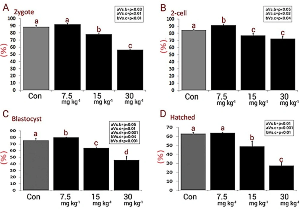
2 in 37 °C. In order to perform IVF, 10-20 oocytes were transferred into each individual drop (500 µL from culture media) and then 10 µL of the sperm medium (containing 3.0-3.6 × 10
6 sperms) was allocated. The percentages of the zygote and pre-implantation 2-cell, blastocyst, and hatched embryos were estimated at 24-h, 4, and 5 days after IVF, respectively (
19).
Evaluation of embryo development
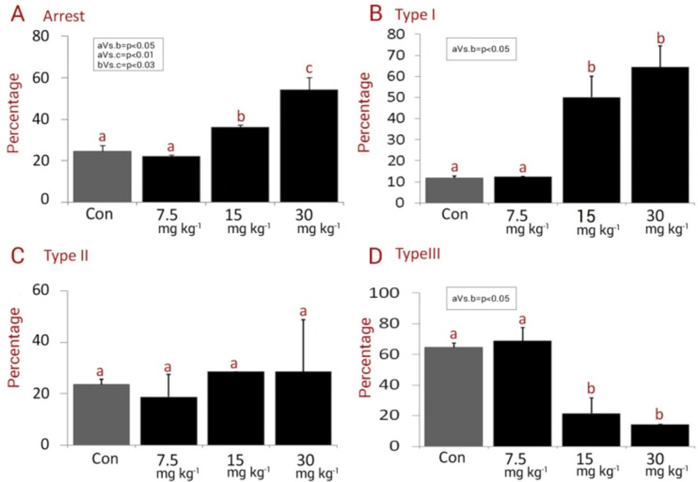
The in-vitro development was evaluated and the intact and fragmented and/or lysed embryos that did not continue developing were recorded as “arrested embryos”. The following type of embryo arrest was considered: Type I: fully lysed, necrotic and/or fragmented embryos. Type II: embryos with partially lysed/ fragmented blastomeres. Type III: embryos with lysed and/or fragmented blastomeres, and embryos with cytoplasmic vesicles. The percentage of each type was assessed in each experimental group and compared between the groups. By quantification of non-compacted, compacted morulae, early blastocysts (with initial blastocele), expanded blastocysts, hatching blastocysts (zona-escaping blastocyst), and hatched blastocysts (extruded or zona-free embryos) the embryo differentiation was examined.
Study limits and strengths
There are some limitations in the current study. Although analyzing the in-vitro fertilization potential and/or pre-implantation embryo development helps us to conclude about the possible effect of NMC on the fertilization status, the effect of NMC on follicular growth, and/or atresia will illustrate the effect of NMC on ovarian physiology and folliculogenesis. Moreover, the effect of NMC on hypophysis-ovary axis-sourced gonadotropins, by assessing the serum levels of FSH and LH, as well as ovarian-sourced hormones, such as estrogen and progesterone should be considered in other studies. However, estimating the GVs, fertilization potential, and pre-implantation embryo development were investigated in the current trial, which first, can be helpful for uncovering the dose effect of NMC on mentioned parameters, and second, can be helpful when the results are possibly considered for human cases.
Statistical analyses
All data were analyzed with One-way ANOVA; a Duncan multiple-comparison test was used to locate differences. For this purpose, the SPSS software (version 11.5) was used. The data are expressed as the mean ± SD, and the P < 0.05 was considered to be statistically significant.