

Introduction
Materials and methods
Results
| Treatment Group | Addition | Cytotoxicity (3 h) | DCF (1 h) |
|---|---|---|---|
| Control | - | 21 ± 2 | 106 ± 5 |
| Nano formulation of DTX | PLGA-DTX(3 nM) | 75 ± 4* | 293 ± 15* |
| +Antioxidantsa | +Mannitol (50 mM) | 49 ± 5† | 140 ± 7† |
| +BHT (50 M) | 45 ± 5† | 138 ± 7† | |
| +MPT pore-sealing | +Carnitine (2 mM) | 51 ± 5† | 152 ± 8† |
| +Cyclosporin (2 M) | 50 ± 3† | 149 ± 7† | |
| +Lysosomotropic | +Choloroquine (100 M) | 39 ± 4† | 128 ± 6† |
| +Methylamine (30 mM) | 43 ± 3† | 127 ± 6† | |
| + Monensin (10 M) | 45 ± 3† | 123 ± 6† | |
| + NADPH-P450 reductase inhibitora | +Diphenyliodonium chloride (50 M) | 28 ± 3† | 107 ± 5† |
| + CYP2E1 inhibitors | +Phenylimidazole (300 M) | 46 ± 5† | 129 ± 6† |
| + 4-Methylpyrazole (500 M) | 44 ± 4† | 126 ± 6† | |
| + CYP3A4 inhibitors | + Troleandomycin (10 M) | 84 ± 3† | 297 ± 10† |
| + Ketoconazole (10 M) | 80 ± 3† | 288 ± 10† | |
| + Hepatoprotectant | +Silymarin (100 M) | 34 ± 4† | 120 ± 6† |
| + Methyl Donors a | +Methionine (1 mM) | 34 ± 3† | 127 ± 6† |
| +Folic acid (100 M) | 32 ± 3† | 123 ± 6† | |
| +Betaine (2 mM) | 39 ± 5† | 130 ± 7† | |
| + Hypomethylator a | +DMSO (150 M) | 92 ± 5† | 360 ± 10† |
| + GSH (5 mM) | 93 ± 4† | 394 ± 15† | |
| + GSH synthesis stimulator a | +Trifluoperazine (15 M) | 86 ± 3† | 312 ± 16† |
| +GSH depleting agent a | +1-Bromoheptane | 39 ± 4† | 151 ± 7† |
| Free Drug | DTX (6 nM) | 72 ± 4* | 277 ± 14* |
| +Antioxidantsa | +Mannitol (50 mM) | 30 ± 3‡ | 140 ± 7‡ |
| +BHT (50 M) | 48 ± 5‡ | 134 ± 7‡ | |
| +MPT pore-sealing | +Carnitine (2 mM) | 48 ± 5‡ | 156 ± 8‡ |
| +Cyclosporin (2 M) | 45 ± 5‡ | 152 ± 8‡ | |
| +Lysosomotropic | +Choloroquine (100 M) | 48 ± 5‡ | 142 ± 7‡ |
| +Methylamine (30 mM) | 50 ± 3‡ | 105 ± 5‡ | |
| + Monensin (10 M) | 53 ± 4‡ | 302 ± 15‡ | |
| + NADPH-P450 reductase inhibitora | +Diphenyliodonium chloride(50 M) | 26 ± 3‡ | 93 ± 5‡ |
| + CYP2E1 inhibitorsa | +Phenylimidazole (300 M) | 52 ± 3‡ | 260 ± 13‡ |
| + 4-Methylpyrazole (500 M) | 50 ± 4‡ | 256 ± 15‡ | |
| + CYP3A4 inhibitorsa | + Troleandomycin (10 M) | 93 ± 3‡ | 302 ± 5‡ |
| + Ketoconazole (10 M) | 98 ± 4‡ | 308 ± 5‡ | |
| + Hepatoprotectant | +Silymarin (100 M) | 63 ± 3‡ | 269 ± 13‡ |
| + Methyl Donors a | +Methionine (1 mM) | 45 ± 5‡ | 136±7‡ |
| +Folic acid (100 M) | 40 ± 4‡ | 130±7‡ | |
| + Betaine (2 mM) | 42 ± 4‡ | 134±7‡ | |
| + Hypomethylator a | +DMSO (150 M) | 89 ± 4‡ | 281±14‡ |
| + GSH (5 mM) | 48 ± 5‡ | 115±6‡ | |
| + GSH synthesis stimulator a | +Trifluoperazine (15 M) | 40 ± 3‡ | 126±6‡ |
| +GSH depleting agent a | +1-Bromoheptane | 80 ± 3‡ | 205±10‡ |
All these agents did not show any toxic effect on hepatocyte at concentrations used (data not shown).
Significant difference in comparison with control hepatocytes (P<0.05).
Significant difference in comparison with PLGA-DTX treated hepatocytes (P<0.05).
Significant difference in comparison with DTX treated hepatocytes (P<0.05).
| Treatment Group | Addition | %ΔΨm | ||
|---|---|---|---|---|
| Incubation time | ||||
| 15 min | 30 min | 60 min | ||
| Nano formulation of DTX | PLGA-DTX (3nM) | 63 ± 3 | 67 ± 3 | 70 ± 4 |
| +Antioxidantsa | +Mannitol (50 mM) | 24 ± 2* | 27 ± 3* | 29 ± 3* |
| +BHT (50 M) | 26 ± 3* | 28 ± 3* | 30 ± 3* | |
| +MPT pore-sealing | +Carnitine (2 mM) | 25 ± 3* | 28 ± 3* | 35 ± 4* |
| +Cyclosporin (2 M) | 22 ± 2* | 26 ± 3* | 33 ± 3* | |
| +Lysosomotropic | + Chloroquine (100 M) | 29 ± 3* | 31 ± 3* | 35 ± 4* |
| + Methylamine (30 mM) | 34 ± 3* | 36 ± 4* | 40 ± 4* | |
| + Monensin (10 M) | 32 ± 3* | 34 ± 4* | 43 ± 4* | |
| + NADPH-P450 reductase inhibitora | +Diphenyliodonium chloride (50 M) | 26 ± 3* | 30 ± 3* | 38 ± 4* |
| + CYP2E1 inhibitorsa | +Phenyl Imidazol (300 M) | 27 ± 3* | 28 ± 3* | 30 ± 3* |
| + 4-Methylpyrazole (500 M) | 25 ± 4* | 30 ± 5* | 34 ± 5* | |
| + CYP3A4 inhibitorsa | + Troleandomycin (10 M) | 75 ± 4* | 79 ± 4* | 87 ± 4* |
| + Ketoconazole (10 M) | 74 ± 4* | 80 ± 4* | 86 ± 4* | |
| + Hepatoprotectant | +Silymarin (100 M) | 27 ± 3* | 30 ± 3* | 32 ± 3* |
| + Methyl Donors a | +Methionine (1 mM) | 28 ± 3* | 29 ± 3* | 31 ± 3* |
| +Folic acid (100 M) | 30 ± 3* | 32 ± 3* | 33 ± 3* | |
| +Betaine (2 mM) | 33 ± 3* | 35 ± 4* | 36±4* | |
| + Hypomethylator a | +DMSO (150 M) | 79 ± 4* | 83 ± 4* | 88 ± 4* |
| + GSH (5 mM) | 76 ± 4* | 79 ± 4* | 85 ± 4* | |
| + GSH synthesis stimulator a | +Trifluoperazine (15 M) | 70 ± 4* | 77 ± 4* | 83 ± 4* |
| +GSH depleting agent a | +1-Bromoheptane | 31 ± 3* | 38 ± 4* | 40 ± 4* |
| Free Drug | DTX(6 nM) | 56 ± 3* | 59 ± 3* | 62 ± 3* |
| +Antioxidantsa | +Mannitol (50 mM) | 26 ± 3† | 28 ± 3† | 31 ± 2† |
| +BHT (50 M) | 20 ± 2† | 22 ± 2† | 23 ± 2† | |
| +MPT pore-sealing | +Carnitine (2 mM) | 40 ± 3† | 46 ± 3† | 56 ± 3† |
| +Cyclosporin (2 M) | 42 ± 3† | 48 ± 3† | 58 ± 4† | |
| +Lysosomotropic | + Chloroquine (100 M) | 56 ± 3† | 58 ± 3† | 63 ± 3† |
| + Methylamine (30 mM) | 54 ± 3† | 57 ± 3† | 61 ± 3† | |
| + Monensin (10 M) | 59 ± 4† | 63 ± 4† | 76 ± 4† | |
| + NADPH-P450 reductase inhibitora | +Diphenyliodonium chloride (50 M) | 42 ± 4† | 48 ± 5† | 53 ± 3† |
| + CYP2E1 inhibitorsa | +Phenyl Imidazol (300 M) | 11 ± 1† | 14 ± 1† | 17 ± 2† |
| + 4-Methylpyrazole (500 M) | 13 ± 3† | 17 ± 3† | 21 ± 4† | |
| + CYP3A4 inhibitorsa | + Troleandomycin (10 M) | 65 ± 5† | 71 ± 3† | 87 ± 3† |
| + Ketoconazole (10 M) | 64 ± 4† | 70 ± 5† | 81 ± 3† | |
| + Hepatoprotectant | +Silymarin (100 M) | 48 ± 4† | 52 ± 4† | 60 ± 5† |
| + Methyl Donors a | +Methionine (1 mM) | 29 ± 3† | 31 ± 3† | 32 ± 3† |
| +Folic acid (100 M) | 26 ± 3† | 28 ± 3† | 29 ± 3† | |
| +Betaine (2 mM) | 27 ± 3† | 29 ± 3† | 30 ± 3† | |
| + Hypomethylator a | +DMSO (150 M) | 80 ± 4† | 84 ± 4† | 89 ± 4† |
| + GSH (5 mM) | 28 ± 3† | 31 ± 3† | 40 ± 4† | |
| + GSH synthesis stimulator a | +Trifluoperazine (15 M) | 30 ± 3† | 37 ± 4† | 46 ± 5† |
| +GSH depleting agent a | +1-Bromoheptane | 75 ± 4† | 77 ± 4† | 86 ± 4† |
All these agents did not show any toxic effect on hepatocyte at concentrations used (data not shown).
Significant difference in comparison with PLGA-DTX treated hepatocytes (P<0.05).
Significant difference in comparison with DTX treated hepatocytes (P<0.05).
| Treatment Group | Addition | % Acridine Orange Redistribution | ||
|---|---|---|---|---|
| Incubation Time | ||||
| 15 min | 30 min | 60 min | ||
| Nano formulation of DTX | PLGA-DTX (3 nM) | 52 ± 3 | 73 ± 4 | 89 ± 4 |
| +Antioxidantsa | +Mannitol (50 mM) | 27 ± 3* | 37 ± 4* | 52 ± 3* |
| +BHT (50 M) | 32 ± 3* | 36 ± 4* | 38 ± 4* | |
| +MPT pore-sealing | +Carnitine (2 mM) | 35 ± 4* | 37 ± 4* | 39 ± 4* |
| +Cyclosporin (2 M) | 33 ± 3* | 35 ± 4* | 41 ± 4* | |
| +Lysosomotropic | +Chloroquine (100 M) | 18 ± 2* | 19 ± 2* | 20 ± 2* |
| +Methylamine (30 mM) | 19 ± 2* | 22 ± 2* | 23 ± 2* | |
| + Monensin (10 M) | 20 ± 2* | 24 ± 2* | 26 ± 3* | |
| + NADPH-P450 reductase inhibitora | +Diphenyliodonium chloride (50 M) | 13 ± 1* | 14 ± 1* | 15 ± 2* |
| + CYP2E1 inhibitorsa | +Phenylimidazole (300 M) | 25 ± 2* | 26 ± 3* | 27 ± 3* |
| + 4-Methylpyrazole (500 M) | 28 ± 3* | 34 ± 3* | 58 ± 3* | |
| + CYP3A4 inhibitorsa | + Troleandomycin (10 M) | 73 ± 4* | 77 ± 4* | 83 ± 4* |
| + Ketoconazole (10 M) | 72 ± 4* | 76 ± 4* | 84 ± 4* | |
| + Hepatoprotectant | +Silymarin (100 M) | 18 ± 2* | 19 ± 2* | 20 ± 2* |
| + Methyl Donors a | +Methionine (1 mM) | 25 ± 3* | 36 ± 4* | 48 ± 5* |
| +Folic acid (100 M) | 20 ± 2* | 34 ± 3* | 40 ± 4* | |
| +Betaine (2 mM) | 23 ± 2* | 30 ± 3* | 39 ± 4* | |
| + Hypomethylator a | +DMSO (150 M) | 70 ± 3* | 82 ± 4* | 93 ± 5* |
| +GSH (5 mM) | 81 ± 4* | 85 ± 4* | 90 ± 5* | |
| + GSH synthesis stimulator a | +Trifluoperazine (15 M) | 77 ± 4* | 84 ± 4* | 89 ± 4* |
| +GSH depleting agent a | +1-Bromoheptane | 30 ± 3* | 33 ± 3* | 35 ± 3* |
| Free Drug | DTX (6 nM) | 44 ± 4 | 67 ± 3 | 72 ± 4 |
| +Antioxidantsa | +Mannitol (50 mM) | 29 ± 3 † | 31 ± 3 † | 35 ± 3† |
| +BHT (50 M) | 31 ± 3 † | 33 ± 3 † | 34 ± 3† | |
| +MPT pore-sealing | +Carnitine (2 mM) | 50 ± 3 † | 56 ± 3 † | 70 ± 3† |
| +Cyclosporin (2 M) | 43 ± 3 † | 57 ± 4 † | 61 ± 4† | |
| +Lysosomotropic | +Chloroquine (100 M) | 20 ± 2 † | 22 ± 2 † | 23 ± 2† |
| +Methylamine (30 mM) | 21 ± 2 † | 24 ± 2 † | 26 ± 3† | |
| + Monensin (10 M) | 22 ± 2 † | 27 ± 3 † | 33 ± 3† | |
| + NADPH-P450 reductase inhibitora | +Diphenyliodonium chloride (50 M) | 14 ± 1 † | 16 ± 2 † | 18 ± 2 † |
| + CYP2E1 inhibitorsa | +Phenylimidazole (300 M) | 20 ± 2 † | 24 ± 2 † | 36 ± 3 † |
| + 4-Methylpyrazole (500 M) | 24 ± 3 † | 26 ± 3 † | 37 ± 4 † | |
| + CYP3A4 inhibitorsa | + Troleandomycin (10 M) | 60 ± 3 † | 67 ± 3 † | 72 ± 4 † |
| + Ketoconazole (10 M) | 64 ± 3 † | 68 ± 3 † | 73 ± 3 † | |
| + Hepatoprotectant | +Silymarin (100 M) | 37 ± 3† | 58 ± 4 † | 63 ± 3† |
| + Methyl Donors a | +Methionine (1 mM) | 22 ± 2 † | 30 ± 3 † | 45 ± 5 † |
| +Folic acid (100 M) | 20 ± 2 † | 28 ± 3 † | 40 ± 4 † | |
| +Betaine (2 mM) | 26 ± 3 † | 35 ± 4 † | 47 ± 5 † | |
| + Hypomethylator a | +DMSO (150 M) | 60 ± 3 † | 71 ± 4 † | 81 ± 4† |
| +GSH (5 mM) | 38 ± 4 † | 41 ± 4 † | 46 ± 5 † | |
| + GSH synthesis stimulator a | +Trifluoperazine (15 M) | 31 ± 3 † | 48 ± 5 † | 57 ± 3† |
| +GSH depleting agent a | +1-Bromoheptane | 77 ± 4 † | 86 ± 4 † | 93 ± 5 † |
All these agents did not show any toxic effect on hepatocyte at concentrations used (data not shown).
Significant difference in comparison with PLGA-DTX treated hepatocytes (P<0.05).
Significant difference in comparison with DTX treated hepatocytes (P<0.05).
| Treatment Group | Addition | Int. GSH (M) | Ext. GSSG (M) |
|---|---|---|---|
| Control | - | 75 ± 4 | 11 ± 1 |
| Nano formulation of DTX | PLGA-DTX(3 nM) | 34 ± 3* | 35 ± 4* |
| +Antioxidantsa | +Mannitol (50 mM) | 54 ± 3† | 21 ± 3† |
| +BHT (50 M) | 50 ± 3† | 22 ± 3† | |
| +MPT pore-sealing | +Carnitine (2 mM) | 51 ± 3† | 23 ± 2† |
| +Cyclosporin (2 M) | 52 ± 3† | 22 ± 3† | |
| +Lysosomotropic | +Chloroquine (100 M) | 54 ± 3† | 24 ± 2† |
| +Methylamine (30 mM) | 53 ± 3† | 23 ± 2† | |
| + Monensin (10 M) | 55 ± 3† | 22 ± 3† | |
| + NADPH-P450 reductase inhibitora | +Diphenyliodonium chloride (50 M) | 58 ± 3† | 21 ± 1† |
| + CYP2E1 inhibitorsa | +Phenylimidazole (300 M) | 59 ± 3† | 25 ± 3† |
| + 4-Methylpyrazole (500 M) | 51 ± 3† | 23 ± 3† | |
| + CYP3A4 inhibitorsa | + Troleandomycin (10 M) | 21 ± 2† | 92 ± 5† |
| + Ketoconazole (10 M) | 20 ± 2† | 95 ± 5† | |
| + Hepatprotectant | +Silymarin (100 M) | 58 ± 3† | 20 ± 2† |
| + Methyl Donors a | +Methionine (1 mM) | 57 ± 3† | 29 ± 3† |
| +Folic acid (100 M) | 60 ± 3† | 21 ± 3† | |
| +Betaine (2 mM) | 59 ± 3† | 24 ± 3† | |
| + Hypomethylator a | +DMSO (150 M) | 23 ± 2† | 41 ± 5† |
| Free Drug | DTX (6 nM) | 43 ± 4* | 30 ± 3* |
| +Antioxidantsa | +Mannitol (50 mM) | 52 ± 3† | 23 ± 3† |
| +BHT (50 M) | 59 ± 3‡ | 24 ± 3‡ | |
| +MPT pore-sealing | +Carnitine (2 mM) | 58 ± 3‡ | 29 ± 3‡ |
| +Cyclosporin (2 M) | 56 ± 3‡ | 24 ± 2‡ | |
| +Lysosomotropic | +Chloroquine (100 M) | 55 ± 3‡ | 23 ± 2‡ |
| +Methylamine (30 mM) | 53 ± 3‡ | 23 ± 2‡ | |
| + Monensin (10 M) | 54 ± 4‡ | 22 ± 3‡ | |
| + NADPH-P450 reductase inhibitora | +Diphenyliodonium chloride (50 M) | 53 ± 4‡ | 20 ± 3‡ |
| + CYP2E1 inhibitorsa | +Phenylimidazole (300 M) | 58 ± 3‡ | 20 ± 3‡ |
| + 4-Methylpyrazole (500 M) | 56 ± 3‡ | 22 ± 3‡ | |
| + CYP3A4 inhibitorsa | + Troleandomycin (10 M) | 31 ± 3‡ | 82 ± 4‡ |
| + Ketoconazole (10 M) | 33 ± 3‡ | 86 ± 4‡ | |
| + Hepatprotectant | +Silymarin (100 M) | 48 ± 5‡ | 28 ± 4‡ |
| + Methyl Donors a | +Methionine (1 mM) | 59 ± 3‡ | 22 ± 3‡ |
| +Folic acid (100 M) | 60 ± 3‡ | 24 ± 3‡ | |
| +Betaine (2 mM) | 63 ± 3‡ | 23 ± 3‡ | |
| + Hypomethylator a | +DMSO (150 M) | 28 ± 3‡ | 70 ± 4‡ |
All these agents did not show any toxic effect on hepatocyte at concentrations used (data not shown).
Significant difference in comparison with control hepatocytes (P<0.05).
Significant difference in comparison with PLGA-DTX treated hepatocytes (P<0.05).
Significant difference in comparison with DTX treated hepatocytes (P<0.05).
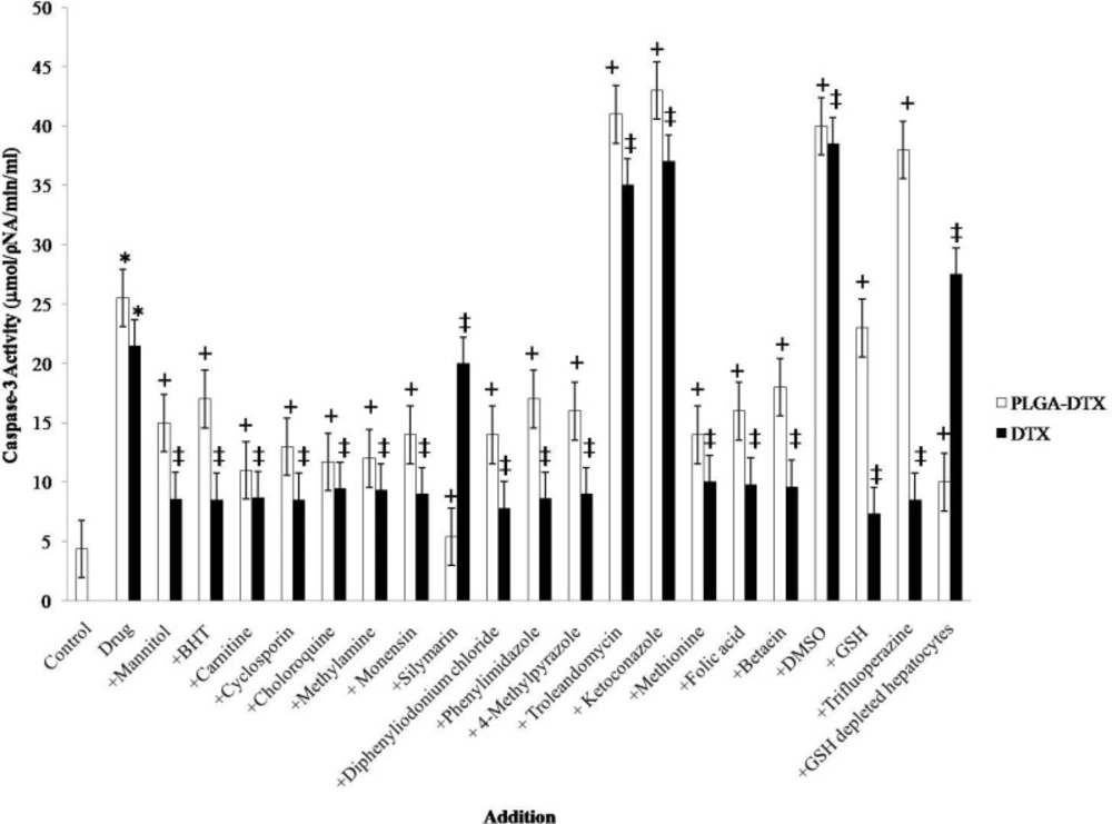
Effect of antioxidants and ROS scavengers, lysosomotropic agents, MPT pore-sealing agents, methyldonor, and NADPH P450 reductase inhibitors on PLGA-DTX and DTX induced hepatocyte Caspase-3 activation.
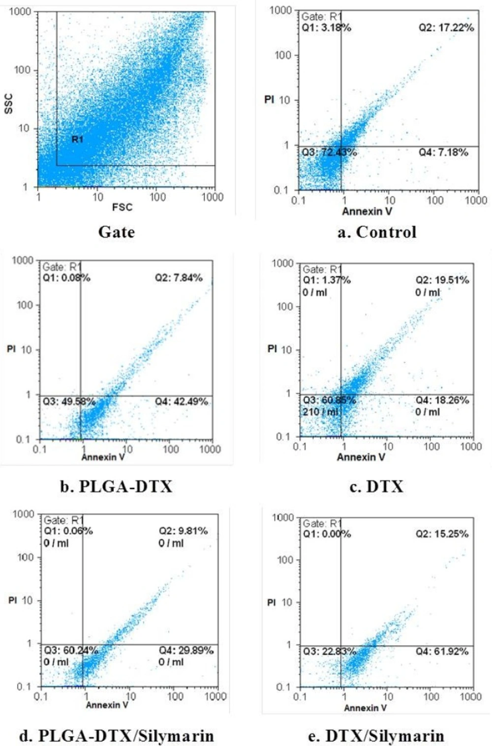
FCM of phospholipid redistribution: Annexin V/Propidium iodide assay. The technique was performed according to Vermes et al. (1995). The samples were analysed for green fluorescence (FITC) and for red fluorescence (PI) by flow cytometry. The assay gives information about the numbers of vital (AV-/PI-) vs. apoptotic (AV+/PI-) cells, and provides also the number of secondary necrotic cells (AV+/PI+).a. Control. b. PLGA_DTX. c. PLGA-DTX/Silymarin. d. DTX. e. DTX/Silymarin. Hepatocytes (106 cells/mL) were incubated in Krebs–Henseleit buffer pH 7.4 at 37 °C for 2 h following the addition of PLGA-DTX & DTX.
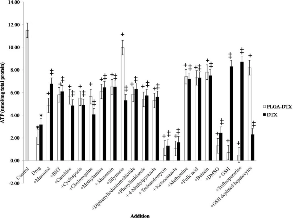
Effect of antioxidants and ROS scavengers, lysosomotropic agents, MPT pore-sealing agents, methyldonor, and NADPH P450 reductase inhibitors on PLGA-DTX and DTX induced hepatocyte ATP depletion.
