Introduction
Leishmaniasis represents a serious threat to the health as one of the most important neglected tropical diseases as designated by the World Health Organization (WHO) (1). Accordingly, about 2 million new cases occur every year and over 12 million people are presently infected (2)
Parasite from the genus Leishmania present a complex life cycle that involves both vertebrate and invertebrate hosts and two developmental stages: promastigotes, the proliferative form found in invertebrate host, and amastigotes, the proliferative form found inside the vertebrate host (3).
The classic treatments of this disease have been compromised by the drugs toxicity and the appearance of new resistance forms.
Therefore, the developing of new treatments strategy has been increased significantly. Natural products seem to be a good solution for those kinds of parasitosis. We have shown in previous study that triterpenic acids isolated form olive leaf extract namely oleanolic and maslinic acids, display inhibitory effects against the promastigotes form of Leishmania amazonensis and infantum (4).
The aim of the present work is to study the selective activity of these compounds against the amasitogotes form of Leishmania amazonensis and Leishmania infantum.
Experimental
Both tritepenic acids, oleanolic and maslinic, have been isolated from olive leaf extract in a previous study (4).

Figure 1
Cytotoxicity effect of triterpenic acid on J774 cell line
Table 1
Anti-amastigote activity, cytotoxicity and selectivity index (SI) of the triterpenic acids oleanolic and maslinic acids
| LC50 (µg/mL) | L.infantum | L.amazonensis | |||
|---|---|---|---|---|---|
| IC50 Anti-amastigote (µg/mL) | SI | IC50 Anti-amastigote (µg/mL) | SI | ||
| Oleanolic acid | 8.103 ± 0.177 | 0.999 ± 0.089 | 8.111 | 3.040 ± 0.505 | 2.665 |
| Maslinic acid | 13.327 ± 0.353 | 2.916 ± 0.066 | 4.570 | 1.417 ±0.401 | 9.405 |
Parasite strains
The activity of both molecules was evaluated against the amastigote form of L. infantum (MHOM/ES/1996/BCN-143) and L. amazonensis (MHOM/BR/77/LTB0016)
Anti-amastigote Activity using the Alamar blue
The anti-amastigote activity was measured according to Jain et al. (2012) (5). Macrophages of the J774 cell line were placed in a 96-well flat bottom plate at a density of 3 × 10⁵/mL in RPMI supplemented with 10% SBF and was incubated overnight at 37 °C in a 5% CO2 environment to allow almost complete differentiation of the cells. 100 µL of stationary phase promastigotes (7 days old culture) were added in a 10:1 ratio and the cultures (3 × 10⁶/mL) and were re-incubated at 37 °C for 24 h. to allow the parasites to infect the differentiated cells. After the incubation, the excess of promastigotes was washed off with the same medium at least 3 times. 100 μL of the culture medium (RPMI-1640 with 10% FBS) were added into each well. In the same time, a serially dilution of the test compounds was made in a 96-deep well plate with the same medium, and then 100 L of this serially-diluted standard were added to each well. The plates were incubated at 37 °C, 5% CO2 for 24 h. After this incubation, we remove the medium from each well and add 30 μL of RPMI-1640 for L.infantum and Schneider for L.amazonensis (with 0.05% SDS) was added to each well. Shake the plate for 30 sec and add 180 μL complete RPMI-1640 (with 10% FBS) or Schneider to each well. Incubate the plates at 26 °C for 48 h. for transformation of rescued amastigote to promastigotes. Add Alamar Blue at 10% into each well of the 96-well plates and incubate them at 26 °C for overnight. After overnight incubation, the plates were read in a spectrofluorimeter at 544 nm excitation, 590 nm emission.
Cytotoxicity against mammalian cells
Cytotoxicity was evaluated after 24 h. incubation of macrophage, the J 774 cell line, with different concentration of the tested molecules. The viability of the macrophages was determined with the Alamar Blue assay. Dose response curves were plotted and the CC50 were obtained. The analyses were performed in triplicate.
Results and Discussion
In a previous paper, we studied the leishmanicidal activity of olive leaf extract on the promastigotes stage of Leishmania infantum and Leishamnia amazonensis (4).
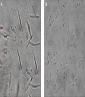
The bio-guided fraction led to the isolation of two triterpenic acids oleanolic and maslinc acids. In the present study, the in vitro activity against amastigote form of Leishmania (L.) infantum and Leishmania (L.) amazonensis was investigated. Figure 1 shows that the isolated molecules from olive leaf extract, exhibited a dose dependent cytotoxicity against macrophage J 774. Meanwhile, within an amount of 5 µg/mL both molecules did not affect the cell development.
As observed in Table 1, the Maslinic acid showed the highest activity, against L. amazonensis, with an IC50 of 1.417 ± 0.401 µg/mL and a selectivity index of 9.405. Meanwhile, the oleanolic acid exhibit a higher activity against L. infantum with an IC50 of 0.999 µg/mL ± 0.089 and selectivity index of 8.111.
Similar results have been reported by Passero etal., (2011) (6), in their study, it was observed that the mix of oleanolic and ursolic acids was very potent against the amastigote form of L.amazonensis with an IC50 of 0.020 ± 0.007 µg/mL. In the same field, Torres-Santos et al. (2004) shows that the oleanolic acid could inhibit the amastigote form of L amazonensis with an IC50 about 11 µg/mL (7).
Yamamoto etal., (2014) have studied, in vivo, the effect of triterpenic fraction purified from Baccharis uncinella leaves on the in- vivo inhibition of Leishmaniaamazonensis, the authors demonstrated that the fraction containing both oleanolic and ursolic acids inhibited the development of skin lesions without affecting the human cells (8).
Conclusion
Despite the fact that both Leishmania species tested in this study are able to cause different forms of leishmaniasis, both molecules tested seems to possess selectivity leishmanicidal properties against both strains.