Silica gel 60 F254 pre-coated aluminium sheets from Merck were used for TLC. The distribution of radioactivity on TLC was determined using a TLC Scanner Mini-Scan, MS.1000. This was equipped with flow count B-FC-1000 and gamma detector MS3200, (Bioscan, Washington, USA). A NaI well counter (Triathler multilabel tester, Hidex, Finland) and a dose calibrator (Atomlab 100, Biodex, NY)
1= F.J.D and S.R contributed equally to this work.
were used to measure low and high levels of radioactivity, respectively. Flow cytometric analysis were performed using a flow cytometer equipped with its accompanying software (FACSCalibur and CellQuestPro, respectively, Becton Dickinson). Size exclusion HPLC (SE-HPLC) analysis was performed using a Merck Hitachi, UV-Vis HPLC, column: TSK-Gel G3000SWXL, 7.8 mm ID, 30 cm L, 5 µM, mobile phase: PBS pH 7.4, flow rate: 0.8 mL/min, sample volume: 20 µL (20 µg). Raji cells, human Burkitt CD20 antigen positive lymphoma cells, Pasture Institute, Tehran, Iran. Anti-human IgG (FC specific)-FITC antibody produced in goat, affinity isolated antibody (Sigma-Aldrich, MO, USA). 90Ycl3 was provided from a 90Sr/90Y electrochemical generator (Pars-isotope, Tehran, Iran). P-SCN-Bz-DOTA was purchased from Macrocyclic, Dallas, TX, USA.
Purification of Rituximab for conjugation
Chimeric anti-CD20 Rituximab as a Pharmaceutical sample (Zytux) from Aryogen Biopharma Co, Karaj, Iran, 100 mg/10 mL was purified and concentrated by centrifugation at 1500 g using Centricon filter (Sartorius, MWCO 30,000). The solution was washed five times with sodium carbonate buffer pH 8.6 (Na2CO3 2 mM, NaHCO3 48 mM, NaCl 150 mM).
Determination of concentration, purity, and integrity of purified Rituximab
The concentration, purity, and integrity of antibody were determined based on following methods.
The concentration was determined by UV absorbance at 280 nm using an extinction coefficient of 1.4 and Bradford assay (1976) using BSA as a standard (
9). The purity was determined by size exclusion chromatography (SE HPLC). The integrity was determined using SDS-PAGE at reducing and non-reducing conditions. Purified antibody was aliquoted and stored at -20 °C for further experiments.
Conjugation of p-SCN-Bz-DOTA to Rituximab
Aliquots of p-SCN-Bz-DOTA (Macrocyclics, Dallas, TX, USA), 2 mg/mL in sodium carbonate buffer pH 8.6, was mixed slowly with aliquots of rituximab solution (20 mg/mL in carbonate buffer pH 8.6) molar ratio Mab:DOTA 1:20 or 1:10. Reaction mixture was incubated at room temperature overnight or 37 °C 1-2 h (
10-
12). The progress of reaction was checked by SE HPLC. The coupling reaction was terminated by centrifugation at 1500 g using Centricone filter (Sartorius, MWCO 30,000) to remove excess DOTA and exchange buffer to 0.25 M ammonium acetate buffer, pH 7. The concentration and integrity of Rituximab-DOTA were determined by UV at 280 nm, SE HPLC, and SDS-PAGE. The DOTA-rituximab was aliquoted and stored at -20 °C for further experiments.
| Tissue | 4 h
| 24 h
| 72 h
|
|---|
| mean±SD (n=3) |
|---|
| Blood | 32.78 ± 0.28 | 10.5 ± 1.34 | 2.96 ± 0.09 |
| Lungs | 34.69 ± 1.21 | 8.92 ± 0.13 | 4.91 ± 0.9 |
| Heart | 21.5 ± 2.07 | 5.71 ± 1.23 | 3.78 ± 0.34 |
| Liver | 25.32 ± 1.16 | 19.13 ± 3.3 | 14.13 ± 5.9 |
| Spleen | 28.67 ± 3.07 | 9.34 ± 1.49 | 18.76 ± 3 |
| Stomach | 2.40 ± 0.28 | 1.71 ± 0.6 | 0.62 ± 0.13 |
| Small intestine | 3.40 ± 0.27 | 1.85 ± 0.2 | 0.97 ± 0.3 |
| Large intestine | 4.51 ± 0.91 | 3.35 ± 0.9 | 2.12 ± 0.3 |
| Kidneys | 17.73 ± 0.87 | 8.2 ± 1.9 | 5.3 ± 0.58 |
| Bone | 6.01 ± 0.07 | 5.13 ± 0.17 | 4.5 ± 0.5 |
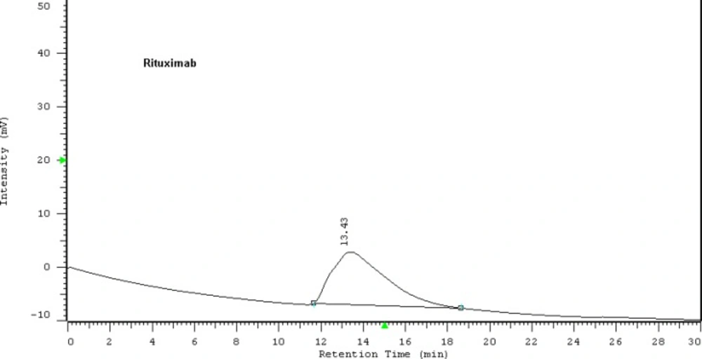
HPLC chromatogram of Zytux solution
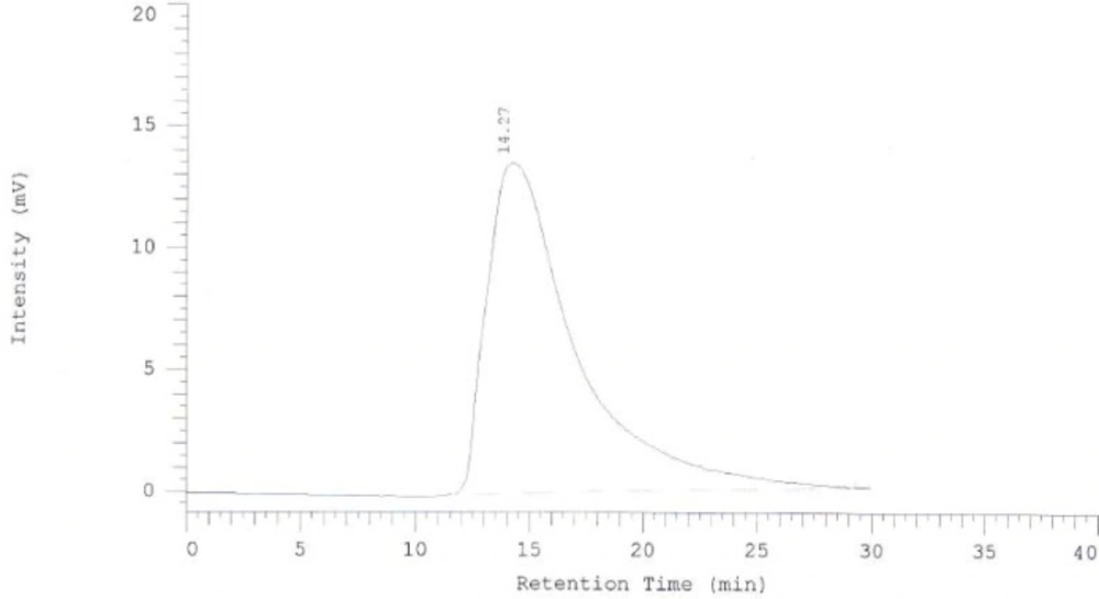
HPLC chromatogram of purified Rituximab
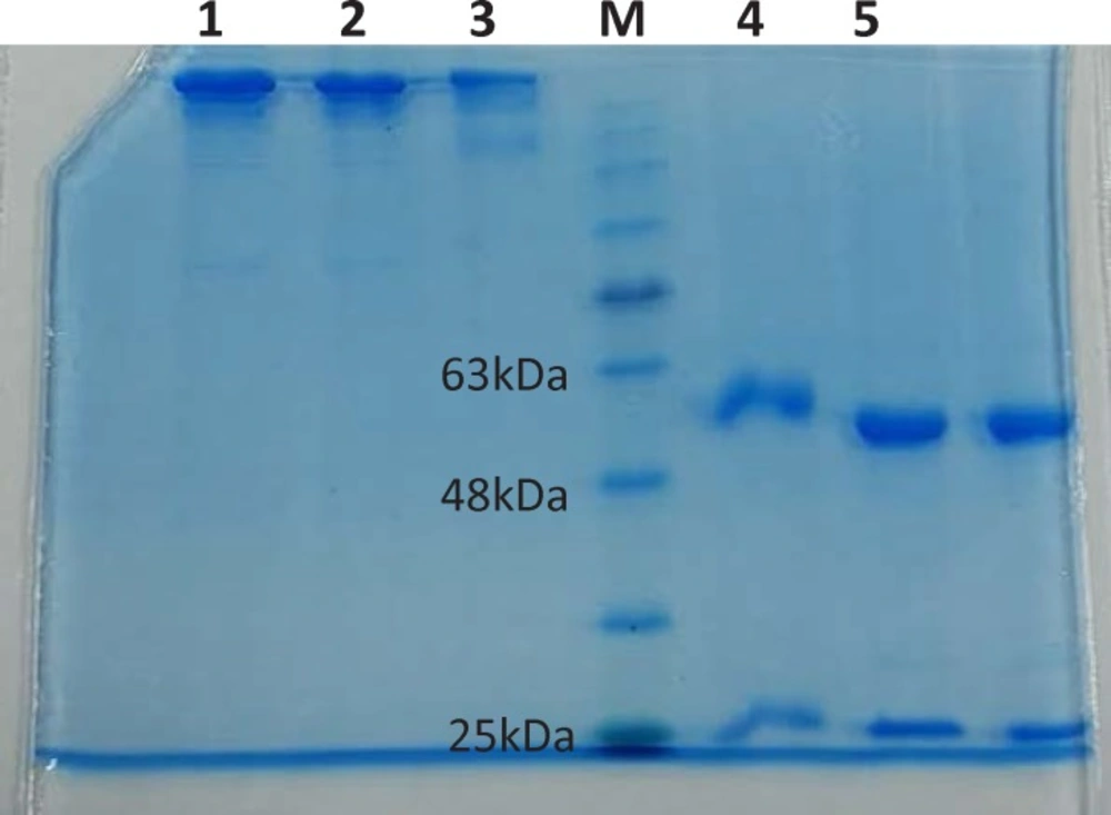
SDS-PAGE of Rituximab in reducing and non-reducing conditions.
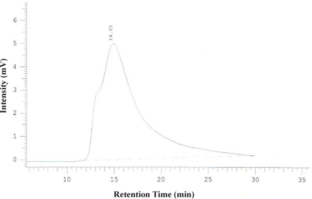
HPLC chromatogram of DOTA-Rituximab after purification using centricone
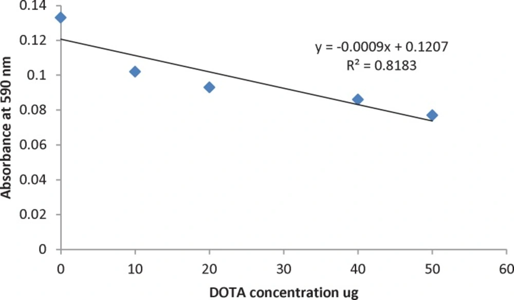
Calibration curve: Absorbance of Pb (II)-AA (III) complex at 590 nm upon adding DOTA ligand.
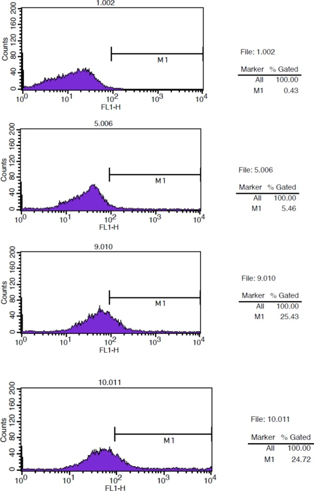
Flow Cytometry studies for determination immunoreactivity of DOTA-conjugated antibody. From up to down. Background: Cells + rituximab. Negative control: Cells + Herceptin + anti-human IgG (FC specific)-FITC.
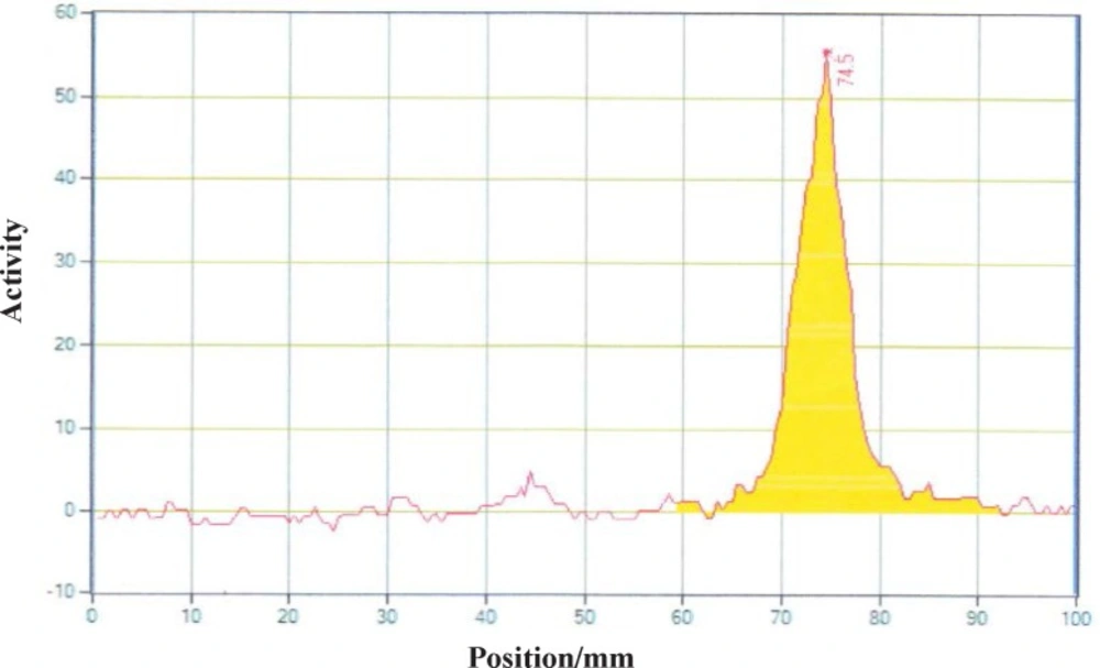
Radiochromatogram of 90YCl3 using TLC-SG, 0.1 M EDTA, 50 mM ammonium acetate pH7, Rf90YCl3=0.75
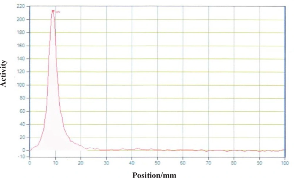
Radiochromatogram of 90Y-DOTA-Rituximab using TLC-SG, 0.1 M EDTA, 50 mM ammonium acetate pH7, Rf90Y-DOTA-Rituximab=0.1
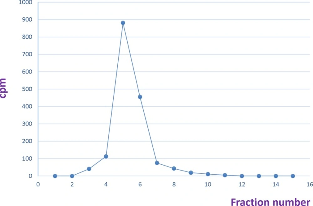
Size exclusion purification of 90Y-DOTA-Rituximab using PD-10 column. Fraction number 4 was considered as radiolabeled antibody
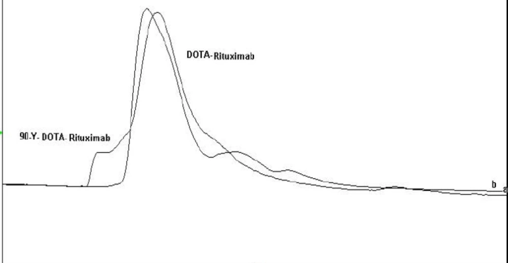
Overlapping HPLC chromatograms of 90Y-DOTA-Rituximab and DOTA-Rituximab.
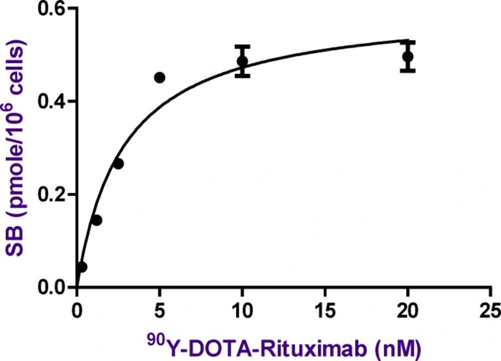
The Saturation curve for the binding of increasing concentrations of 90Y-DOTA-Rituximab in Raji cells. The amount of radioactivity bound to the cells, measured in cpm by gamma counter has been converted to nmol of 90Y-DOTA-Rituximab per cell in the incubation mixture. The values shown are the Mean±SD of three independent determinations
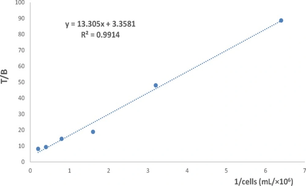
The immunoreactive fraction of 90Y-DOTA-Rituximab
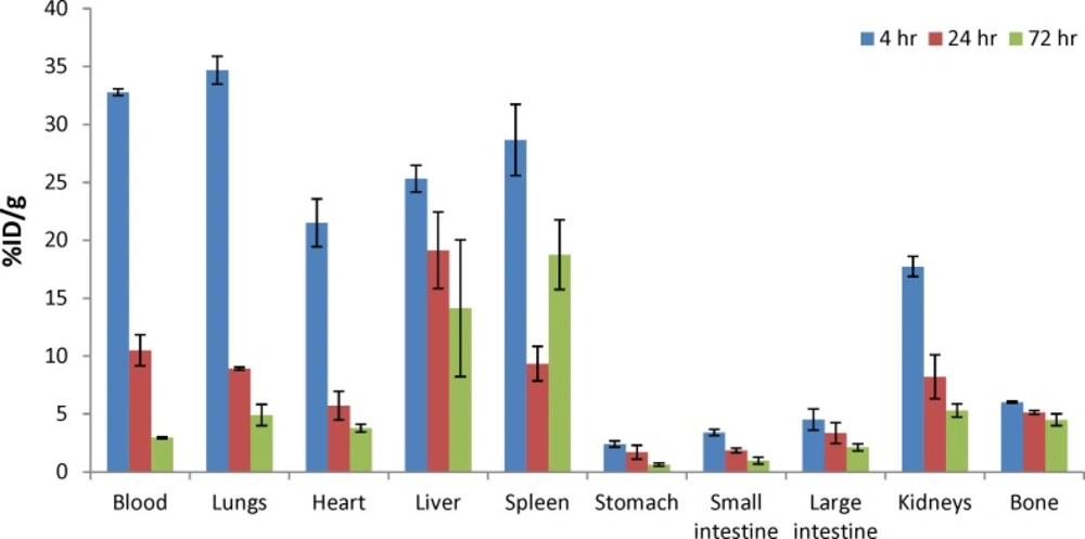
Biodistribution study of 90Y-DOTA-Rituximab in normal mice at 4, 24, and 48 hr post injection (n=3). Radioactivity is shown in terms of %ID/g organ. The values shown are the Mean±SD of three independent determinations
Determination the number of DOTA conjugated per Rituximab molecule
A complex was prepared of Arsenazo (III) [AA(III), Sigma-Aldrich] and Pb (II) (Atomic absorption standard solution, 1000 ppm Pb (II) in HNO3, Merck) in 0.15 M ammonium acetate buffer, pH 7 [10 µM AA(III) and 2 µM Pb (II)]. The complex was stored in the dark at 4 °C.
A stock solution of DOTA (2 mg/mL in bicarbonate buffer) was prepared. For calibration curve, 850 µL AA (III)-Pb (II) complex, 50 µL NaCl 1 mg/mL, 0-25 µL DOTA solution, and 125-150 µL of 0.15 M ammonium acetate buffer, pH7 were incubated in the dark at room temperature for 20 min. The volume of each sample was 1050 µL. Absorbance of samples was read at 590 nm. Calibration curve was plotted from absorbance at 590 nm versus DOTA concentration.
To determine the ratio of DOTA to antibody, 850 µL AA (III)-Pb (II) complex, 50 µL NaCl 1 mg/mL, 100 µL DOTA-rituximab, and 50 µL of 0.15 M ammonium acetate buffer, pH7 were incubated in the dark at room temperature for 20 min (
13-
14).
Immunoreactivity of the DOTA-rituximab
The immunoreactivity of the DOTA-rituximab was determined by flow cytometry. The binding of DOTA-rituximab and unconjugated rituximab to CD20 positive Raji cells was compared. Raji cells were grown in RPMI-1640 medium supplemented 10% heat inactivated fetal bovine serum (FBS). Cells were centrifuged, washed 2X with PBS (1% BSA), re-suspended in PBS (1% BSA), and aliquoted in Eppendorf tubes pre-coated with 1%BSA in PBS (1×10⁶ cells/tube). Cells were incubated at 4 ºC for 2 h with 100 µg rituximab (positive control), 100 µg Herceptin (negative control) and 100 µg DOTA-rituximab (test solution). Cells were washed twice with PBS (1% BSA), re-suspended in 1 mL PBS (1% BSA) and incubated with 100 µL/well anti-human IgG (FC specific)-FITC (Sigma-Aldrich) diluted 1:100 in PBS (1% BSA) in the dark at 4 ºC for 30 min. Cells were washed twice with PBS (1% BSA), re-suspended in 1 mL PBS (1% BSA) and analyzed with FACSCalibur (Becton Dickinson) using cellquest software. Raji cells and Raji cells with rituximab were used as background for autofluorescense.
Stability studies of DOTA-Rituximab
The integrity of DOTA-Rituximab stored at -20 °C in 0.25 M ammonium acetate buffer, pH 7 was determined by SE HPLC and SDS-PAGE at 1, 3, and 6 months after preparation. Each aliquot was labelled with 90YCl3 and radiochemical purity was determined by radio TLC.
90Y labeling of DOTA-rituximab
90-Yttrium as yttrium chloride in 0.05 N HCl with Radioactivity concentration of 45.45 mCi/mL (1.68 GBq/mL), and 2 ppm 90Sr from a electrochemical 90Sr/90Y generator (ITD: isotope technology Dresden) was provided by Pars Isotope Co, Tehran, Iran.
In the first step, the pH of Yttrium chloride was adjusted to 5-5.5 by using ammonium acetate buffer 0.5 M pH 7. Different amounts of DOTA-rituximab (10 µg-1 mg) was incubated with 1 mCi
90YCl
3, pH5.5. The experiment was done at 37, 40, 42, 45, and 50 °C. The progress of reaction was checked by Radio-TLC. The final volume of reaction mixture was 0.5 mL using ammonium acetate buffer, pH7. The reaction mixture was purified by disposable PD-10 desalting column Sephadex G-25, (GE Healthcare life sciences) with PBS contains 2% BSA, pH7.4 as eluent. 15 fractions each 0.75 mL were collected and analyzed by Radio-TLC, TLC-SG as stationary phase and 0.1 M ammonium acetate pH 7, 50 mM EDTA as mobile phase. The fractions contained radiolabeled antibody (fractions 4-6) pulled together and analyzed by SE HPLC UV. 0.75 mL eluate fractions were collected using a fraction collector. The activity in each fraction was measured. A graph of activity against number of fractions was obtained (
7,
10,
14-
15).
Stability studies of 90Y-DOTA-Rituximab in human plasma
The stability of 90Y-DOTA-Rituximab was studied in human plasma. Briefly, 50 µCi (1 µCi/ µL) of labeled antibody was added to 450 µL of fresh human plasma and incubated for 4, 24, 48, and 72 h at 37 °C. At different time points, aliquots of the sample were removed and analysed by Radio-TLC.
Stability studies of 90Y-DOTA-Rituximab in PBS
The stability of 90Y-DOTA-Rituximab was studied in PBS. Briefly, purified fraction of radiolabelled antibody in PBS was kept at at 4 °C and room temperature. At different time points (4, 24, 48, and 72 h), aliquots of the sample were removed and analysed by Radio-TLC.
Saturation binding studies of 90Y-DOTA-Rituximab
A binding assay was performed in triplicate in the presence of increasing amounts of radiolabeled rituximab using Raji cells. 500 L of cell suspension in PBS pH7.4 (1×10
6 cells) was added to 1 mL Eppendorf tubes (pre-incubated with 1% BSA in PBS at 4 °C, at least 30 min before experiment) and incubated with increasing concentrations of radiolabeled antibody (0.03-30 nM, specific activity 300 Ci/mmole) for 2 h at 4 °C with continuous rotation. At the end of incubation times, the mixture was centrifuged (1500 g, 10 min), the cell pellet was washed with cold 1% BSA in PBS (3X) and the radioactivity of pellet was measured as total binding (TB). For each radiolabeled rituximab concentration, nonspecific binding (NSB) was determined by incubation of cells with excess amount of unlabeled rituximab (100X of maximum concentration of radiolabeled antibody) (
16-
22).
Immunoreactivity of 90Y-DOTA-Rituximab
The immunoreactive fraction of
90Y-DOTA-Rituximab was determined based on method of Lindmo
et al. (
22-
24). The study was performed in Eppendorf tubes (pre-incubated with 1% BSA in PBS at 4 °C, at least 30 min before experiment). A series of increasing concentration of cells (5×10
6-0.156×10
6) were incubated with 200 fold dilution of the saturation concentration of
90Y-DOTA-Rituximab for 2 h at 4 °C with continuous rotation. For each cell concentration, non-specific binding was determined by incubation of cells with 100 µg of cold antibody 30 min before adding radiolabeled antibody. At the end of incubation times, the mixture was centrifuged (1500 g, 10 min), the cell pellet was washed with cold 1% BSA in PBS (3X) and the radioactivity of pellet was measured. The immunoreactive fraction was determined using a double reciprocal of the number of total binding (TB) over specific binding (SB) counts against the reciprocal of the number of cells. The reciprocal of the Y-intercept equals the immunoreactive fraction (
22-
24).
Biodistribution studies in normal Balb/C mice
Balb/C adult mice (6-8 week old) were used and obtained from the breeding facility of the Department of Pharmacology and Toxicology, School of Pharmacy, Shahid Beheshti University of Medical Sciences. All animal studies were conducted in accordance with the guidelines established by the Shahid Beheshti University of Medical Sciences. 100 µCi (50 µg) of radiolabeled antibody (1 µCi/1µL in saline, specific activity: 2 µCi/µg 74 MBq/mg) was injected via the tail vein of normal mice. The animals were sacrificed at 4, 24, and 72 h post injection (n = 3 for each time point). Interested organs and tissues were separated, weighted, and counted. The results were reported as percentage of injected dose per gram of organ (%ID/g).