Compound 1, white crystals with mp 180-184°C, showed the molecular formula of C
30H
50O
2 based on positive EI-HR-MS m/z 442.3784 (calc. for C
30H
50O
2: 442.3811, Δ 6.10 ppm) ), in accordance with the number and the multiplicity of
13C-NMR spectra (
Table 1).
| 13C | 1 | 2 |
|---|
| 1 | 31.66t | 31.97t |
| 2 | 30.45t | 30.38t |
| 3 | 78.89d | 78.85d |
| 4 | 40.48s | 40.49s |
| 5 | 47.19d | 47.1d |
| 6 | 21.14t | 21.14t |
| 7 | 28.12t | 28.17t |
| 8 | 47.99d | 48.02d |
| 9 | 20.41s | 19.98d |
| 10 | 26.15s | 25.78s |
| 11 | 26.04s | 26.04t |
| 12 | 35.61t | 32.88t |
| 13 | 45.29s | 45.29s |
| 14 | 48.8s | 48.81s |
| 15 | 32.02t | 34.95t |
| 16 | 26.55t | 26.46t |
| 17 | 52.26d | 52.26d |
| 18 | 18.03q | 18.07q |
| 19 | 29.89t | 29.93t |
| 20 | 36.01d | 36.35d |
| 21 | 18.37q | 18.23q |
| 22 | 31.53t | 35.57t |
| 23 | 28.12d | 31.31t |
| 24 | 76.74d | 156.96s |
| 25 | 149.77s | 33.8s |
| 26 | 111.33t | 22.02t |
| 27 | 17.27q | 19.34q |
| 28 | 19.36q | 18.31q |
| 29 | 25.48q | 14.03q |
| 30 | 14.02q | 25.45q |
| 31 | - | 105.91t |
The IR spectrum confirmed presence of hydroxyl group (3373 cm
-1), double bond absorption (1650 and 756 cm
-1), C-O functions (1219, 1095 and 1026 cm
-1), and cyclopropane C-H (3018 cm
-1) together with C-H stretch bonds (2916 and 2848 cm
-1).
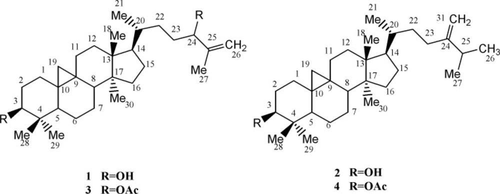
1H-NMR revealed five singlet methyls at δ
H 1.70 (s, Me
27), 0.94 (s, 6H: Me
18, Me
30), 0.87 (s, Me
28) and 0.78 (s, Me
29), one secondary methyl group at 0.84, and a pair of doublets in the up-field area (δ
H 0.30,
J = 4.2 Hz and 0.52,
J = 4.2 Hz), characteristic of cycloartane cyclopropane ring. A double doublet carbinolic proton at δ
H 3.25 (dd,
Jax,ax = 11.1 ,
Jax,eq= 4.5 Hz, H
3), with reference to its axial and α-orientation, assigned the hydroxyl group as 3
β-OH. Using HMBCs, the downfield carbinolic proton at δ
H 3.99 (t,
J = 6.3 Hz, H
24), showed connectivity with a pair of olefinic protons at δ
H 4.80 (d,
J = 1.2 Hz) and 4.90 br-s (each one H), suggesting a terminal methylene. As a whole, the six-degree of unsaturation and the
13C-NMR data (
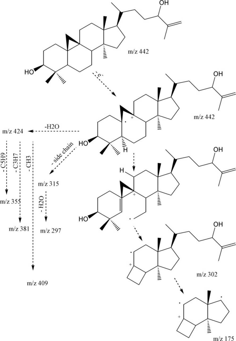
Table 1), suggested the presence of a double bond and, therefore, a pentacyclic skeleton. EI-MS fragmentation pattern, supported m/z 355.3018 [C
25H
39O]
+ and 302.2616 [C
21H
34O]
+, typical ions of 4,4’ dimethyl 9,19 cycloesterols (
Figure 2).
EI-Mass fragmentation pattern of cycloart-25-en-3β,24-diol
The presence of the monounsaturated side chain was confirmed by the
m/z 313.2502 [C
22H
33O]
+, 315 [C
22H
35O]
+ and 297.2587 [C
22H
33]
+. In addition, 381.3153 [M-H
2O-C
3H
7]
+ together with 355.3018 [M-H
2O-C
5H
9]
+, fragmented due to the elimination of parts of side chain (during a Mc Lafferty process), inferred presence of hydroxyl group in side-chain as is shown in
Figure 2 (
11). Regarding to these findings and the published data (
11), compound 1 was identified as cycloart-25-en-3
β,24-diol.
HR-EI-MS identified compound 2, as C
31H
52O with molecular ion peak at m/z 440.4015 (calc. for C
31H
52O: 440.4018, Δ 0.7 ppm). The IR spectrum confirmed absorption of hydroxyl group (3311 cm-1), double bond peak (1640 and 771 cm
-1), C-O functions (1219 cm
-1) and C-H stretching at 3020 (cyclopropane ring), 2916 and 2848 cm
-1. Six degree of unsaturation suggested a double bond (
Table 1) and consequently five rings in the molecule. The resonances encompassed thirty-one carbons including seven methyls, twelve methylenes, six methines and six quaternary carbons.
1H-NMR revealed five singlet methyls at δ
H 1.01 (d,
J = 3 Hz, Me
27), 0.99 (d,
J = 3 Hz, Me
26), 0.94 (s, 6H: Me
18, Me
30 ), 0.88 (s, Me
28), 0.86 (d,
J = 6 Hz, Me
21) and 0.79 (s, Me
29), a pair of doublets in the up-field area at δ
H 0.30 and 0.53 (
J = 4.25 Hz) indicative of cyclopropane ring characteristic of cycloartanes. A doublet of doublet proton at δ
H 3.26 (dd,
Jax,ax = 11.0 ,
Jax,eq= 4.0 Hz, H3), indicative of equilateral β orientated hydroxyl-group, and one pair of olefinic protons δ
H 4.64 (d,
J = 0.5 Hz) and 4.69 (br-s) related to exocyclic terminal methylene. Based on these data, compound 2 was determined as 24-methylene-cycloartan-3
β-ol (
12), confirmed by the EI-MS fragments m/z 425 [M-CH
3], 407 [425-H
2O], 315 [M-side chain], 297[315-H
2O], 300[C
22H
36] and 175 [300-sidechain].
Compound 3 obtained by the acetylation of 1, was identified as 3β, 24-O-diacetyl-cycloart-25-en through EI-MS molecular ion peak m/z 526 [M]+, 466 [M-CH3COOH]+, 423[466-CH3CO], 406[466-CH3COOH]+ and 1H-NMR spectrum. The IR spectrum supported absorptions at 3020 (cyclopropane ring), 2936, 2868, 1736 (esteric carbonyl), 1650, 1456, 1373, 1244, 1026 and 758 cm-1 without hydroxyl group peak at [3500-3300 cm-1]. Likewise, the structure of compound 4 after acetylation of 2, was confirmed as 3β-O-acetyl-24-methylene-cycloartan on the bases of EI-MS molecular ion peak m/z 482 [M]+ and 422 [M-COOH]+, IR and NMR spectra. IR spectrum showed absorption at 3018 (Cyclopropane ring), 2916, 2848, 1736 (Esteric carbonyl), 1642, 1456, 1373, 1244, 1026 and 758 cm-1 without any peak at hydroxyl area [3600-3200 cm-1] and the signals of δH 4.64 (d, J = 0.5 Hz) and 4.69 br-s, each one H related to external methylene, 3.26 (dd, Jax,ax = 11.0 , Jax,eq= 4.0 Hz, H3) geminal to oxygenated carbon, 2.03 (s, acetate methyl), 1.01 (d, J = 3 Hz, Me27), 0.99 (d, J = 3 Hz, Me26), 0.94 (s, 6H:Me18, Me30 ), 0.88 (s, Me28), 0.86 (d, J = 6 Hz, Me21), 0.79 (s, Me29) together with 0.30 (d, 4.25 Hz) and 0.53 (J = 4.25 Hz) of cyclopropane ring were observed in 1H-NMR spectrum.
Proliferation assay
The anti-proliferation effect of the test compounds was determined by measuring the PHA-induced T-cell proliferation by determining radioactive thymidine incorporation. Comparison of pasitive, negative controls were included to assiss the activity of test compounds. cycloart-25-ene-3
β, 24-diol (
1) showed dose-dependent decrease in lymphocyte proliferation with IC
50: 12.1 ± 0.6 μg/mL. This result was in conformity with another study by Smith-Kielland (
14) which showed cytotoxic activity against Ehrlich ascites tumor cells in mice. Likewise, 24-methylene-cycloartan-3
β-ol (
2), presented dose dependent inhibitory effect with IC50: 10.4 ± 0.1 μg/mL agreed with other published data supporting pain-relieving activity, and anti-inflammatory effect by TPA-induced ear oedema in mice (
15). Masking free OH groups of 1 and 2 by acetylation, anti-proliferative effect decreased significantly (IC
50 > 50 μg/mL). On the other hand, in the case of 3 with two acetyloxy groups (3-OAc and 24-OAc), proliferation of PBLs increased by 23-25% at the low concentration (0.5 μg/mL) in comparison with PHA (5 μg/mL) as positive control. However of the higher concentration 50 and 5 μg mL− 1 of 66-70% and 36-39% increas in prolification were absorb. These results suggested that the proliferation stimulatory activity on PBLs is related to the presence of 24-OAc function while anti-proliferation effect induced by free 3-OH group (
Figure 3).
Proliferation assay on peripheral blood lymphocytes of cycloartanes in Euphorbia aellenii. cycloart-25-en-3β,24-diol (1), 24-methylene-cycloartan-3β-ol (2), 3β, 24-O-diacetyl-cycloart-25-en (3), 3β-O-acetyl-24-methylene-cycloartan (4), T-cells were stimulated by phytohemagglutinin (PHA) in the presence of three different concentrations of compounds. Significance differences between the means of compounds as compared to the control (PHA +ve) were calculated by using one-way ANOVA at p = < 0.05
Docking results
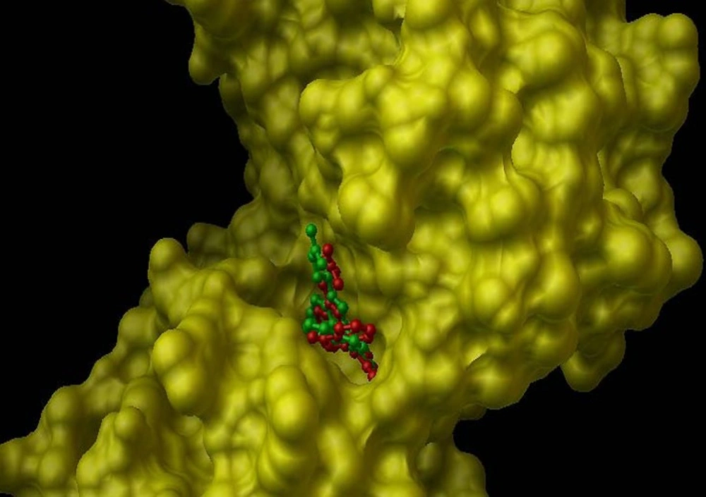
In the internal validation phase of docking, bis(indolyl) maleimide was docked onto the PKC and the lowest energy pose for docking is shown in
Figure 4. Superimposing the experimental and predicted conformations, the RMSD was achieved as 1.02 A°, considered as a successful docking (
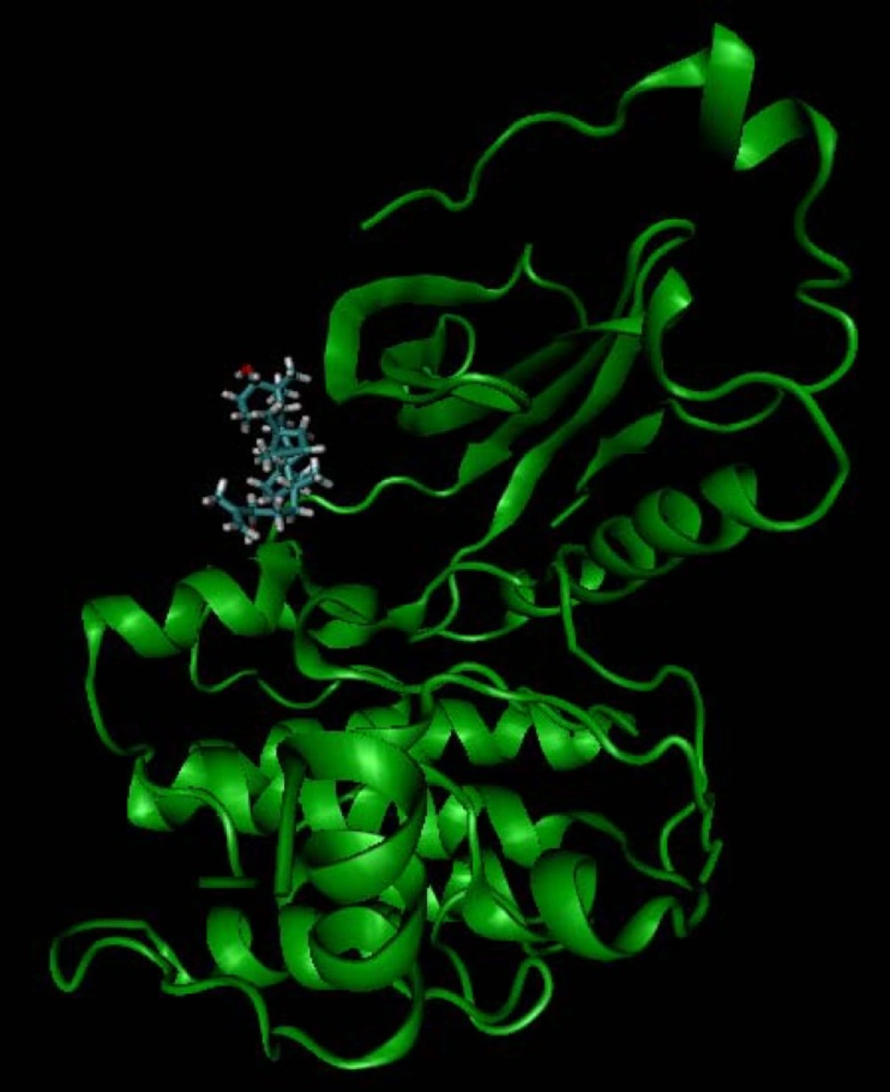
16) of such ligands with PKC. Thereafter, the proposed mechanism of the action was validated by docking the compounds (
1-
4) in the binding site (
Figure 5). All of four compounds tended to accept similar orientations, and docked into the active site of PKC. In compounds (
1-
2) forming hydrogen-bond interactions between 3-OH and Pro-532, could explain the antiproliferative effect of T-cell derived PKC
in-vitro. Therefore, based on this structure-function study, the presence of 3-OH could be correlated with the ability to deactivate PKC and inhibited T-cell proliferation. Likewise, hydrogen bond interaction between 24-O-acetyl group (
3) and Asp-330 of PKC active site could be responsible for induction of lymphocyte proliferation derived PKC
in-vitro.
Internal validation phase result. PKC active site structure rendered as solvent-excluded surface (SES) and conformational comparsion of bisindolylmaleimide from crystal structure (green structure) with that from AutoDock model (red structure).
Docking simulation result of cycloart-25-en-3β,24-diol (1).