Preparation of TFG extract
Fenugreek seed was obtained from local grocery (Tehran) in June, and was systemically identified at the Department of Botany of Shaheed Beheshti University (Voucher number: 2005-46). Then, seeds were washed, dried under shade at room temperature, and finely powdered with a grinder. Thereafter, 100 g of powder was suspended in 1: l of methanol for 48 h in dark (percolation method). The extract was then filtered and concentrated to obtain the solid residue which was and refrigerated until further use. The yield of this process was 19.5% (w/w). Fenugreek extract of lower concentrations of Fenugreek extract were prepared by dilution of the stock with cold and sterile 0.9% saline solution.
Animal experiments
Male albino Wistar rats (Pasteur’s institute, Tehran, Iran) weighing 245-285 g (10-12 weeks old) were housed in an air-conditioned colony room at 23 ± 1 °C and supplied with standard pellet diet and tap water ad libitum. Procedures involving animals and their care were conducted in conformity with NIH guidelines for the care and use of laboratory animals.
The animals (n = 30) were randomly divided into four experimental groups: vehicle-treated control (VC, n = 7), extract-treated control (EC, n = 7), vehicle-treated diabetic (VD, n=7), and extract-treated diabetic (ED, n = 9). Diabetes was induced by a single intraperitoneal injection of streptozotocin (STZ, 60 mg/Kg dissolved in cold 0.9% saline) immediately before test. Control and extract-treated control animals received normal saline solution and alcoholic extract of fenugreek extract (200 mg/Kg, IP) respectively. This dose was chosen on the basis of our pilot dose-response studies. The extract was administered one other day to extract-treated diabetic animals from day 3 after diabetes induction. Serum glucose level and body weight were measured one week before and also four weeks after the experiment. Diabetes was verified by a non-fasting serum glucose level higher than 250 mg/dL using glucose oxidation method (glucose oxidase kit, Zistchimie, Tehran, Iran). All of the abovementioned were treatments continued for one month.
Experimental procedure
The routine protocol was applied as described before (
8-
9). Briefly, after being anesthetized, descending thoracic aorta of the animal was carefully excised and placed in a petri dish filled with cold Krebs solution containing (in mM): NaCl 118.5, KCl 4.74, CaCl
2 2.5, MgSO
4 1.18, KH
2PO
4 1.18, NaHCO
3 24.9, and glucose 10.0 (
8). The aorta was then cleaned of excess connective tissue and fat and cut into rings of approximately 4 mm in length. One ring of each pair was left intact, and in the other ring, endothelium was mechanically removed by gently rotating it on a glass micropipette. Aortic rings were suspended between the bases of two triangular-shaped wires. One wire was attached to a fixed tissue support in a 50 mL isolated tissue bath containing Krebs solution (pH 7.4) maintained at 37°C and continuously aerated with a mixture of 5% CO
2 and 5% O
2. The other end of each wire was attached by a cotton thread to a F60 isometric force transducer, which was connected to A/D board of IBM-compatible computer. Recording and analysis of data was performed using the software Physiograph I (Behineh Arman Co., Tehran, Iran). The rings were allowed to equilibrate for 60 min under a resting tension of 2 g before experiments were begun. During equilibration period, the rings were washed every 30 min. Successful removal of the endothelium was confirmed by loss of acetylcholine (10
-5 M)-induced relaxation in preconstricted rings by NA (10
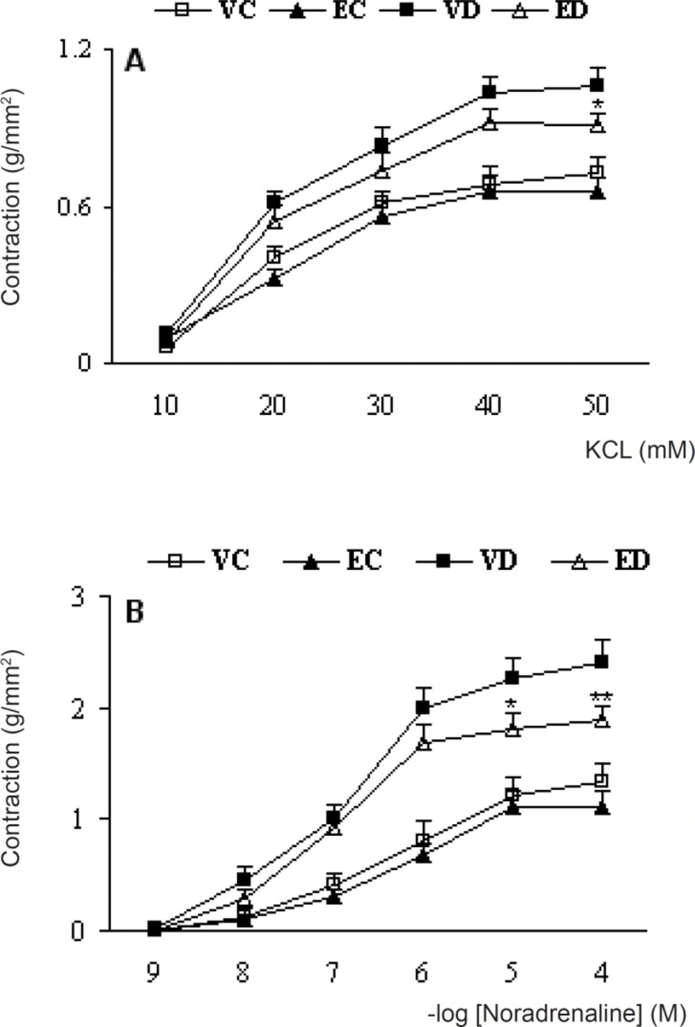
-6 M). Concentration-response curves were obtained with KCl and thereafter with NA in aortic rings with or without endothelium. In this regard, KCl (10-50 mM) and NA (10
-9-10
-4 M) were added in a cumulative manner until a maximum response was achieved.
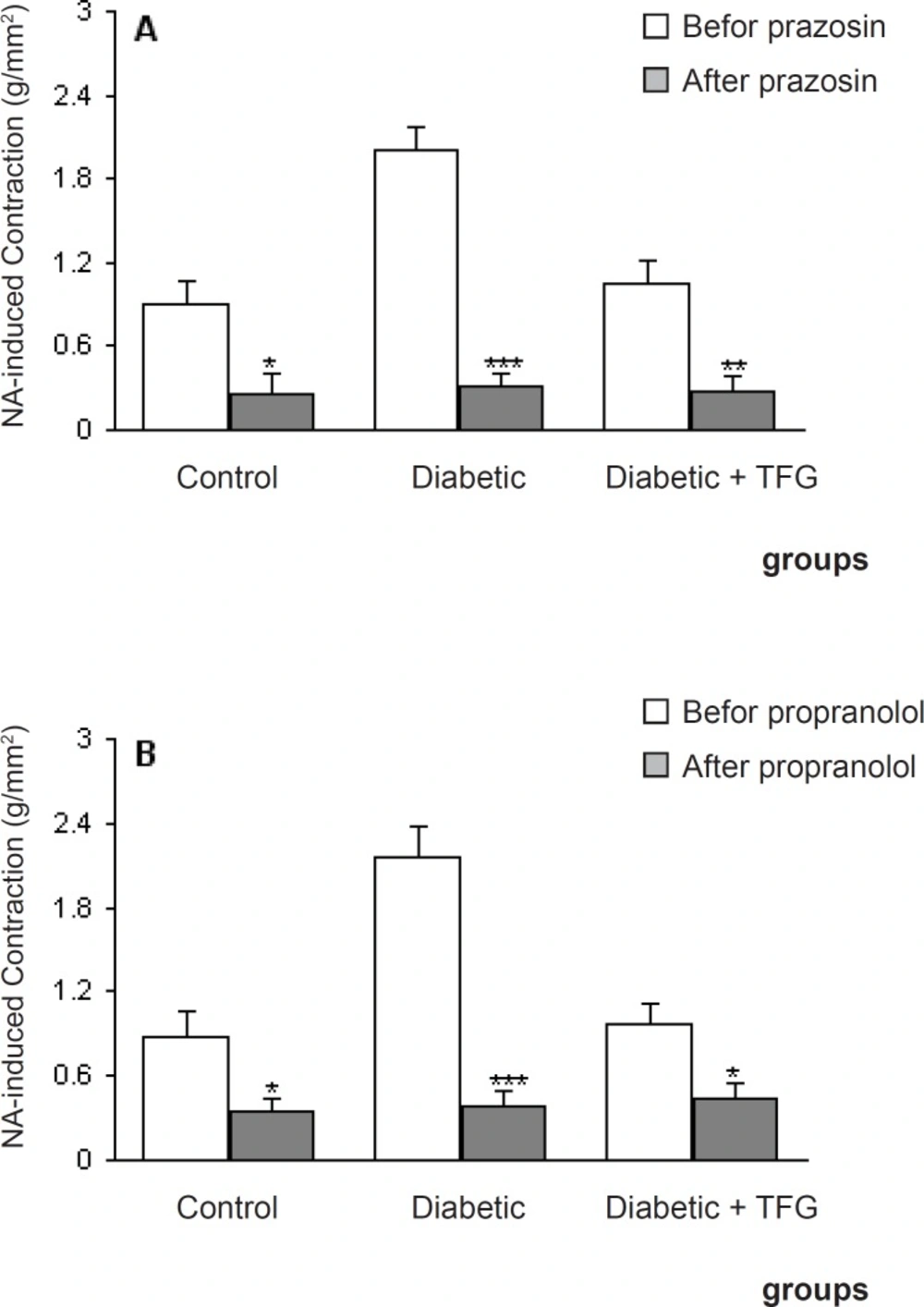
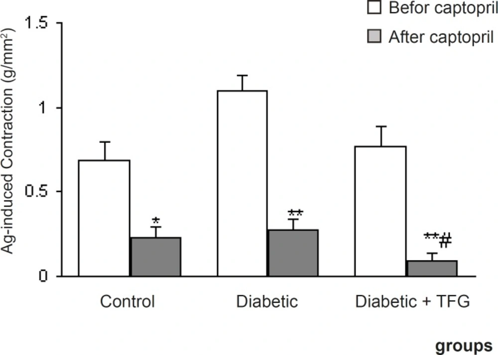
To determine the involvement of adrenergic and angiotensinergic systems, rings were incubated 1 min before the experiments with α1-adrenoceptor blocker, prazosin (10 nM) and β-adrenoceptor blocker propranolol (10 nM) and then contractile response to NA at a submaximal concentration (1 μM) were carried out. Meanwhile, some rings were incubated 5 min before the experiment with angiotensin-converting enzyme inhibitor captopril (10 μM) and then angiotensin I (100 nM)-induced contraction was recorded.
After each experiment, aortic rings were dried at 45°C for 5 min, weighed, and cross-sectional area (CSA) were calculated using the following formula: Cross-sectional area (mm
2) = weight (mg) × [length (mm) × density (mg/mm
3)]
-1. The density of the preparations was assumed to be 1.05 mg/mm
3 (
10).
Drugs and chemicals
Noradrenaline, acetylcholine-HCl, prazosin, and SNP were purchased from Sigma Chemical (St. Louis, Mo., USA). Streptozotocin was obtained from Pharmacia and Upjohn (USA). All other chemicals were purchased from Merck (Germany) and Temad (Tehran, Iran). STZ was freshly dissolved in 0.9% saline solution. Prazosin was dissolved in DMSO.
Data and statistical analysis
All values were given as means ± SEM. Contractile responses to NA, KCl, and Ag I were expressed as grams of tension per cross-sectional area of tissue. Statistical analysis was carried out using student’s paired t-test and one-way analysis of variance (ANOVA) followed by Tukey post-hoc test. A statistical p-value less than 0.05 was considered significant.