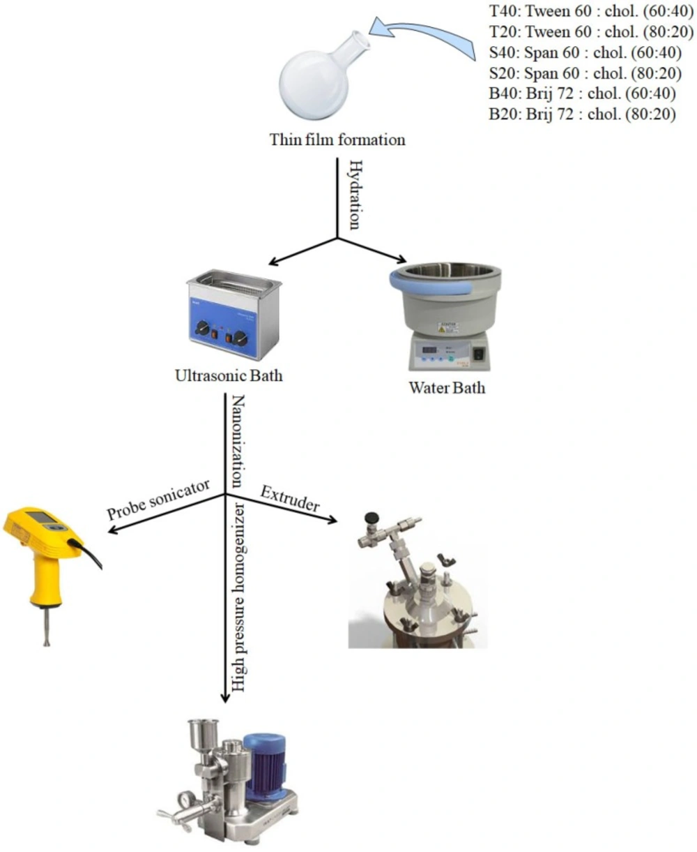
To investigate the effect of surfactant type, cholesterols content, and nanosizing method on the particle size and PDI of niosomes, various formulations were prepared using thin film hydration method. Composition of the prepared niosomes was shown in
Table 2.
For each formulation the hydration process was carried out in either the presence or absence of bath sonication treatment and the effect of hydration method on size and PDI of the obtained niosomes were reported in
Table 3. The results indicated that employing bath sonication during hydration process did not appear to have any significant effect on the vesicle size, while it had a significant effect on decrease in PDI for all niosomal formulations. The decrease in the PDI values indicates that the homogeneity of niosome size increases with applying bath sonication during the hydration process. These data emphasize the usefulness of bath sonication during the hydration process to obtain niosomes with a low PDI. In agreement with our findings, Lachataignerais
et al. reported similar results about decrease of PDI with employing bath sonication (
21). Therefore, bath sonication was applied during thin film hydration process in all further experiments.
Effect of surfactant type on particle size of niosomes
The effect of surfactant type on the particle size and PDI was presented in
Table 4. As shown the type of surfactant significantly influenced the particle size of the niosomes. Based on the results, the size of niosomes showed a regular increase with an increase of the surfactant HLB values. Among the nonionic surfactants employed in the present study, Tween 60 has the highest HLB value of 14.9 and contains a lower hydrocarbon chain volume in comparison with the hydrophilic surface area while the relevant values for both Span 60 and Brij 72 are much lower, namely 4.7 and 4.9, respectively (
Table 1). As it is clear from
Table 4, the particle size of Tween 60 niosomes was about 48%, and 40% larger than those of Span 60 and Brij 72 niosomes (
P < 0.05), indicating that at the same cholesterol content, niosomes composed of surfactants with a lower HLB value are expected to have smaller particle size than those with higher HLB values (
22).
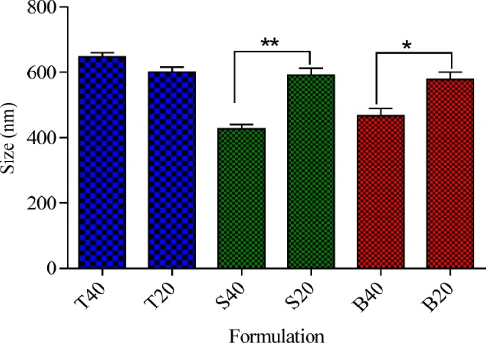
Effect of cholesterol percentage on the particle size of niosomes
Cholesterol is one of the main components of niosomes that can influence their physicochemical characteristics and stability (
23). Particle size is an important characteristic of vesicles from the pharmaceutical viewpoint. To study the effect of cholesterol content on the size of niosomes, a series of formulations containing two cholesterol percentages of 20% and 40% were prepared and the results were presented in
Figure 2. As it can be observed irrespective of surfactant type, cholesterol was found to have significant effect on the particle size of the niosomes. However, the influence of cholesterol percentage on the size of niosomes was markedly dependent on the type of nonionic surfactant. For Tween 60 niosomes, an increase in the cholesterol percentage from 20 to 40% did not have any significant effect on the particle size (
P > 0.05), while for the Brij 72 and Span 60 niosomes, increasing the amount of cholesterol caused a significant decrease in the average diameter of the particles. As shown in
Figure 2, increasing cholesterol amount from 20 to 40 % caused about 20 and 27% decrease in the vesicle size of Brij 72 and Span 60 niosomes (591 ± 5.08
vs. 426 ± 2.18 and 578 ± 5.79
vs. 466 ± 7.32, respectively). This observation may be justified by the fact that the addition of cholesterol can enhance the bilayer hydrophobicity, leading to a decrease in the surface free energy and therefore decrease of particle size (
22,
24). As regards for the Tween based formulations, it seems that because of the high hydrophilicity (high HLB), of this nonionic surfactant, the increase in the percentage of cholesterol was not enough to affect the hydrophobicity of bilayer, and therefore, no significant changes were observed in particle size of the relevant vesicles.
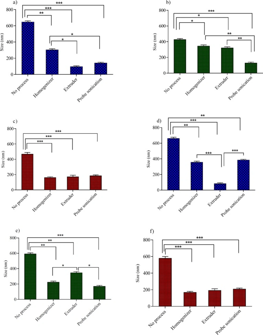
The effect of size reducing methods on the particle size of niosomes
In the process of niosomes preparation, often a size reducing method must be incorporated into the production procedure. A reduction in the vesicle size may be achieved by a number of methods; however, niosome composition is expected to play a critical role in the ability of the desired downsizing method. These considerations promoted us to examine the effects of three downsizing methods (probe sonicator, extruder and high pressure homogenizer) on the vesicle size and PDI of niosomes with various compositions. The obtained results for the particle size and PDI were shown in
Figures 3 and
4, respectively.
For Tween 60 based formulations (
Figures 3a, 3d,
4a and 4d), all the employed downsizing methods had significant effect on decreasing the size and PDI of the vesicles when compared with the untreated niosomes (
P < 0.05). However, extrusion was found to be the most efficient method for reducing size and PDI of the Tween 60 niosomes. The nanoparticles obtained were smaller than 100 nm in size and showed obviously lower PDI values (
Figures 4a and 4d), indicating the homogeneity of the vesicles (Z-average: 95 ± 2.56 nm, PDI: 0.189 ± 0.001 for T40 and Z-average: 82 ± 5.23 nm, PDI: 0.212 ± 0.004 for T20). Extrusion is used as the common approach for downsizing of liposomes (
18). It has been reported that the gel-fluid transition temperature (T
c) of the phospholipid composition along with the adjusted process temperature have impact on the efficiency of extrusion method (
25). At lower temperatures than the T
c, the rate of extrusion is usually slow, but at higher temperatures the rate is higher. The inability of extrusion below the phase transition temperature can be related to the much higher viscosity of gel-state membranes and their decreased deformability (
26). The extrusion temperatures employed in the present study were all above the gel-fluid transition temperatures of the surfactants used.
The higher performance of extrusion for size reducing of T40 and T20 niosomes could be attributed to the lower T
c value of Tween 60, which leads to formation of relatively fluid vesicles and consequently easier passage of vesicles through the polycarbonate filters of extruder than other formulations. The lack of efficiency of extrusion in the particle size reduction of Span 60 based formulation (S40 and S20) confirms the aforementioned suggestion further. Span 60 is solid at room temperature and has the highest T
c value (53 °C,
Table 1) among the surfactants used in this study. As regards Span 60 niosomes, irrespective of Chol percentage, treating the vesicles with a probe sonicator led to better results (Z-average: 178 ± 5.49 nm, PDI: 0.261 ± 0.004 for S40 and Z-average: 170 ± 8.21 nm, PDI: 0.307 ± 0.005 for S20) (
P < 0.05; compared with no process) (
Figures 3b, 3e,
4b and 4e).
For B40 and B20 formulations (Brij 72 based niosomes), as shown in
Figures 3c, 3f,
4c, and 4f, the performance of all mentioned size reduction methods was almost similar and resulted in the same particle size and PDI values (about 200 nm and 0.2, respectively). The linear structure of Brij 72 and its intermediate T
c value may justify the observed data (
Table 1).
Stability testing
Niosomes composed of 40% Chol, including T40, S40, and B40, which were downsized using extrusion, probe sonication, and high pressure homogenization, respectively were undertaken for stability studies. The reason for selection of formulations with a higher Chol content was that it can increase the microviscosity of the membrane by abolishing the gel-to-liquid phase transition of the surfactant bilayer, and therefore, results in a more stable and hydrophobic bilayer (
27). For this purpose, niosomes were stored at temperatures of 4 °C and 25 °C for 28 day and particle size and PDI of the samples were analyzed after 7, 21 and 28 day storage. As shown in
Tables 5 and
6, all formulations, except the Brij based niosomes, were stable at 4 °C over a 28 day period of time. While when stored at 25 °C, the size of all tested niosomes was increased significantly over a 21 day period. Increase in size may be related to the fusion and aggregation of vesicles during storage time. The current results showed that T40 and S40 were stable when maintained at 4 °C for at least 28 day. However, B40 formulation was unstable and became very turbid after 21 day incubation at both temperatures of 4 °C and 25 °C, more likely due to aggregation and fusion of the vesicles.
The exact reasons for instability of the Brij based niosomes are not clear and further investigations are required to clarify the issue more.