MNPs Characterization
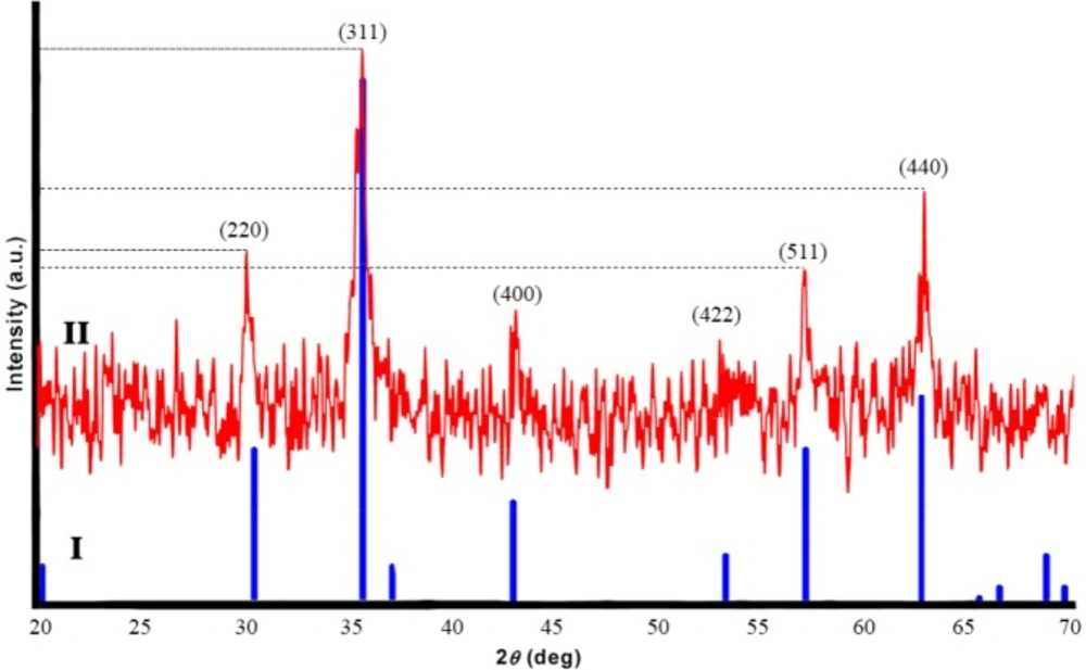
XRD Analysis
Figure 3 shows XRD patterns of pure Fe
3O
4 (I, Blue) and synthesied Fe
3O
4 (II, Red). Six characteristic peaks for Fe
3O
4 corresponding to (220), (311), (400), (422), (511), and (440) were observed in the sample (
40). These peaks reveal that the resulting nanoparticles were pure Fe
3O
4.
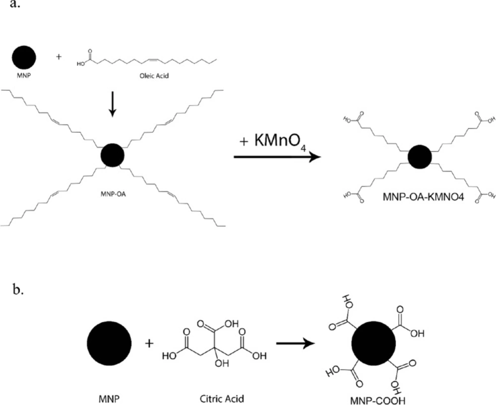
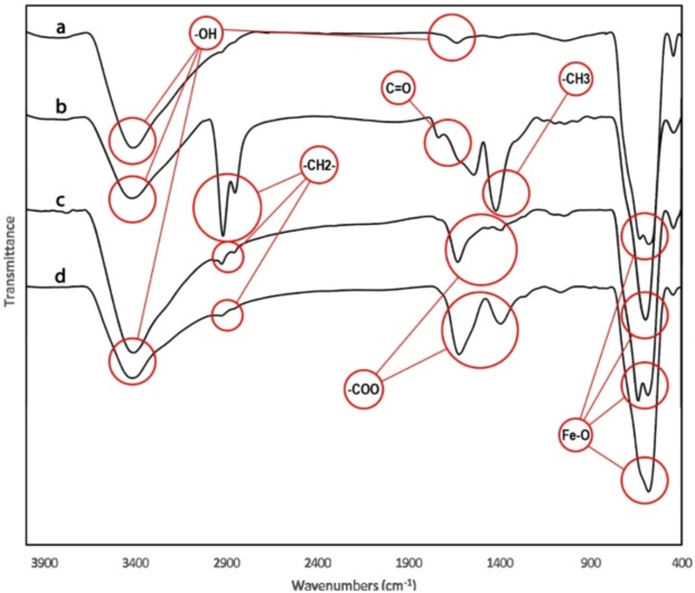
FT-IR Analysis of functionalized MNPs
The MNPs was further characterized by FTIR to confirm the chemical binding. FT-IR spectra of MNPs shown in
Figure 4a cursory inspection of the spectra shows two absorption bands below 1000 cm
-1 as a common feature of all the ferrites. Absorption in this region is not restricted to this class of compounds but occurs in the spectra of most metal oxides (
41). In all spectra the band at 580 cm
-1 corresponds to the vibration of the Fe–O bonds in the crystalline lattice of Fe
3O
4 (
42). During preparation of Fe
3O
4 nanoparticles by the chemical co-precipitation, their surfaces were readily covered with hydroxyl groups in an aqueous environment, the characteristic bands of hydroxyl groups, 1630 and 3405 cm
-1, appear in the FTIR spectrum
Figure 4a (
43,
44). The bands at 2852 and 2922 cm
-1 are attributed to the asymmetric CH
2 stretch and the symmetric CH
2 stretch in oleic acid, respectively, and the band at 1409 cm
-1 corresponds to the CH
3 umbrella mode of oleic acid and the band at 1710 cm
-1, corresponds to stretching vibration of C = O in oleic acid (
Figure 4b). After using KMnO
4 on MNP functionalized with oleic acid the product nanoparticles (MNP-OA-KMNO4) were analyzed with FTIR. Two bands at 1457 and 1523 cm
-1 appeared as seen in
Figure 4c and were attributed to the asymmetric (–COO) and symmetric (–COO) stretch vibration band, that confirmed the carboxyl group on it. FTIR of MNP-COOH shown in
Figure 4d that two bands at 1457 and 1523 cm
-1 confirmed carboxylation of MNPs.
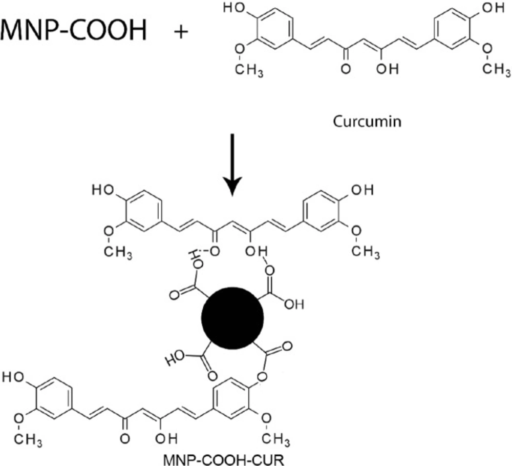
Curcumin Loading
To investigate the effect of solvent and activator on Curcumin loading, the Curcumin on the functionalized MNPs was released through treatment with acetone, quantified, and compared. Loading obtained in DMF (CUR1, CUR2 and CUR3) was very low, the produced nanoparticles analyzed by FT-IR and almost no change was observed. The amount of loaded Curcumin less than the resolution of the device could be the reason for this observation. The release data of these samples (CUR1, CUR2 and CUR3) were collected in
Table 1. According to these results, the most Curcumin loading was related to MNP-CA1, so this sample was selected for further analyses. In the following of the text MNP-COOH representes MNP-CA1.
In order to increasing the Curcumin loading, DMSO, ethanol, water, ethanol/water as the solvent and DCC, EDC/NHS as activator were tested. The results of these tests are shown in
Table 2 (CUR4, CUR5, CUR6, CUR7 and CUR8) and illustrate an increased amount of Curcumin loaded on the MNPs using a mixture of water and ethanol as solvent (CUR8). This sample collected for further analyzed with FT-IR.
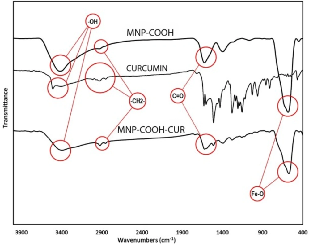
FT-IR Analysis of MNP-COOH-CUR
Figure 5 shows the FTIR spectra of MNPs-COOH, Curcumin and MNPs-COOH-CUR. The absorption peaks around 563 or 580 cm
-1 in
Figure 5 are the characteristic absorption of Fe-O bond of magnetite nanoparticles (
41). The absorption bands at 2920-2850 cm
-1 ascribed to the C-H stretching vibrations, which is indicative of citric acid and Curcumin on MNPs. A large and intense band around 3400 cm
-1 is assigned to the structural OH groups on the citric acid, Curcumin and MNPs (
45). The 1600 cm
-1 peak is assignable to the C = O vibration (symmetric stretching) from the COOH group of citric acid and indicate ester linkage between citric acid and Curcumin.
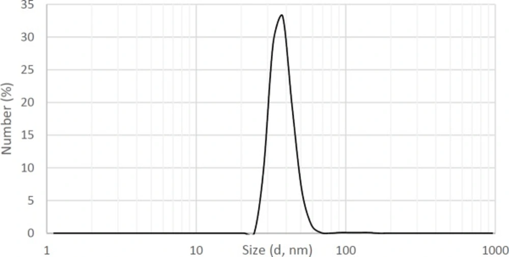
DLS Analysis
As MNPs are widely used in medical and diagnostic processes, the properties of these particles in the fluid environment are important and valuable to study. Hydrodynamic and rheological properties of MNPs dramatically depend on morphology and surface charge. The study of these parameters performed using DLS analysis.
As seen in
Figure 6, the average hydrodynamic diameter of particles is about 37 nm. Particles with diameters between 7 to 3500 nm have been used commercially in medical science (
1).
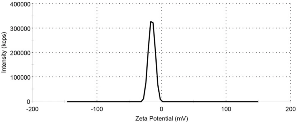
The zeta potential result of the MNPs-COOH shown in
Figure 7 and
Table 3. Due to the presence of carboxyl group on MNP, the value of the zeta potential is negative.
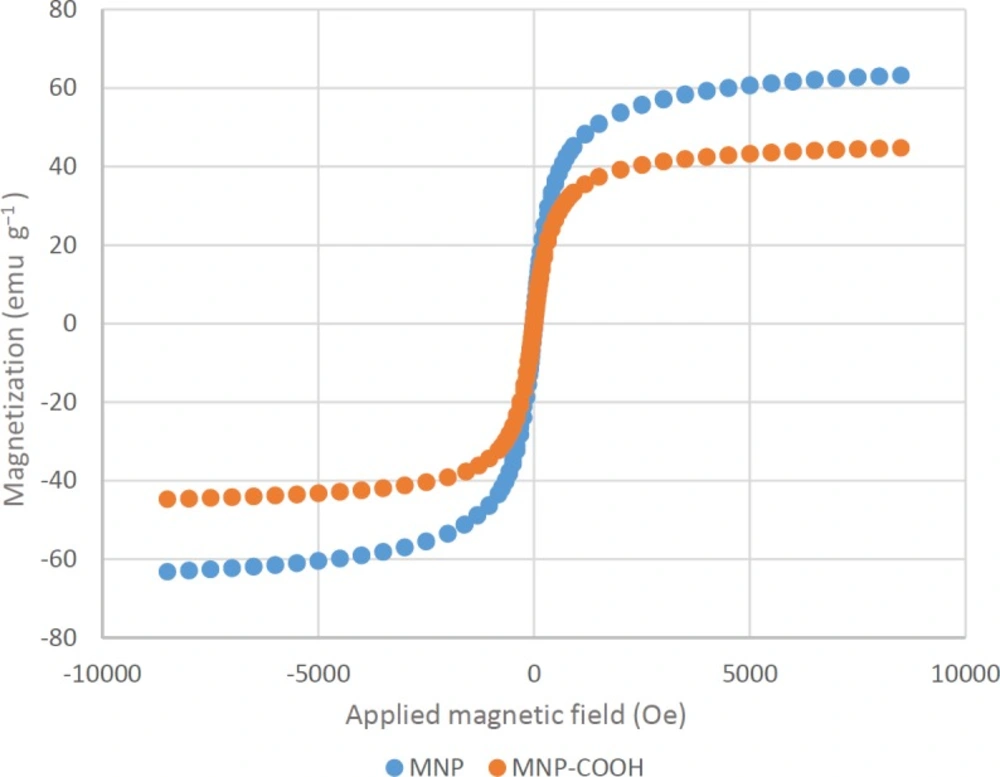
Magnetic properties of MNPs
The magnetic properties of MNPs were characterized by vibrating sample magnetometer (VSM). It is important to characterize the magnetic properties of MNPs before their use as a contrast agent for MRI.
Figure 8 shows that the VSM curves of both MNPs and MNP-COOH rapidly approach a saturation magnetization. The absence of hysteresis in their magnetic profiles suggest that these nanoparticles are superparamagnetic. The saturation magnetization (Ms) of Fe3O4 particles` is in the range of 60–80 emu g
−1 and is lower than that of bulk Fe3O4 (about 90 emu g
−1). This discrepancy in Ms can be due to the difference in particle size, as it has been reported in the literature that saturation magnetization of Fe3O4 nanoparticles decreases with reducing particle size (
46). The significant decrease in Ms value of MNP-COOH is attributed to the existence of a magnetically inactive surface layer and some diamagnetic contribution from the citric acid (
47).
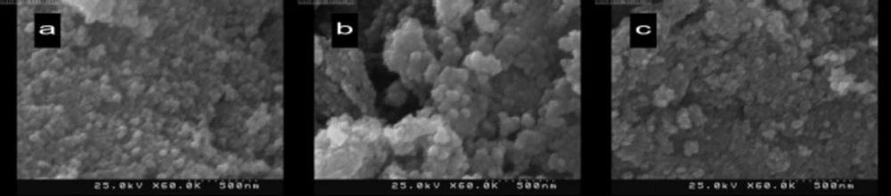
. SEM Analysis
The agglomerate sizes of the MNPs are determined by a scanning electron microscope (SEM) (KYKY EM - 3200) shown in
Figure 9. The average size of the agglomerated MNPs (a), MNPs-COOH (b), and MNPs-COOH-CUR (c) were 55, 71, and 77 nm, respectively. The increasing trend of agglomerate sizes of samples from 55 to 77 nm asserts the conjugation of Curcumin to functionalized MNPs.
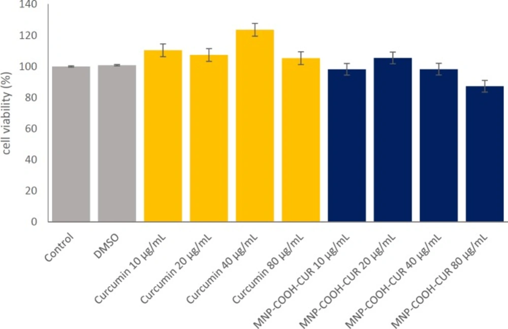
Cell viability test of nanoparticles
The anticancer cytotoxic activities of MNPs were evaluated by cell viability. It was previously reported that Iron Oxide nanoparticles have no significant cytotoxicity up to 100 μg/mL (
48,
49). The cytotoxicity assay carried out with MDA-MB-231 cells at different concentrations of Curcumin in solution or Curcumin was loaded in MNPs for a day by MTT assay and the results are shown in
Figure 10. For the Curcumin in concentrations below 80 μg/mL the cell viability increased showing the cell protective property of Curcumin, which has been confirmed in literature (
50). It found that there is no significant difference in cell viability of the MNP-COOH-CUR in the cells treated with a concentration of 40 μg/mL compared to the cells treated without nanoparticles. At a concentration of 80 μg/mL MNP-COOH-CUR, the cell viability decreased and the anticancer ability was shown. However, the comparative cytotoxicity activity between Curcumin and MNP-COOH-CUR demonstrates better therapeutic efficacy of MNP-COOH-CUR than that of Curcumin, validating the effectiveness of MNP-COOH-CUR. The authors propose that the MNP-COOH-CUR will demonstrate its anticancer properties by delivering CUR more efficiently to the cancer cells. The control cancer cells were not affected at ultrastructural level and did not show any changes in cellular integrity.
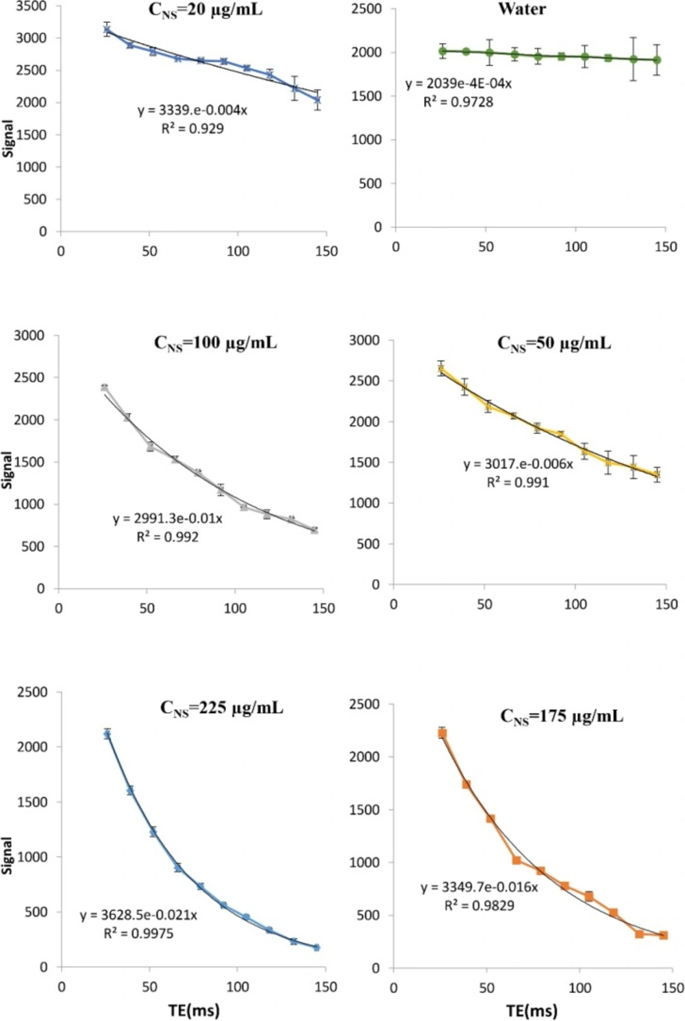
In-vitro MRI test
Engineered MNPs have the potential for simultaneous diagnosis and therapy in one formulation, unlike traditional contrast agents or drugs (
30). We have evaluated our iron oxide formulations (MNP-COOH-CUR) for
in-vitro MRI agent characteristics as a contrast agent and shown the effects of MNP on the relaxation time of environment. The traverse relaxation signal intensities (T2) of the pure water and water with concentration of 20, 50, 100, 175, and 225 mg/mL of MNPs versus TE (echo time) are shown in
Figure 12. Signal intensity is dependent on echo time (TE) and formulated by the following equation.
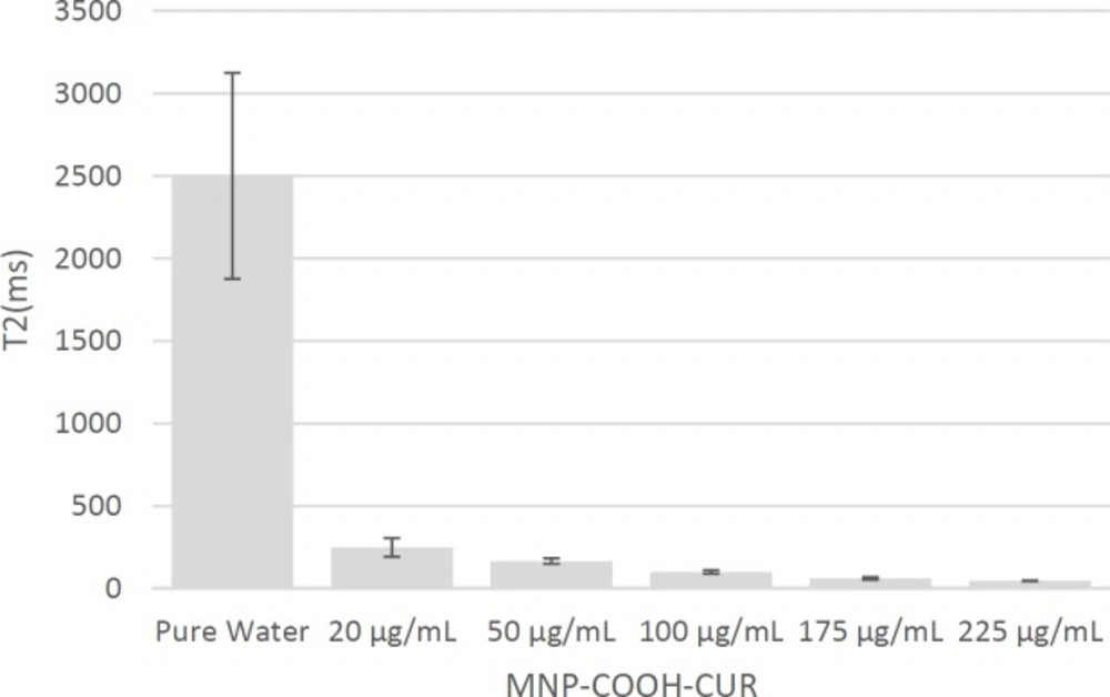
Using this function and regression with respect to the data, T2 values obtained for different concentrations, are shown in
Table 4 and
Figure 11. These values show that the presence of MNPs led to a sharp reduction in the time T2 and an increased concentration of MNPs exaggerateds this effect.