Chemical compounds identification
By using step fractionation, two pale yellowish amorphous powders were isolated from
A. gigantea aerial parts extract. Molecular weights of these compounds (compound
1 and compound
2) according to the exact mass analysis were determined 300.26, and 344.31 g/mol, respectively. Based on primary prediction from the
1H and
13C NMR (
Table 1) data, it was estimated that these compounds might have flavonoid backbone. For compound
1, the
1H NMR spectrum showed the presence of four substitutions on A and C ring of flavonoid core structure. Based on the
13C NMR data, due to the presence of five aromatic protons at δ 6.20 (H-6, 1H, d, J = 2 Hz), 6.62 (H-8, 1H, s), 7.48 (H-2’, 1H, d, J = 2 Hz), 6.94 (H-5’, 1H, d, J = 8.5 Hz), 7.52 (H-6’, 1H, dd, J = 8.5, 2 Hz), protons pic at δ 6.46 (H-3, 1H, d, J = 2 Hz) and singlet in δ 3.95, a mono methoxylated flavonoid structure was considered for compound
1. The analysis of HMBC data of this compound showed the following correlations: (
a) H-8 with C-6, C-7, C-9, C-10 in A ring, (
b) H-3 with C-2, C-4, (
c) H-2’ with C-3’, C-4’, C-2, and C-6’ in B ring, (
d) H-5’ with C- 3’, C-4’, and C-1’ in B ring, and © C-5 of methoxyl group with H-5 in A ring. In addition to the pervious NMR informations, NOSEY and TOCSY data were reveled following correlation in compound 1 chemical structure: (from NOSEY spectrum) (a) H-6 with H-8, and vice versa, H-8 with H-6, (b) H-5’ with H-6’ and H-2’, (c) H-2’ with H-5’. (From TOCSY spectrum) (a) H-3 with H-2’, (b) H-6’ with H-3, and H-5’ (
Table 1).
In addition to these finding, by using the correlation between various carbons and protons in structure with TOCSY, COSY, DEPT, HSQC, and NOSEY techniques compound
1 structure was clarified as luteolin 5-methyl ether (supporting information was uploaded as a supplementary file) (
29).
For compound
2, the
1H and
13C NMR showed similar pattern like the compound
1 (Table 1). Some important chemical shifts suggesting a flavone backbone are δ 154.4, 133.7, 95, 117.3, 122.4, and 152.8 ppm in
13C NMR. The proton chemical shifts are 6.67 (H-3, 1H, s), 6.58 (H-8, 1H, s), 7.49 (H-2’, 1H, d, J = 2 Hz), 7.11 (H-5’, 1H, d, J = 8.0 Hz), and 7.63 (H-6’, 1H, dd, J = 8.5, 2 Hz) in
1H NMR. Three singlets at δ 3.92, 3.91, and 3.87 represents the methoxy group in the structure. In addition to 1D NMR data, 2D NMR data confirmed the predicted three methoxylated flavone structure for compound
2. The analysis of HMBC data of compound
2 showed the following correlations: (
a) H-8 with C-10, C-6, C-9, C-7, C-4 in A ring, (
b) H-3 with C-1’, C-2, C-10, C-4, (
c) H-2’ with C-3’, C-4’, C-2, and C-6’ in B ring, (
d) H-5’ with C- 3’, C-4’, and C-6’ in B ring, © H-6, H-7, and H-3’ of methoxyl groups with C-6, C-7, and C-2, respectively, in A ring (
Table 1). For compound
2, NOSEY and TOCSY spectrum showed very similar pattern to the compound
1. Following correlations were identified for this compound: (from NOSEY spectrum) (
a) H-3 with H-6’, (
b) H-2’ with H-6’, (
c) H-5’ with H-6’, and (
d) H-6’ with H-2’, and H-5’. (From TOCSY spectrum) (
a) H-2’ with H-5’, and H-6’ (
b) H-6’ with H-2’, (
c) H-5’ with H-2’. Based on these information in addition to the TOCSY, COSY, HSQC, and NOSEY data (
Table 1 and the supporting information uploaded as a supplementary file compound 2 was identified as cirsilineol (
30-
33)
.In addition to these flavonoids, chemical compositions of the volatile oils of
A. gigantea aerial parts and seed were identified. According to the GC-MS data the main constitutions of these volatile oils belong to the simple phenolic category which include coniferyl alcohol (18.80%) and eugenol (12.19%) in aerial parts and seeds, respectively (
Table 2).
Molecular modelling results
In this study, we calculated the free binding energy of luteolin 5-methyl ether (
1) and cirsilineol (
2) to four Homo sapiens AhR which are listed in
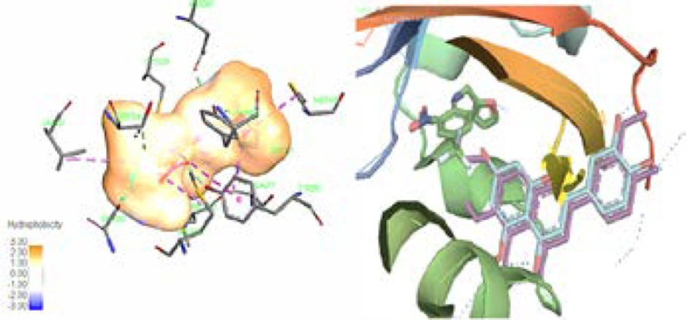
Table 3. The better sub form of enzyme was chosen based on the matching of our compounds with active box for future analysis. Computer aided analysis indicated that besides the importance of the His248 and Tyr307 amino residues of 3H82 active box for creating hydrogen bond for its ligand (020), hydrophobic environment which include Phe244, Phe254, Phe280, Tyr307, Met309, and Leu319 amino acids side chains play notable role in interaction between ligand and enzyme (
34). It is evident that like other specific ligands of this enzyme, luteolin 5-methyl ether (
1) and cirsilineol (
2) have also shown direct hydrophobic interactions with this hydrophobic box residues, which mostly involve the “A & C-ring” of these flavonoids (
35). Although Tyr281 could show the π-π interaction with aromatic “C-ring” of these compounds, it could also act as important residues in active box especially in the presence of “C-ring hydroxyl groups” of the flavonoids (
Figure 2) (
36). Our finding showed that in this series of enzyme, the backbone of chromen-4-one is probably surrounded by the hydrophobic environment. In contrast, “B-ring” of the flavonoids could also play an important role in hydrophilic interaction especially by His248 and Tyr307 in active box of 3H82 (
37). Based on previous finding, the flavonoids or their derivatives with the 7- and 8-carbons in C-ring, 1-pyran and a B-ring, have shown affinity for the hydrophobic pocket of AhR which confirmed our
in-silico finding (
Figure 2) (
36).
Working on the cytotoxicity profiles characterization of the methoxylated and other lipophilic flavonoids on carcinogenic cells led to the identification of some structure-activity relationships (SAR) of these compounds (
34,
35). Based on SAR studies, hydroxyl substituents at C-3′ and C-5 and methoxyl groups at C-4′ have the most effect on this potency in flavones (
38). Recent laboratory studies have demonstrated the important role of AhR/CYP systems in cancer initiation, resistance, and progression (
37,
39). As AhR/CYP chemical inhibitors, these classes of compounds have shown high activity in some
in-vitro assay(
40). AhR receptors are ligand-activated factors which regulate various cell functions such as differentiation, proliferation as well as reproduction(
41). AhR activation by different ligand, such as benzo[a]pyrene (BaP) or 7, 12-dimethybenz[a]anthracene (DMBA), could initiate this activated transcriptional factor for regulating the number of genes such as the cytochrome P450 1B1 (CYP1B1) and CYP1A1(
42). Induction of these cytochromes is a well-known mechanism for the development of epoxide and diol-epoxide carcinogenic intermediates which finally form DNA adducts and triggered tumor initiation (
43). Therefore, inactivation of AhR/ cytochrome system using the chemical inhibitor could be considered as a valuable point for cancer chemoprevention and chemotherapy development (
31,
44).
In summary, molecular models showed that AhR-flavones could be considered as the suitable target for more studies for finding lead structures with good anticancer potential. Based on molecular molding, it could be predicted that adding the hydrophobic group at B-ring of flavones structures creates more tendency for the AhR active box. In contrast, adding the polar group to B-ring of these structures often caused decrease in the tendency for the AhR active box. Methylation of the 7-OH, and 3’ -OH in flavone backbone (in cirsilineol) possibility amplified interaction between compound and AhR active box compared to the less methoxylated flavone, luteolin 5-methyl ether. However, according to the pervious investigation methylation of 6-OH in A ring of flavone backbone could decrease tendency for active box but in the present investigation this structural change in flavone backbone didn’t attenuate the interaction with active box.
Overall, according to the
in silico result in the present study, methoxylated flavones isolated from
A. gigantea are able to interact with AhR and probably play agonistic, antagonistic or even neutral effect for cancer cells (
Table 3 &
Figure 2).
MTT assay results
The widespread of the flavonoids class of chemical compounds in plants and their useful potential in cancer prevention have attracted huge attention to the screening of their anticancer potential. Although many of these class of compounds don’t have high toxicity against normal cells, numerous investigations showed their cytotoxic potential against various carcinogenic cell lines (
33). These anticancer potentials of the various flavonoids have attracted huge attention for finding natural or synthetic flavonoids related compounds as lead anticancer compounds.
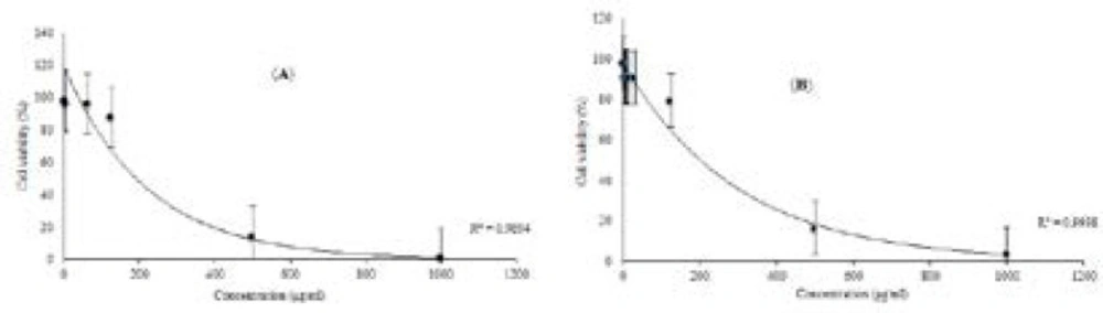
Therefore, in the following
in-silico assay, cytotoxic potential of luteolin 5-methyl ether and cirsilineol was tested against the 4T1 breast carcinoma cell line which is well known for AhR oncogenic functions in carcinogenicity(
45). The cytotoxicity and antiproliferative activity of luteolin 5-methyl ether (1) and cirsilineol (2) against 4T1 cell are shown in
Figure 3. At concentration of 500 μg/mL, both compounds showed cytotoxic effect against 4T1 cell. The IC
50 values on cytotoxicity were 428.24 ±3.21 and 412.7±3.02 μg/mL for luteolin 5-methyl ether and cirsilineol, respectively. Thus, it can be concluded that these flavonoids did not exhibit significant cytotoxicity against 4T1 breast carcinoma cell line.
In many cases, results from
in-silico experiments cannot be directly extrapolated to
in-vitro effects. This opposed
in-vitro results could be explained by previous investigation outputs, many of the flavone based structures such as 6, 2’, 4’-trimethoxyflavone could exhibit significant antagonistic effect on AhR activity but other related compounds of this class such as β-naphthoflavone, quercetin, kaempferol, diosmin, and diosmetin exhibited agonistic activity for AhR (
46,
47).
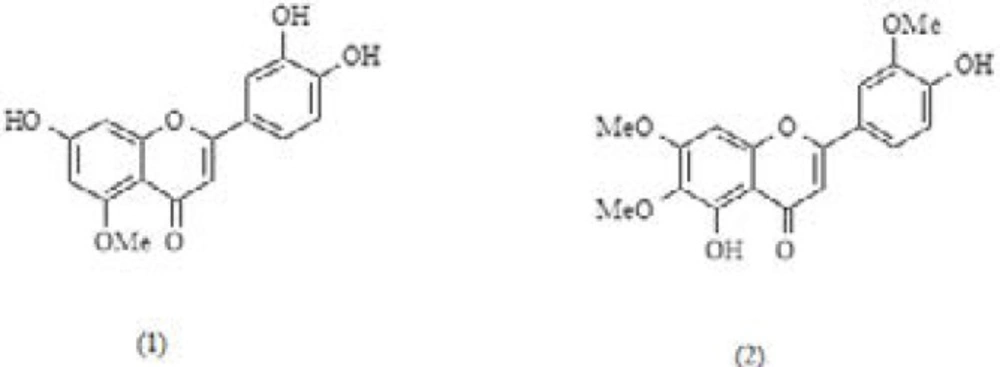
Luteolin 5-methyl ether (1) and cirsilineol (2) chemical structures isolated from A. gigantea aerial parts
(Leaft) The ligand (020) in center of the binding pocket of 3H82, amino acids of the active box, and hydrophobicity map of binding pocket. (Right) The same view of binding pocket of 3H82 with ligand (020), luteolin 5-methyl ether, and cirsilineol. Chromen-4-one and C-ring of flavones clearly surrounded by the hydrophobic environment. Dotted line (dark blue) in B-ring showed potential of this part of flavones structures for hydrophilic interaction. All panels are shown in the same orientation. Active site analysis was carried out using Pymol & Discovery studio visualizer software
Effects of luteolin 5-methyl ether (a) and cirsilineol (b) on the viability of 4T1 cell for 24 h, respectively. 4T1 cell was treated with various indicated concentrations of two compounds, separately. MTT assay was used for cell viability determination, this results were expressed as mean±S.D. of three separate experiments. Luteolin 5-methyl ether and cirsilineol were shown very close activity, the IC50 values on cytotoxicity were 428.24 ±3.21 and 412.7±3.02 μg/mL. Significant differences are indicated by **p < 0.01
| Position | 1H NMR | | 13 C NMR** | | TCOSY | | HMBC | | COSY | | NOESY | |
|---|
| δ H mult. (J in Hz) | | | | | | (H→C) | | (H→H) | | | |
|---|
| Com. 1 | Com. 2 | Com. 1 | Com. 2 | Com. 1 | Com. 2 | Com. 1 | Com. 2 | Com. 1 | Com. 2 | Com. 1 | Com. 2 |
|---|
| 1 | - | - | - | - | - | - | - | - | - | - | - | - |
| 2 | - | - | 165.5 | - | - | - | - | - | - | - | - | - |
| 3 | 6.46, d (2) | 6.67, s | 105.8 | - | - | - | 2, 4 | 1’, 2, 4, 10 | - | - | - | 6’ |
| 4 | - | - | 183.4 | 184.9 | - | - | - | - | - | - | - | - |
| 5 | - | - | 159.5 | - | - | - | - | - | - | - | - | - |
| 6 | 6.20, d (2) | - | 100.0 | 133.7 | 8 | - | - | - | - | - | - | - |
| 7 | - | - | 166.1 | 154.4 | - | - | - | - | - | - | - | - |
| 8 | 6.62,s | 6.58, s | 95.1 | 95 | 6 | - | 7, 9, 10, 6 | 10, 6, 9, 7, 4 | - | - | - | - |
| 9 | - | - | 158.3 | 159.4 | - | - | - | - | - | - | - | - |
| 10 | - | - | 103.7 | 105.7 | - | - | - | - | - | - | - | - |
| 1’ | - | - | 124.1 | 124.4 | - | - | - | - | - | - | - | - |
| 2’ | 7.48, d (2) | 7.49, d (2) | 110.3 | 111.6 | 5’ | 6’, 5’ | 3’, 4’, 2, 6’ | 6’, 3’, 4’, 2 | - | 6’ | 3 | 6’ |
| 3’ | - | - | 149.7 | 151.6 | - | - | - | - | - | - | - | - |
| 4’ | - | - | 152.8 | 153.8 | - | - | - | - | - | - | - | - |
| 5’ | 6.94, d (8.5) | 7.11, d (8) | 117.3 | 113.7 | 6’, 2’ | 2’ | 3’, 4’, 1’ | 6’, 3’, 4’ | 6’ | 6’ | - | 6’ |
| 6’ | 7.52, dd (8.5,2) | 7.63, dd (8, 2) | 122.4 | 121.2 | 5’ | 2’ | 4’ | 2’, 4’ | 5’ | 5’, 2’ | 3, 5’ | 5’, 2’ |
| (in 5) OCH3 | 3.95, s | - | 56.3 | - | - | - | 5 | - | - | - | - | - |
| (in 6) OCH3 | - | 3.92, s | - | 56.1 | - | - | - | 7 | - | - | - | - |
| (in 7) OCH3 | - | 3.91, s | - | 56.3 | - | - | - | 6 | - | - | - | - |
| (in 3’) OCH3 | | 3.87 s | - | 61.2 | - | - | - | 2 | - | - | - | - |
| Compound name | Aerial parts | Seed | | |
|---|
| | | | | |
|---|
| | HD (%) c | HD (%) c | RI a | RI b |
|---|
| | | | | |
| 1 | 2-ethyl-1-hexanol | - | 1.1 | 1032 | 1028 |
| 2 | 1-methyl-2-pyrolidinone | 3.7 | 0.8 | 1034 | 1030 |
| 3 | 2-ethenyl-1,3,3-trimethyl-cyclohexane | 0.8 | - | - | 1132 |
| 4 | 2, 3-dihydro-Benzofuran | 5.1 | - | 1226 | 1221 |
| 5 | Ascaridole epoxide | 0.8 | 3.2 | - | 1245 |
| 6 | Thymol | - | 8.1 | 1292 | 1289 |
| 7 | Eugenol | - | 15.1 | 1356 | 1354 |
| 8 | α- Copaene | - | 0.5 | 1377 | 1375 |
| 9 | Chavibetol | 7.1 | - | 1392 | 1387 |
| 10 | 6-methoxy-2-methyl-tetracyclo[5.3.1.0(2,6).0(8.11)undecan-4-ol | - | 0.9 | - | 1389 |
| 11 | β-Cubebene | - | 0.6 | 1390 | 1392 |
| 12 | Vanillin | 8.1 | - | 1405 | 1400 |
| 13 | β- Caryophyllene | - | 5.4 | 1420 | 1421 |
| 14 | 1,4-Dimethoxy-2,3-dimethylbenzene | 3.1 | - | - | 1442 |
| 15 | γ-muurolene | - | 0.6 | 1477 | 1479 |
| 16 | 4-(5,5-dimethyl-1-oxaspiro[2.5]oct-4-yl)3-buten-2-one | 0.5 | - | 1493 | 1489 |
| 17 | Cis-β-Guaiene | - | 0.7 | 1491 | 1490 |
| 18 | β-Ionone | 0.5 | - | 1503 | 1500 |
| 19 | γ-Cadinene | - | 4.6 | 1514 | 1518 |
| 20 | 4a-acetoxy-5,5,8a-trimethyl-octahydrobenzo[b]pyran | - | 0.5 | - | 1521 |
| 21 | 3-(2,2,3,3-tetramethylcyclopropylidenmethylidene)-4-methyl-hexanoic acid | - | 0.6 | - | 1529 |
| 22 | Dihydroactinidiolide | 6.2 | 3.2 | 1532 | 1534 |
| 23 | Trans-4-Propenylsyringol | 0.4 | - | - | 1536 |
| 24 | Spatulenol | - | 0.6 | 1576 | 1578 |
| 25 | 4-(Acetyloxy)-3-methoxy-methylester benzoic acid | 0.7 | - | - | 1660 |
| 26 | Coniferyl alcohol | 18.8 | - | - | 1682 |
| 27 | Acorenone | 0.5 | - | 1693 | 1690 |
| 28 | 4-hydroxy-3,5,5-trimethyl-4-(3-oxo-1-butenyl)-2-cyclohexen-1-one | 0.5 | - | - | 1787 |
| 29 | Dehydrovomifoliol | 4.7 | - | 1796 | 1793 |
| 30 | Decahydro-1,5,5,8a-tetramethyl-[1S-(1α,3β,3aβ,4α,8aα)]-1,4-methanoazulen-3-ol | - | 2.1 | - | 1827 |
| 31 | 2-(2,2,6-trimethyl-7-oxa-bicyclo[4.1.0]hept-1-yl)-propenyl ester acetic acid | 15.1 | - | - | 1832 |
| 32 | Methyl linoleate | - | 6.8 | 2096 | 2092 |
| 33 | Octadecanoic acid | - | 10.4 | 2158 | 2154 |
| 34 | 3',4',5'-O-trimethyltricetin | 1.5 | - | - | 2521 |
| 35 | α-tocopherol | 3.1 | 1.1 | 3111 | 3108 |
| 36 | Stigmasterol | - | 0.8 | 3170 | 3165 |
| 37 | β-Sitosterol | - | 0.5 | 3203 | 3200 |
| | | | | |
| | | | | |
| | 81.2 | 69.8 | | |
| AhR receptor | PDB ligand | | | luteolin 5-methyl | | | cirsilineol | | |
|---|
| EFEB (kcal/mol) | rmsd l.b. | rmsd u.b. | EFEB (kcal/mol) | rmsd l.b. | rmsd u.b. | EFEB (kcal/mol) | rmsd l.b. | rmsd u.b. |
|---|
| 5UFP | -7.0 | 0.000 | 0.000 | -6.3 | 0.880 | 2.030 | -6.8 | 1.436 | 2.132 |
| 5TBM | -10.2 | 0.000 | 0.000 | -6.3 | 0.988 | 2.082 | -7.5 | 1.150 | 2.570 |
| 4XT2 | -9.0 | 0.000 | 0.000 | -6.7 | 1.018 | 2.112 | -7.3 | 1.133 | 2.422 |
| 3H82 | -7.1 | 0.000 | 0.000 | -6.3 | 1.071 | 2.149 | -7.1 | 1.854 | 1.871 |