
1. Background
Hypertension is recognized as one of the most common health problems worldwide and is a major contributor to cardiovascular disease-related morbidity and mortality (1). Prolonged elevation of blood pressure subjects the myocardium to sustained mechanical and biochemical stress, which can ultimately lead to hypertensive heart disease and the development of heart failure (2, 3). Key characteristics of hypertensive heart disease include increased oxidative stress, mitochondrial dysfunction, inflammation, and apoptosis, which can ultimately lead to cardiomyocyte death (4, 5).
Among the core mediators of cardiac damage in hypertension, Ang II triggers a series of noxious events, including increased ROS production, proinflammatory cytokine production, and activation of apoptosis. Cellular dysregulation, including mitochondrial depolarization, reduced mitochondrial membrane potential (ΔΨm), and activation of the death-associated proteins BAX and caspase-3, has been commonly reported. In addition to these classical markers of Ang II-induced cellular dysregulation, events associated with the execution phase of apoptosis, including PARP cleavage, have also been reported. PARP cleavage is a robust quantitative marker of cellular stress.
Sirtuins are members of the NAD+-dependent histone deacetylase enzyme family and are important in modulating homeostasis and stress responses in the cardiovascular system (6). SIRT1 has emerged as a key modulator of oxidative, proinflammatory, and mitochondrial responses (7, 8). SIRT1 exerts protective effects against apoptosis, enhances antioxidant defenses, and maintains mitochondrial health by deacetylating downstream targets p53, FOXO3, and NF-κB. FOXO3 function is closely associated with its subcellular distribution, as nuclear availability is required for transcriptional regulation of antioxidant and stress-adaptive programs; therefore, quantification of FOXO3 nuclear versus cytoplasmic localization provides mechanistic insight into the state of stress responsiveness (9-13).
Previous studies, including Hori et al. (14), showed that pharmacological modulation of SIRT1 regulates FOXO and p53 activity under oxidative stress; however, these studies were largely conducted in non-cardiac or acute stress models and did not address Ang II-mediated hypertensive pathology. This study investigated the effects of SIRT1 inhibition under hypertensive stress, an aspect that remains incompletely understood. Given the broad evidence that SIRT1 activation provides cardioprotection, its pharmacological blockade may disrupt redox balance and impair mitochondrial stability, thereby increasing the magnitude of inflammatory and apoptotic cascades.
2. Objectives
Therefore, this study used EX-527, a selective SIRT1 inhibitor, to examine its effects on Ang II–induced myocardial injury in H9c2 cardiomyoblasts, AC16 cardiomyocytes, and cardiac fibroblasts (CFB). By assessing cell viability, ROS generation, mitochondrial membrane potential, apoptosis, and inflammatory gene expression, together with changes in the SIRT1/p53/FOXO3a signaling axis, including p53 acetylation and FOXO3 nuclear availability as determined by nuclear–cytoplasmic fractionation, this study aimed to elucidate the mechanistic consequences of SIRT1 inhibition and clarify its potential risk in hypertensive myocardial injury.
3. Methods
3.1. Reagents and Antibodies
Angiotensin II (Ang II) and EX-527 (a selective SIRT1 inhibitor) were purchased from Sigma-Aldrich (St. Louis, MO, USA). The 5,5′,6,6′-tetrachloro-1,1′,3,3′-tetraethylbenzimidazolylcarbocyanine iodide (JC-1) mitochondrial membrane potential assay kit and the 2′,7′-dichlorodihydrofluorescein diacetate (DCFH-DA) probe for ROS detection were obtained from Beyotime Biotechnology (Shanghai, China). DAPI, primary antibodies against SIRT1, p53, acetyl-p53, FOXO3a, PARP and cleaved PARP (c-PARP), BAX, Bcl-2, and cleaved caspase-3, as well as HRP- or Alexa Fluor-conjugated secondary antibodies, were procured from Cell Signaling Technology (Danvers, MA, USA) or Abcam (Cambridge, UK). Antibodies used for nuclear and cytoplasmic fraction validation, including Lamin B1 (nuclear marker) and GAPDH or β-tubulin (cytoplasmic marker), were used to assess fraction purity. All reagents were freshly prepared and diluted in sterile PBS or culture media immediately before use to minimize degradation.
3.2. Cell Culture
Rat cardiomyoblasts (H9c2), human cardiomyocytes (AC16), and primary cardiac fibroblasts (CFB) were cultured in DMEM supplemented with 10% fetal bovine serum (FBS), 100 U/mL penicillin, and 100 µg/mL streptomycin at 37 °C in a humidified 5% CO_2 incubator (15). Cells were maintained below passage 15 to preserve phenotypic stability. The culture medium was replaced every 48 h, and cells were routinely screened for mycoplasma contamination using PCR-based detection.
H9c2 and CFB cells were used as the primary experimental models for viability, apoptosis, and inflammatory gene-expression analyses to capture responses in both cardiomyocyte-like and fibroblast compartments relevant to hypertensive remodeling. AC16 human cardiomyocytes were included specifically to validate oxidative stress and mitochondrial membrane potential phenotypes in a human cardiac cell context. However, a full, endpoint-matched comparison across all three cell types was not performed in the present study. Accordingly, apoptosis, inflammatory transcriptional responses, and p53/FOXO3a mechanistic endpoints were not assessed in AC16 cells, whereas ROS and ΔΨm analyses were not performed in CFB cells. Therefore, the dataset should be interpreted as providing cross-model support for selected phenotypes rather than complete mechanistic concordance across all cellular systems.
3.3. Experimental Design
Cells were seeded in appropriate plates and allowed to reach ~70% confluency before treatment. The study was designed as a four-arm, parallel-group in vitro intervention framework to distinguish the independent and combined effects of Ang II-induced hypertensive stress and pharmacological SIRT1 inhibition. The group structure was defined a priori to enable the following mechanistic contrasts: (i) control vs. Ang II to determine the injury phenotype attributable to hypertensive stimulation; (ii) control vs. EX-527 to determine the basal effect of SIRT1 inhibition in the absence of Ang II; (iii) Ang II vs. Ang II + EX-527 to test whether pharmacological SIRT1 inhibition exacerbates Ang II-induced injury; and (iv) EX-527 vs. Ang II + EX-527 to assess whether Ang II imposes an additional stress burden beyond SIRT1 inhibition alone.
The concentrations of Ang II (1 μM) and EX-527 (10 μM), and the 24 h exposure period, were selected based on prior cell-based literature and preliminary in-house optimization experiments aimed at identifying a treatment window that would produce a consistent injury phenotype while retaining sufficient viable cells for mechanistic assays. Specifically, these conditions were selected because they reproducibly induced measurable changes in viability, oxidative stress, mitochondrial membrane potential, apoptosis-related signaling, and inflammatory transcripts without causing near-complete cell loss that would compromise downstream analyses. Thus, the selected exposure conditions were intended to represent a biologically informative and experimentally stable intervention window rather than a maximal-toxicity setting. However, formal dose–response and time-course experiments were not performed in the present study; therefore, the current design does not determine whether these conditions are optimal, threshold-level, or part of a broader concentration- and time-dependent response profile. The following four experimental groups were established in each applicable assay:
• Control: untreated
• Ang II: 1 μM angiotensin II for 24 h
• EX-527: 10 μM EX-527 for 24 h
• Ang II + EX-527: cotreatment with 1 μM angiotensin II and 10 μM EX-527 for 24 h
All treatments were performed in serum-reduced media (1% FBS) to minimize confounding growth factor effects. Group allocation was based on predefined treatment conditions rather than random assignment, as this was a controlled cell-culture intervention study. The same intervention structure was applied across experiments, although endpoint-specific analyses were performed according to the designated cell model: H9c2 and CFB cells were used for viability, apoptotic, and inflammatory analyses, whereas AC16 cells were used to validate oxidative stress and mitochondrial membrane potential phenotypes in a human cardiomyocyte context. Each treatment condition within each assay was evaluated using three independent biological experiments (n = 3), with each biological replicate representing a separate cell-culture experiment initiated, treated, and analyzed independently on different occasions. Within each biological experiment, replicate wells or measurements were averaged first to generate a single value per condition before statistical analysis. Unless otherwise specified, this replication framework was applied consistently across the figures presented in the study.
The unit of analysis for all statistical comparisons was the independent biological experiment, not the individual well, field, lane, or technical replicate. Technical replication was used only to improve measurement reliability within a biological experiment and was not treated as an independent n for hypothesis testing.
Assay-specific application of the replication structure was as follows: MTT, ROS, JC-1, qPCR, and densitometric quantification of immunoblot-based endpoints were each derived from three independent biological experiments per treatment group. For experiments involving more than one cell type, the same n = 3 biological replicate structure was maintained separately within each cell model.
Outcome prioritization was defined before analysis. Primary outcomes were selected to capture the principal injury phenotype relevant to the central hypothesis that pharmacological SIRT1 inhibition worsens Ang II-induced cardiac stress; these included viability, ROS generation, and mitochondrial membrane potential. Secondary outcomes were selected to evaluate downstream injury consequences at apoptotic and inflammatory levels, including PARP cleavage, BAX, Bcl-2, cleaved caspase-3, and transcript levels of TNF-α, IL-6, and NF-κB. Exploratory mechanistic outcomes were included to assess pathway-level associations and consisted of SIRT1 expression, p53 acetylation, and FOXO3a subcellular localization.
3.4. Cell Viability Assay
Cell viability was determined using the MTT assay. Cells were seeded at a density of 1 × 10⁴ cells/well in 96-well plates (100 µL medium per well). After treatment, cells in 96-well plates were incubated with MTT solution (0.5 mg/mL) for 4 h at 37 °C. Formazan crystals were dissolved in DMSO, and absorbance was recorded at 570 nm using a microplate reader (BioTek Instruments, USA). Results were expressed as a percentage of control viability (16). Blank wells containing only medium and MTT were used to account for background readings. These treatment sets were selected to ensure robust oxidative, mitochondrial, apoptotic, and inflammatory processes within a single comparative window across different assays.
3.5. Apoptotic Injury Quantification by PARP Cleavage Index
To evaluate apoptosis during the execution phase, PARP cleavage was determined in H9c2 and CFB cells treated with different agents using Western blotting. Band intensities of total PARP and cleaved PARP (c-PARP) were quantified and normalized to GAPDH (or β-actin). The PARP cleavage index for each set of treated samples was calculated by dividing c-PARP by total PARP and normalizing to the control samples.
3.6. Intracellular Reactive Oxygen Species Measurement
Intracellular ROS levels were measured using DCFH-DA fluorescence. Measurements were performed according to the standard protocol (17). Cells were seeded at a density of 1 × 10⁵/well in 24-well plates with coverslips and treated with the drugs. Cells were then incubated with 10 μM DCFH-DA at 37 °C for 30 minutes. After washing with PBS, cells were imaged by fluorescence microscopy. Fluorescence was quantified using ImageJ software.
3.7. Mitochondrial Membrane Potential
To evaluate ΔΨm according to standard procedures (18, 19), JC-1 dye was used. Cells were plated at a density of 1 × 10⁵ per well in 24-well plates, incubated with the dye at a concentration of 5 μg/mL, and analyzed after 30 minutes at 37 °C. Red fluorescence indicated polarized mitochondria, whereas green fluorescence indicated depolarized mitochondria.
3.8. Western Blotting
Cells were lysed in RIPA buffer supplemented with protease and phosphatase inhibitors. For each condition, protein was extracted from approximately 1 × 10⁶ cells. Protein concentrations were quantified, and 30 μg of protein was loaded onto SDS-PAGE gels and transferred to PVDF membranes. Membranes were blocked in 5% BSA and incubated overnight at 4 °C with primary antibodies against SIRT1, total p53, acetyl-p53, FOXO3a, PARP and cleaved PARP (c-PARP), BAX, Bcl-2, and cleaved caspase-3. After washing, HRP-conjugated secondary antibodies were applied for 1 h at room temperature. Bands were detected using enhanced chemiluminescence (Bio-Rad) and analyzed densitometrically using ImageJ (16).
3.9. Nuclear-Cytoplasmic Fractionation and FOXO3a Localization
To quantify FOXO3a subcellular distribution, nuclear and cytoplasmic fractions were prepared after treatment using a standard fractionation protocol. Briefly, cells were harvested, washed in ice-cold PBS, and lysed in hypotonic buffer to release cytoplasmic contents, followed by centrifugation to separate cytoplasmic supernatants from nuclear pellets. Nuclear pellets were resuspended in nuclear extraction buffer, and protein concentrations were determined for each fraction. Equal amounts of nuclear and cytoplasmic proteins were resolved by SDS-PAGE and immunoblotted for total FOXO3a. Lamin B1 and GAPDH (or β-tubulin) were used to confirm nuclear and cytoplasmic fraction purity, respectively. FOXO3a nuclear enrichment was quantified as the nuclear/cytoplasmic FOXO3a ratio based on densitometry.
3.10. Real-Time Quantitative Polymerase Chain Reaction
Total RNA was isolated using TRIzol reagent (Invitrogen). RNA concentration and purity were confirmed (A_260/A_280 = 1.8 - 2.0) using a NanoDrop spectrophotometer. Reverse transcription was performed using 1 μg RNA with a commercial cDNA synthesis kit (Thermo Fisher). qPCR was conducted using SYBR Green master mix on a QuantStudio 5 system (Applied Biosystems). Primer sequences for TNF-α, IL-6, NF-κB, and GAPDH (internal control) were designed and validated using Primer-BLAST. The 2-ΔΔCt method was used to calculate relative mRNA expression levels. Inflammatory readouts in the present study were restricted to transcript-level assessment of TNF-α, IL-6, and NF-κB-related signaling markers by qPCR. No protein-level quantification of TNF-α or IL-6 and no direct assessment of NF-κB activation status, such as p65 phosphorylation or nuclear translocation, were performed. Therefore, these measurements were interpreted as indicators of a proinflammatory transcriptional response rather than definitive evidence of cytokine protein production or pathway activation.
(20). For qPCR, RNA was isolated independently from each biological replicate experiment (n = 3 per group). Each cDNA sample was analyzed in triplicate technical PCR reactions, and the mean Ct value from these technical replicates was used to calculate a single expression value for that biological replicate. Statistical analysis was then performed on the resulting biological replicate values rather than on individual technical-replicate Ct measurements.
3.11. Statistical Analysis
Data are presented as mean ± standard deviation (SD) from three independent biological experiments unless otherwise indicated. The statistical unit of analysis was the independent biological replicate for each treatment condition within each assay. For assays involving technical replicates, these were averaged within each biological replicate before inferential analysis.
Group comparisons within each assay were performed using one-way ANOVA followed by Tukey’s post hoc multiple-comparison test to compare the control, Ang II, EX-527, and Ang II + EX-527 conditions. Before parametric testing, data distribution and variance assumptions were evaluated using standard diagnostic assessments of residual normality and homogeneity of variance. Given the relatively small biological sample size, these assumption checks were interpreted cautiously in conjunction with data distribution patterns.
No data points were excluded as outliers post hoc. All biologically independent observations generated under the predefined experimental conditions were included in the final analysis.
Because the study involved multiple mechanistically related endpoints, statistical analyses were interpreted on an assay-by-assay basis rather than within a single pooled hypothesis-testing framework. Tukey’s post hoc procedure was used to control for multiple pairwise comparisons within each individual assay. However, no additional global multiplicity correction across all endpoints was applied; therefore, mechanistic findings should be interpreted with appropriate caution as part of an integrated experimental framework rather than as isolated confirmatory tests.
A P value < 0.05 was considered statistically significant. Graphs and statistical analyses were performed using GraphPad Prism 9.0 software.
4. Results
4.1. SIRT1 Inhibition Exacerbates Ang II-Induced Cytotoxicity and Apoptotic Injury in Cardiac Cells
To investigate the role of SIRT1 in hypertension-induced myocardial injury, we evaluated the effects of pharmacological SIRT1 inhibition on cardiomyocyte and cardiac fibroblast viability under Ang II-induced stress. Two cell models were selected for these functional studies: rat cardiomyoblasts (H9c2) and cardiac fibroblasts (CFB), which are targets of hypertensive remodeling. Cells were exposed to Ang II at 1 μM for 24 h with or without the SIRT1 inhibitor EX-527 at 10 μM. Cell viability was measured using the MTT assay (Figure 1A), and apoptotic damage was assessed by the cleavage index of total PARP: c-PARP/total PARP (Figure 1B).
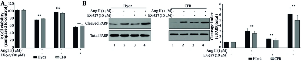
Figure 1.
SIRT1 inhibition exacerbates Ang II–induced cytotoxicity and apoptotic injury in H9c2 cardiomyoblasts and cardiac fibroblasts (CFB). Cells were assigned to four treatment groups: control, Ang II (1 μM, 24 h), EX-527 (10 μM, 24 h), and Ang II + EX-527 (cotreatment for 24 h). (A) Cell viability was assessed by MTT assay in H9c2 and CFB cells. Ang II significantly reduced viability relative to control, and cotreatment with EX-527 further decreased viability. (B) Apoptotic injury was evaluated by Western blot–based quantification of PARP cleavage and expressed as the cleaved PARP/total PARP (c-PARP/total PARP) cleavage index in H9c2 and CFB cells. Ang II increased PARP cleavage, and this effect was further amplified by EX-527 cotreatment. Data are presented as mean ± SD from three independent biological experiments (n = 3). Statistical analysis was performed by one-way ANOVA followed by Tukey’s post hoc test. A P value < 0.05 was considered statistically significant. Abbreviations: Ang II, angiotensin II; EX-527, selective SIRT1 inhibitor; PARP, poly(ADP-ribose) polymerase; c-PARP, cleaved PARP; CFB, cardiac fibroblasts.
In both cell types, Ang II alone induced a significant reduction in cell viability compared with the control group (P < 0.05). Coadministration of EX-527 with Ang II further reduced cell viability compared with Ang II alone (P < 0.05). EX-527 alone caused a mild reduction in cell viability, which was less pronounced than that induced by Ang II alone or by Ang II combined with EX-527 (Figure 1A).
Next, we analyzed PARP cleavage as an execution-phase marker of cell damage (Figure 1B). In both H9c2 and CFB cells, Ang II increased the PARP cleavage index (c-PARP/total PARP) compared with the control group. Notably, EX-527 markedly amplified Ang II-induced PARP cleavage, yielding the highest cleavage index in the Ang II + EX-527 group, whereas EX-527 alone produced only a modest elevation above baseline (Figure 1B).
4.2. SIRT1 Inhibition Enhances Ang II-Induced Oxidative Stress and Mitochondrial Dysfunction
To examine the role of SIRT1 in regulating oxidative stress under hypertensive conditions, we assessed intracellular ROS generation and mitochondrial membrane potential in cardiac cells treated with Ang II (1 μM) with or without EX-527 (10 μM). H9c2 rat cardiomyoblasts and AC16 human cardiomyocytes were used to validate these findings across cardiac cell models. ROS generation was evaluated using DCFH-DA fluorescence, and mitochondrial membrane potential was evaluated using JC-1 fluorescence. As shown in Figure 2, DCFH-DA fluorescence indicated increased intracellular ROS generation in Ang II-treated H9c2 and AC16 cells compared with controls (P < 0.05) (Figure 2A). In cells cotreated with EX-527, ROS generation was further elevated compared with Ang II treatment alone (P < 0.05). EX-527 alone also increased ROS generation. Confocal microscopy showed diffuse DCFH-DA fluorescence, indicating ROS accumulation (Figure 2B). Control cells showed minimal green fluorescence with well-defined nuclear outlines (Figure 2B1). Ang II-treated cells showed increased cytoplasmic green fluorescence, consistent with increased ROS levels (Figure 2B2). EX-527 alone produced moderate fluorescence (Figure 2B3). Ang II + EX-527-treated cells showed the highest level of green fluorescence, which was sufficiently diffuse that nuclear outlines were no longer clearly discernible, confirming marked oxidative damage (Figure 2B4).
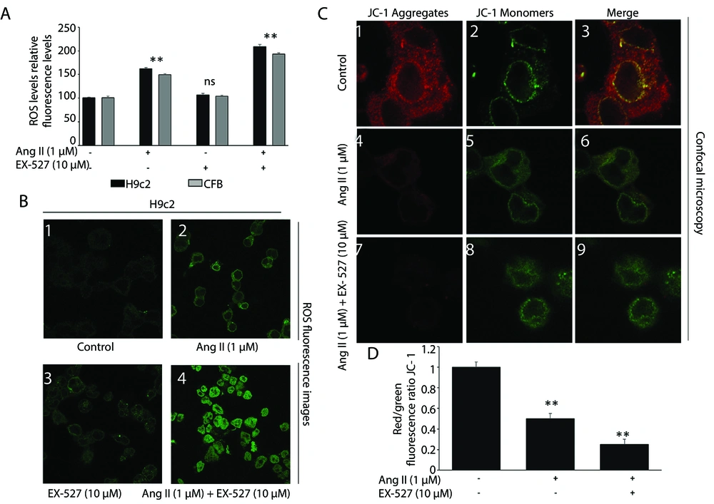
Figure 2.
SIRT1 inhibition enhances oxidative stress and mitochondrial depolarization in H9c2 and AC16 cardiomyocytes. Cells were assigned to four treatment groups: control, Ang II (1 μM, 24 h), EX-527 (10 μM, 24 h), and Ang II + EX-527 (cotreatment for 24 h). A, Intracellular reactive oxygen species (ROS) levels were assessed by DCFH-DA fluorescence in H9c2 and AC16 cells. Ang II increased ROS accumulation relative to control, and cotreatment with EX-527 further enhanced this response. B, Representative fluorescence images of DCFH-DA staining showing intracellular ROS accumulation under the indicated treatment conditions. C, Mitochondrial membrane potential (ΔΨm) was assessed by JC-1 staining. Loss of red fluorescence and predominance of green fluorescence indicate mitochondrial depolarization. (D) Quantitative analysis of the JC-1 red/green fluorescence ratio confirmed a significant decline in ΔΨm after Ang II treatment, with further reduction in the Ang II + EX-527 group. Data are presented as mean ± SD from three independent biological experiments (n = 3). Statistical analysis was performed by one-way ANOVA followed by Tukey’s post hoc test. A P value < 0.05 was considered statistically significant. Abbreviations: ROS, reactive oxygen species; DCFH-DA, 2′,7′-dichlorofluorescein diacetate; JC-1, cationic mitochondrial membrane potential dye; ΔΨm, mitochondrial membrane potential (** P < 0.05)
Significant differences in mitochondrial membrane potential (ΔΨm) were also observed by JC-1 staining, supporting a role for SIRT1 in maintaining mitochondrial integrity under hypertensive stress (Figure 2C). In control cells, JC-1 dye was mainly aggregated in polarized mitochondria and displayed strong red fluorescence throughout the cytoplasm (Figure 2C1-3), indicating preserved mitochondrial polarization. In Ang II-treated cells, red fluorescence decreased and green fluorescence increased, reflecting JC-1 monomers and mitochondrial membrane depolarization (Figure 2C4-6). When cells were treated with Ang II and EX-527, depolarization was further enhanced (Figure 2C7-9). This cell population showed intense green fluorescence with very low or undetectable red fluorescence and extensive fragmentation or diffuse patterns in the mitochondrial network. The red/green fluorescence ratio declined in Ang II-exposed cells (P < 0.05) and decreased further in cells exposed to Ang II and EX-527 (P < 0.05 vs Ang II alone) (Figure 2D). These findings underscore the combined effect of SIRT1 inhibition and Ang II in disrupting mitochondrial integrity. Collectively, the JC-1 data support the hypothesis that SIRT1 has a protective function in cardiac mitochondria and that its inhibition aggravates mitochondrial dysfunction under Ang II-induced hypertensive conditions.
4.3. SIRT1 Inhibition Intensifies Ang II-Induced Apoptotic Signaling and Proinflammatory Gene Expression
To evaluate whether SIRT1 inhibition affects apoptotic and inflammatory pathways involved in hypertensive myocardial injury, proapoptotic and antiapoptotic protein levels and proinflammatory mRNA responses were assessed in H9c2 and CFB cells treated with Ang II (1 μM), EX-527 (10 μM), or both for 24 h. Western blot analysis of BAX, Bcl-2, and caspase-3 levels, as well as real-time PCR of TNF-α, IL-6, and NF-κB mRNAs, is presented in Figure 3. Western blot analysis confirmed a significant elevation of BAX and caspase-3 levels and a simultaneous reduction in Bcl-2 expression levels after treatment of H9c2 and CFB cells with Ang II compared with the control group (P < 0.01) (Figure 3A, lane 1 vs. lane 2). Notably, cotreatment with EX-527 significantly increased BAX and cleaved caspase-3 levels and further suppressed Bcl-2 expression (Figure 3A: lanes 1 - 3 vs. 4). Cells treated with EX-527 alone showed mildly increased levels of BAX and cleaved caspase-3 and mildly reduced Bcl-2 expression compared with controls, although to a lesser magnitude than that observed with Ang II (Figure 3A: lane 1 vs. lane 3). These findings support the importance of SIRT1 activity in sustaining apoptotic homeostasis and indicate that its suppression increases programmed cell death under hypertension-related stress. Quantitative analysis of TNF-α, IL-6, and NF-κB-related transcripts by qPCR showed significant upregulation relative to controls (P < 0.05; Figure 3B–D), indicating a proinflammatory transcriptional response in H9c2 and CFB cells exposed to Ang II. Cotreatment with EX-527 further increased the mRNA levels of these markers compared with Ang II alone (P < 0.05), suggesting that pharmacological SIRT1 inhibition amplifies inflammatory gene-expression responses under hypertensive stress conditions. However, because these inflammatory mediators were assessed only at the transcript level, the present data do not directly establish corresponding increases in cytokine protein abundance or definitive activation of the NF-κB pathway. Therefore, the inflammatory findings should be interpreted as evidence of enhanced proinflammatory transcriptional signaling rather than conclusive proof of full inflammatory pathway activation. Notably, EX-527 alone provoked a mild, non-significant increase in these markers relative to the control group, underscoring the baseline anti-inflammatory function of SIRT1 even in the absence of hypertensive stimuli. Collectively, these findings demonstrate the dual protective role of SIRT1 in regulating both apoptosis and inflammation in cardiac cells under hypertensive conditions. Its inhibition by EX-527 amplifies Ang II-induced injury by promoting mitochondria-dependent apoptosis and proinflammatory gene activation.
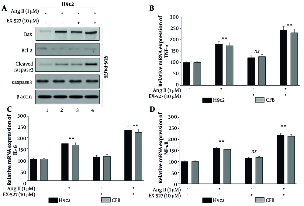
Figure 3.
SIRT1 inhibition intensifies apoptotic and inflammatory signaling in H9c2 cardiomyoblasts and cardiac fibroblasts (CFB). Cells were assigned to four treatment groups: control, Ang II (1 μM, 24 h), EX-527 (10 μM, 24 h), and Ang II + EX-527 (cotreatment for 24 h). A, Apoptosis-related proteins were analyzed by Western blotting in H9c2 and CFB cells. Ang II increased BAX and cleaved caspase-3 and reduced Bcl-2 expression, whereas EX-527 cotreatment further strengthened this proapoptotic profile. B–D, Relative mRNA expression of TNF-α, IL-6, and NF-κB-related transcripts was measured by qPCR in H9c2 and CFB cells. Ang II increased these inflammatory transcriptional markers, and cotreatment with EX-527 further augmented their expression. These data represent transcript-level inflammatory responses and do not directly indicate cytokine protein abundance or definitive NF-κB pathway activation. Data are presented as mean ± SD from three independent biological experiments (n = 3). Statistical analysis was performed by one-way ANOVA followed by Tukey’s post hoc test. A P value < 0.05 was considered statistically significant. Abbreviations: BAX, Bcl-2-associated X protein; Bcl-2, B-cell lymphoma 2; TNF-α, tumor necrosis factor alpha; IL-6, interleukin 6; NF-κB, nuclear factor kappa B; qPCR, quantitative polymerase chain reaction; CFB, cardiac fibroblasts (** P < 0.05).
4.4. SIRT1 Inhibition Modulates p53 Acetylation and Impairs FOXO3a Nuclear Localization Under Hypertensive Stress
To provide mechanistic insight into how SIRT1 suppresses Ang II-driven myocardial injury, we examined the SIRT1-p53-FOXO3a signaling axis. SIRT1, total p53, and acetylated p53 (Ac-p53) were quantified by Western blotting, and FOXO3a subcellular distribution was determined by nuclear-cytoplasmic fractionation followed by immunoblotting of total FOXO3a, with Lamin B1 and GAPDH used as nuclear and cytoplasmic markers, respectively. Western blotting showed that Ang II decreased SIRT1 protein abundance compared with the control group (P < 0.05) (Figure 4A, lane 1 vs lane 2). Correspondingly, total p53 increased and its acetylated form (Ac-p53) was elevated, consistent with induction of the p53 stress pathway under hypertensive stimulation (Figure 4A, lane 1 vs lane 2). This response was further exacerbated in the Ang II + EX-527 group, in which SIRT1 expression was lowest and Ac-p53 was strongly upregulated compared with Ang II alone (P < 0.05) (Figure 4A, lane 2 vs lane 4), supporting the established role of SIRT1 as a negative regulator of p53 acetylation. EX-527 alone produced a modest increase in total and acetylated p53 (Figure 4A, lane 1 vs lane 3), indicating that SIRT1 inhibition is sufficient to perturb p53 regulation even in the absence of Ang II. Collectively, these findings indicate that loss of SIRT1 activity enhances p53 acetylation and strengthens p53-linked stress signaling.
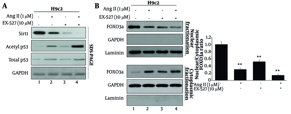
Figure 4.
SIRT1 inhibition augments p53 acetylation and reduces nuclear FOXO3a availability under Ang II stress. Cells were assigned to four treatment groups: control, Ang II (1 μM, 24 h), EX-527 (10 μM, 24 h), and Ang II + EX-527 (cotreatment for 24 h). A, Western blot analysis of SIRT1, total p53, and acetylated p53 (Ac-p53) showing reduced SIRT1 expression and increased p53 acetylation following Ang II treatment, with the strongest Ac-p53 signal observed in the Ang II + EX-527 group. B, Nuclear–cytoplasmic fractionation followed by immunoblotting of FOXO3a showing reduced nuclear FOXO3a and increased cytoplasmic FOXO3a under Ang II stress, with further suppression of nuclear FOXO3a availability after cotreatment with EX-527. Quantification is expressed as the nuclear/cytoplasmic FOXO3a ratio. Lamin B1 and GAPDH were used as nuclear and cytoplasmic markers, respectively. Data are presented as mean ± SD from three independent biological experiments (n = 3). Statistical analysis was performed by one-way ANOVA followed by Tukey’s post hoc test. ** P-value < 0.05 was considered statistically significant. Abbreviations: Ac-p53, acetylated p53; FOXO3a, forkhead box O3a; Ang II, angiotensin II; EX-527, selective SIRT1 inhibitor; GAPDH, glyceraldehyde-3-phosphate dehydrogenase.
Next, we quantified FOXO3a subcellular localization as a downstream effector of SIRT1 modulation (Figure 4B). In control cells, FOXO3a displayed a balanced nucleo-cytoplasmic distribution, whereas Ang II reduced nuclear FOXO3a and increased cytoplasmic FOXO3a, indicating impaired nuclear availability during hypertensive stress. Notably, cotreatment with EX-527 further reduced nuclear FOXO3a enrichment and produced the lowest nuclear-to-cytoplasmic FOXO3a ratio compared with Ang II treatment alone (Figure 4B). Collectively, these findings show that pharmacological SIRT1 inhibition by EX-527 under Ang II stress is accompanied by enhanced p53 acetylation and reduced nuclear FOXO3a, supporting an association between perturbation of the SIRT1–p53–FOXO3a signaling axis and the aggravated injury phenotype observed in this model. However, because the present study relied on pharmacological inhibition alone and did not include genetic or pathway-reconstitution experiments, these data should be interpreted as mechanistically suggestive rather than definitive proof of causal pathway mediation. In addition, canonical FOXO3a transcriptional targets were not measured in the present study; therefore, the functional linkage between altered FOXO3a localization and downstream antioxidant gene output remains inferential.
4.5. Proposed Mechanistic Model of SIRT1 Inhibition in Hypertension-Induced Myocardial Injury
Figure 5 presents a schematic flowchart showing the mechanistic cascade derived from this study, illustrating how SIRT1 inhibition contributes to the exacerbation of Ang II-induced myocardial injury. Under physiological conditions, SIRT1 functions as a central regulatory node that helps maintain cellular integrity through the deacetylation of p53 and the translocation of FOXO3a to the nucleus. This preserves mitochondrial integrity, limits oxidative stress, suppresses apoptosis, and restrains proinflammatory gene expression. However, Ang II induces the downregulation of SIRT1 expression, leading to hyperacetylation of p53, reduced nuclear availability of FOXO3a, increased production of ROS, and decreased mitochondrial membrane potential. These events contribute to an exaggerated apoptotic signaling cascade, including upregulation of BAX and cleaved caspase-3 and suppression of Bcl-2, as well as increased expression of proinflammatory mediators such as TNF-α, IL-6, and NF-κB. Co-exposure to the SIRT1 inhibitor EX-527 significantly enhances all of these changes, inducing overt oxidative damage, mitochondrial dysfunction, and structural injury in cardiomyocytes. This model highlights the role of the SIRT1/p53/FOXO3a pathway in cellular resistance to hypertensive stress, as well as the contribution of pharmacological inhibition to myocardial injury.
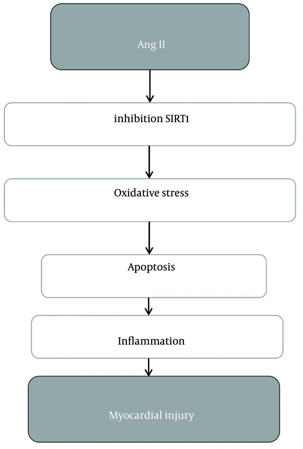
Figure 5.
Proposed mechanistic model of pharmacological SIRT1 inhibition in Ang II–induced myocardial injury. This schematic summarizes the working model derived from the present in vitro findings obtained in H9c2, AC16, and CFB cell systems. Under Ang II stress, reduced SIRT1 activity is associated with increased p53 acetylation, impaired FOXO3a nuclear availability, elevated ROS accumulation, loss of mitochondrial membrane potential (ΔΨm), enhanced apoptotic signaling, and increased proinflammatory transcriptional responses. Co-exposure to EX-527 is proposed to amplify these injury-related changes, culminating in aggravated myocardial injury phenotypes. This figure represents a mechanistic interpretation of the experimental data and should be regarded as a proposed model rather than definitive proof of causal pathway mediation. Abbreviations: Ang II, angiotensin II; EX-527, selective SIRT1 inhibitor; ROS, reactive oxygen species; ΔΨm, mitochondrial membrane potential; Ac-p53, acetylated p53; FOXO3a, forkhead box O3a; CFB, cardiac fibroblasts.
5. Discussion
Given the already substantial global burden of chronic and infectious diseases, hypertension represents an additional major burden on global health (21-24). In this study, pharmacological SIRT1 inhibition with EX-527 aggravated Ang II-induced injury across cardiac cell models, supporting a protective role for basal SIRT1 activity in maintaining redox balance, mitochondrial integrity, and stress-adaptive signaling while limiting apoptotic and inflammatory responses.
One strength of the present study is the inclusion of multiple cardiac-relevant cell systems, which allowed us to examine whether the injury-amplifying effect of SIRT1 inhibition could be observed beyond a single in vitro model. H9c2 cardiomyoblasts and cardiac fibroblasts were used for viability, apoptotic, and inflammatory transcriptional analyses, whereas AC16 human cardiomyocytes were used primarily to validate oxidative stress and mitochondrial membrane potential phenotypes in a human cardiac context. However, because the complete endpoint panel was not examined in parallel across all three models, the present findings should not be interpreted as demonstrating fully matched mechanistic equivalence among H9c2, AC16, and CFB cells. Rather, the data provide model-consistent evidence for selected injury phenotypes, with broader mechanistic generalization requiring more comprehensive side-by-side validation across all cellular systems.
From a functional perspective, our viability assay data suggest that SIRT1 activity buffers Ang II-induced cytotoxic stress. Ang II caused a significant reduction in cell survival, which was further enhanced by SIRT1 inhibition, whereas EX-527 alone had relatively mild effects. This finding aligns with the extensive literature showing that SIRT1 activation or overexpression suppresses pathological hypertrophy, oxidative injury, and cardiomyocyte loss (25-30). For example, cardiac SIRT1 overexpression inhibited hypertrophy and apoptosis in vivo (25). SIRT1 activation by resveratrol improves hypertrophic cardiac performance in hypertension while reducing oxidative injury (26).
Similarly, loss of SIRT1 through genetic deficiency or suppression has been shown to worsen multiple types of myocardial damage, including drug-induced injury and ischemia-reperfusion injury, strengthening the model in which SIRT1 functions as an endogenous defensive node during cardiac stress (28-30). The present study extends these observations by showing that pharmacological inhibition of SIRT1 worsens damage in both cardiomyocytes and fibroblasts in an Ang II model of hypertension.
Oxidative stress, together with mitochondrial dysfunction, is a common early feature of Ang II-initiated cardiovascular damage, and our findings suggest that SIRT1 plays a critical role in controlling this process. We found that Ang II treatment generated substantial ROS formation and mitochondrial membrane depolarization, both of which were magnified by the SIRT1 inhibitor EX-527. These data suggest that SIRT1 suppresses oxidative damage and preserves mitochondrial membrane polarization during hypertensive stress. Previous studies suggest that SIRT1 can modulate oxidative stress through effects on NADPH oxidase-associated pathways and mitochondrial antioxidant regulation. In the present study, however, upstream ROS sources were not directly dissected; therefore, we cannot determine whether the excess ROS observed under Ang II + EX-527 arose predominantly from NADPH oxidases, mitochondria, or combined sources (31-34). In hypertensive models, reduced SIRT1 expression has been associated with exacerbated oxidative damage and worsening mitochondrial structure and function. Conversely, SIRT1 activation improved enzymatic activity within the mitochondrial population while suppressing ROS generation. Our results agree with these perspectives, supporting, at the cellular level, that SIRT1 inhibition during Ang II + EX-527 treatment severely disrupts redox balance. The role of FOXO3a is relevant to the interpretation of our findings. Based on prior literature, SIRT1-dependent FOXO3a activity has been linked to the transcription of antioxidant genes such as MnSOD and catalase. However, these downstream FOXO3 target genes were not measured in the present study; thus, the observed reduction in nuclear FOXO3a under Ang II + EX-527 should be interpreted as consistent with, rather than direct proof of, impaired antioxidant transcriptional output.
Regarding cell-death execution, the present data indicate that SIRT1 counteracts mitochondrial apoptosis under hypertensive stress conditions. Ang II enhanced apoptotic signaling, as indicated by increased BAX, reduced Bcl-2, and enhanced caspase-3 activation. Increased cleavage of poly(ADP-ribose) polymerase further indicated heightened execution-phase cardiomyocyte stress under hypertensive conditions, an effect amplified by EX-527. This finding is mechanistically consistent with previous observations indicating that SIRT1 deacetylates and functionally regulates the tumor suppressor p53 (11). Indeed, previous investigations indicate that EX-527 increases p53 acetylation and apoptosis under oxidative stress conditions (34). Furthermore, SIRT1 activation has been associated with decreased cardiomyocyte loss and blunted fibrotic remodeling under hemodynamic stress, suggesting that the protection observed here in both myocytes and fibroblasts may also be relevant to broader remodeling phenotypes (33, 35). Overall, our assessment of both PARP cleavage and canonical BAX/Bcl-2/caspase-3 profiling reinforces the notion that SIRT1 inhibition increases the likelihood of irreversible apoptotic injury during Ang II stress.
Another important mediator of hypertensive remodeling is inflammation. In the present study, transcriptional analysis showed that Ang II increased the mRNA expression of TNF-α, IL-6, and NF-κB-related markers, and that EX-527 further enhanced these responses. These findings support the interpretation that SIRT1 activity restrains proinflammatory gene-expression programs under hypertensive stress. However, because inflammatory endpoints were measured only at the mRNA level, the data do not directly confirm corresponding cytokine protein production or definitive NF-κB pathway activation. Accordingly, our findings should be interpreted as evidence of enhanced inflammatory transcriptional signaling rather than as complete validation of inflammatory effector activation. This interpretation is nevertheless consistent with prior literature showing that SIRT1 can negatively regulate NF-κB-associated inflammatory responses in stressed cardiac cells (36, 37).
Mechanistically, our pharmacological data implicate the SIRT1/p53/FOXO3a axis as a biologically plausible pathway associated with the Ang II injury response and its exacerbation by EX-527, but they do not establish a fully causal, pathway-level mechanism. Rather, the present findings support the interpretation that worsening injury phenotypes under pharmacological SIRT1 inhibition occur in parallel with increased p53 acetylation and impaired FOXO3a nuclear availability, consistent with disruption of a stress-adaptive signaling axis. Definitive mechanistic assignment will require genetic gain- and loss-of-function strategies, rescue experiments, and pathway reconstitution studies to determine whether modulation of p53 and FOXO3a is necessary and sufficient for the observed injury amplification. Our data clearly show that Ang II decreases SIRT1 protein levels, accompanied by increases in both total and acetylated p53 levels. EX-527 treatment potentiated the accumulation of acetyl-p53, suggesting that SIRT1 may act as a negative regulator of acetylation. These findings on the SIRT1/p53 axis are supported by previous studies reporting that inhibition of SIRT1 increases acetyl-p53 and disrupts the FOXO response, particularly with regard to oxidative stress (14); this is further supported by evidence implicating the SIRT1/FOXO3a pathway in antioxidant defense responses within the cardiomyocyte. Importantly, we quantitatively measured the subcellular localization of FOXO3a, extending beyond the qualitative, impressionistic assessment afforded by immunocytochemistry-based imaging techniques.
From a pharmacological perspective, these findings have two clear implications. First, they reinforce SIRT1 as a protective target in hypertensive cardiac injury, particularly in contexts dominated by Ang II signaling, oxidative stress, and mitochondrial dysfunction. Second, they raise a cautionary note regarding SIRT1 inhibition in cardiovascular settings: while SIRT1 blockade may be explored in other therapeutic domains, our data suggest that it may carry cardiovascular risk in diseases characterized by oxidative stress and inflammatory activation. These implications are especially relevant given the growing interest in sirtuin modulators and NAD+-linked pathways as druggable nodes. Recent work has also emphasized the broader clinical and mechanistic importance of cardiovascular risk, vascular dysfunction, and chronic disease-associated stress responses across diverse pathological contexts (38-42). Recent studies further support the broader relevance of stress-responsive cardioprotective mechanisms by showing that modulation of the SIRT1/p53 axis, regulation of DNA damage- and cell death-related pathways, and pharmacological attenuation of cardiac stress responses can all influence myocardial injury outcomes in experimental and clinical settings (43-45).
Certain limitations should be considered. The study was performed in vitro and therefore does not fully capture the complexities of neurohormonal regulation, hemodynamic load, immune cell contributions, or paracrine interactions between fibroblasts and cardiomyocytes present in the hypertensive heart. Although this study validated important phenotypes in rat and human cardiomyocytes and included fibroblasts as a remodeling-relevant cell type, in vivo studies are required to confirm translational significance. Another limitation is that the intervention design used a single concentration of Ang II, a single concentration of EX-527, and a single exposure duration of 24 h. Although these conditions were selected based on prior literature and preliminary optimization to provide a reproducible and analyzable injury window, the absence of formal dose–response and time-course studies limits interpretation of the pharmacological interaction. Accordingly, we cannot exclude the possibility that the observed effects reflect a condition-specific response window, nor can we determine whether lower or higher concentrations, or shorter or longer exposures, would yield different magnitudes or patterns of injury. Future studies should incorporate concentration-gradient and temporal profiling to define the dynamic range, optimal intervention window, and potential toxicity boundaries of Ang II and EX-527 in these cardiac cell models. Finally, the current study was designed using one Ang II concentration (1 μM), one EX-527 concentration (10 μM), and one time point (24 h). While these conditions were selected to induce robust and reproducible injury phenotypes, no dose-response or time-course analyses were performed. Therefore, future studies should define concentration dependence, more precisely determine the toxicity window, and investigate whether ROS accumulation and mitochondrial depolarization precede the subsequent amplification of apoptotic and inflammatory signaling cascades. Additionally, the inflammatory response was restricted to the transcript levels of TNF-α, IL-6, and NF-κB. Validation at the protein level using ELISA or immunoblotting, and direct measurement of NF-κB activation using p65 phosphorylation or translocation, were not performed and should be considered in future studies. In addition, although reduced nuclear FOXO3a was observed, canonical downstream FOXO3 target genes such as MnSOD, catalase, or other antioxidant/stress response mRNAs were not examined; therefore, FOXO3 transcriptional activity was not functionally verified. In addition, upstream sources of ROS were not functionally elucidated, and it is not possible to distinguish between mitochondrial and NADPH oxidase sources of ROS based on these data. In addition, not all mechanistic endpoints were assessed across all three cell models. H9c2 and CFB cells were used for viability, apoptosis, and inflammatory transcriptional analyses, whereas AC16 cells were used primarily to validate oxidative stress and mitochondrial dysfunction in a human cardiomyocyte context. As a result, the present dataset does not provide a fully parallel, cross-model demonstration of the complete injury phenotype or the proposed SIRT1–p53–FOXO3a-associated mechanism. Therefore, the mechanistic interpretation should be viewed as model-supported rather than universally established across all cardiac cellular systems examined here.
5.1. Conclusions
In conclusion, our findings show that pharmacological SIRT1 inhibition aggravates Ang II-induced redox imbalance, mitochondrial depolarization, apoptotic execution, and inflammatory activation, and that these effects are accompanied by enhanced p53 acetylation and impaired FOXO3a nuclear availability. These data support a pharmacologically defined association between SIRT1 inhibition and aggravated myocardial injury that is consistent with involvement of the p53–FOXO3a axis, but they do not yet provide definitive causal proof of pathway mediation.