


1. Background
2. Objectives
3. Materials and Methods
3.1. Cell Preparation and Labeling
3.2. Cell Phantom
3.3. Animal Experiments
3.4. Direct Implantation into Normal Rat Brains
3.5. Internal Carotid Artery (ICA) Infusion in Normal Rats and Rat Models with Cerebral Infarction
3.6. Phantom MRI
3.7. In Vivo Rat Brain MRI
3.8. MRI Analysis
3.9. Tissue Extraction and Pathology
4. Results
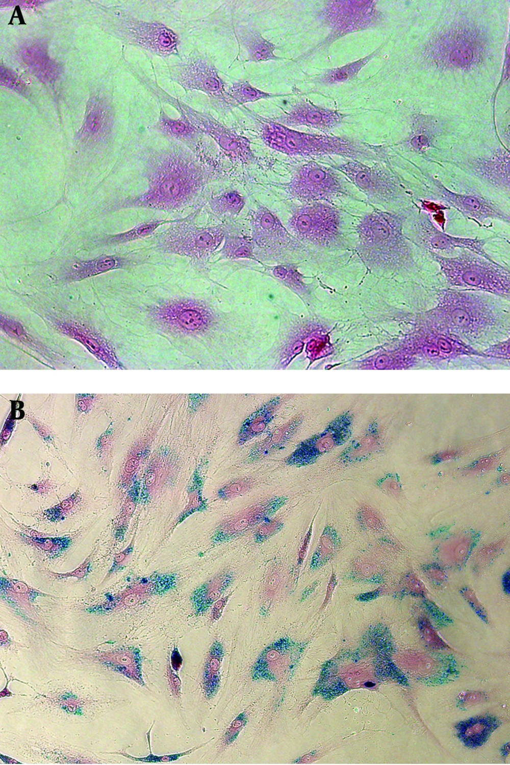
Prussian blue staining of unlabeled human bone marrow. Derived mesenchymal stem cells (hMSCs). A, superparamagnetic iron oxide (SPIO)-labeled hMSCs. B. All hMSCs are sufficiently labeled with SPIO nanoparticles without any cell damage in the medium containing 50 µg Fe/mL of SPIO nanoparticles and 750 ng/mL poly L-lysine (magnification, × 200).
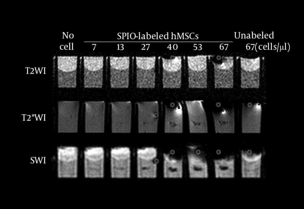
MRI of the phantom with eight wells. Each well has a 15-µL lump of superparamagnetic iron oxide-labeled human bone marrow-derived mesenchymal stem cells (SPIO-hMSCs) in different concentrations: no cells; 7, 13, 27, 40, 53, and 67 SPIO-hMSCs/µL; and 67 unlabeled hMSCs/µL. T2-weighted imaging (T2WI) does not detect SPIO-hMSCs even in the highest concentration of 67 SPIO-hMSCs/µL. T2*-weighted imaging (T2*WI) shows a dark region for cell lumps in 15 µL of 27, 40, 53, and 67 SPIO-hMSCs/µL. On susceptibility-weighted imaging (SWI), cell lumps in 15 µL of 13, 27, 40, 53, and 67 SPIO-hMSCs/µL show dark regions. SWI detects a lump of a lower concentration of 13 SPIO-MSCs/µL compared with T2*WI, which detects a concentration of 27 SPIO-MSCs/µL. The dark regions on SWI are more intense and larger in size than those on T2*WI for the same lump. The bullseyes indicate air bubble artifacts.
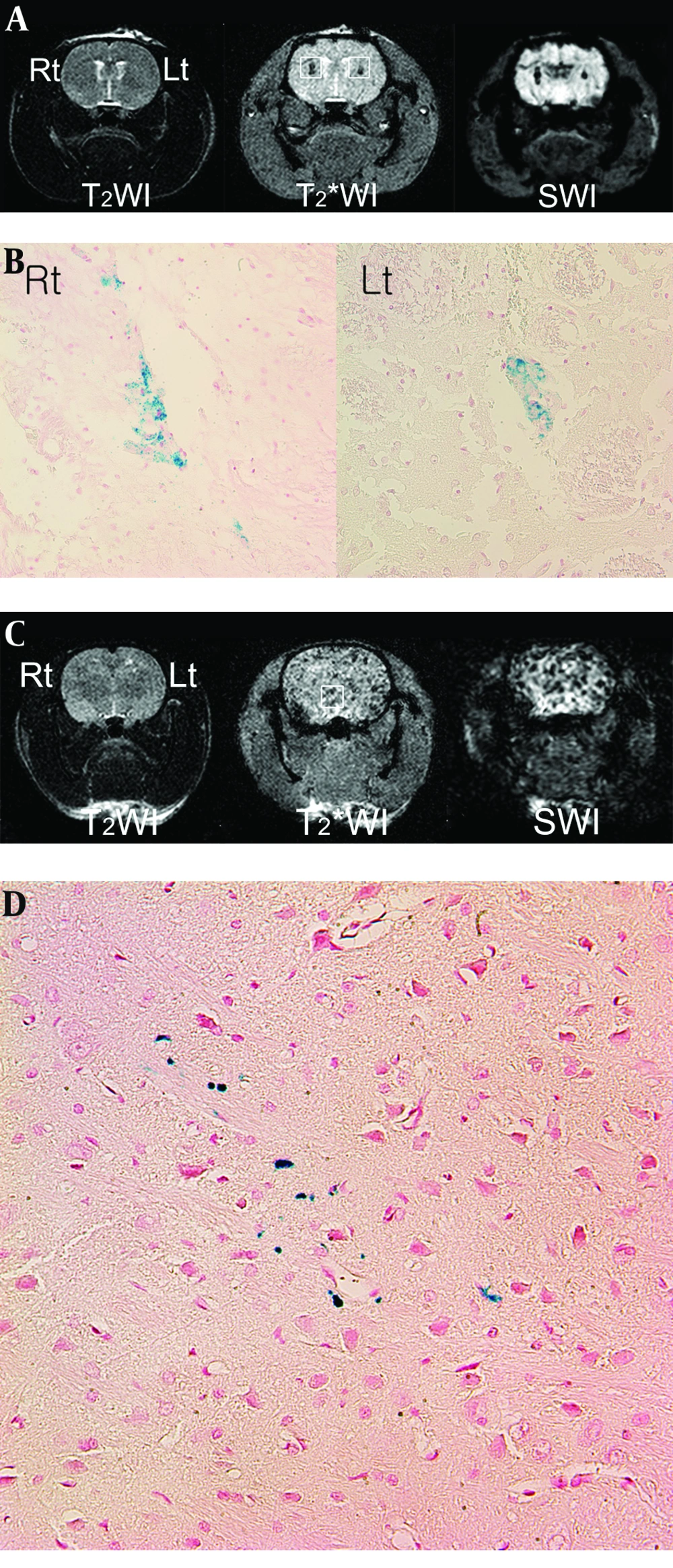
Superparamagnetic iron oxide-labeled human bone marrow. Derived mesenchymal stem cell (SPIO-hMSC) detection using magnetic resonance imaging (MRI) 1 h after direct implantation (A) and 24 h after intra-arterial implantation (C) in the normal rat brain T2-weighted imaging (T2WI) does not detect SPIO-hMSCs (A, C). On T2*-weighted imaging (T2*WI) and susceptibility-weighted imaging (SWI), the dark region in the right caudate putamen implanted with 3 µL of 333 SPIO-hMSCs/µL is larger than that in the left caudate putamen implanted with 3 µL of 167 SPIO-hMSCs/µL (A, squares). The dark regions are larger and more prominent on SWI than on T2*WI (A). On normal rat brain T2*WI and SWI, numerous foci of dark regions are dispersed throughout the brain (C, square), probably because the rat has a patent circle of Willis. On Prussian blue staining, the dark regions are confirmed to be SPIO-hMSCs (B, D) (magnification, × 400).
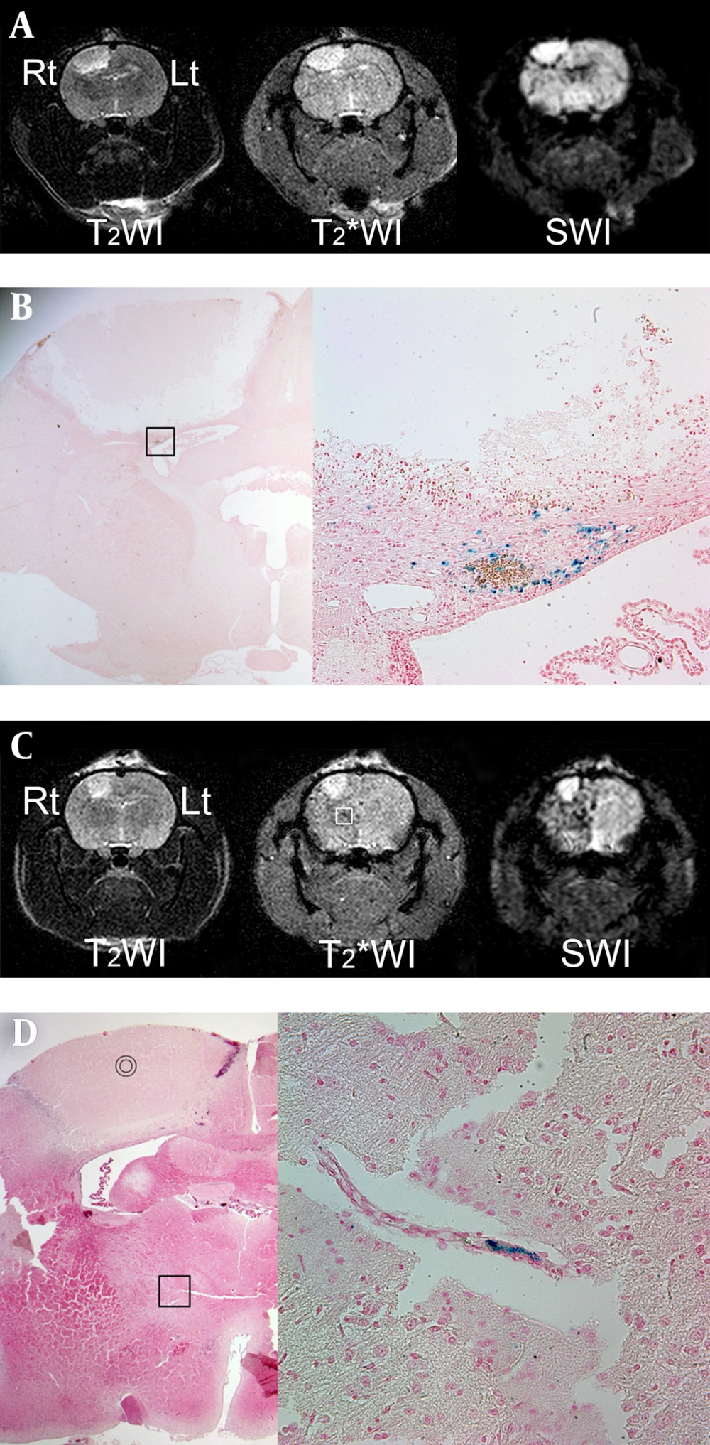
MRI performed 24 h after intra-arterial superparamagnetic iron oxide-labeled human bone marrow-Derived mesenchymal stem cell (SPIO-hMSC) infusion in rats with photothrombotic cerebral infarction. Focal and oval cerebral infarctions are induced in the right frontoparietal lobe, with high signal intensity on T2-weighted imaging (T2WI), T2*-weighted imaging (T2*WI), and susceptibility-weighted imaging (SWI) (A, C). Dark regions surrounding the cerebral infarct are confirmed as engrafted SPIO-hMSCs by Prussian blue staining (magnification, × 12.5 and × 400) (B; square). Other foci of dark regions are dispersed in the right hemisphere, particularly on SWI, and they are not remarkable even on T2*WI (C, square). The bullseyes indicate cerebral infarction on hematoxylin and eosin staining (magnification, × 12.5) (D). The foci of dark regions are identified as SPIO-hMSCs plugging the cerebral artery by Prussian blue staining (magnification, × 400; D; square).
| No. | Location of the Infarction | Dark Regions Around the Infarct a,b | Remark | ||
|---|---|---|---|---|---|
| T2WI | T2*WI | SWI | |||
| 1 | Rt. frontoparietal | − | + | ++ | |
| 2 | Rt. frontoparietal | − | +/− | +++ | Numerous dark foci in the right hemisphere |
| 3 | Rt. frontoparietal | +/− | + | ++ | |
| 4 | Rt. frontoparietal | +/− | + | ++ | Numerous dark foci and consequent changes in the right hemisphere |
| 5 | Rt. frontoparietal | − | +/− | ++ | |
| 6 | Rt. frontoparietal | − | +/− | + | |
aGrades of dark regions around each cerebral infarct: strong and continuous distribution (+++); complete, but not remarkable or remarkable, but not complete (++); not complete and unremarkable (+); only marks (+/−); and no marks (−).
bDark regions surrounding the cerebral infarcts on magnetic resonance imaging performed 24 h after intra-arterial infusion of superparamagnetic iron oxide-labeled human bone marrow-derived mesenchymal stem cells (SPIO-hMSCs) are most prominent on susceptibility-weighted imaging (SWI), followed by T2*-weighted imaging (T2*WI) and T2-weighted imaging (T2WI) in six rats. The trend analysis is statistically significant (P = 0.002).


