


1. Background
2. Objectives
3. Patients and Methods
3.1. Patients
3.2. Mammography Interpretation
3.3. Biopsy Procedures
3.4. Image-Histology Correlation
3.5. Data and Statistical Analysis
4. Results
| Total | 6-months follow-up (n = 194) | Without 6-months follow-up (n = 68) | P value | |
|---|---|---|---|---|
| Age, y | 47.7 ± 9.0 | 47.4 ± 8.7 | 48.7 ± 9.8 | 0.515 |
| Pathologic diagnosis on ST-VABb | 0.022 | |||
| Specific | 20 (7.6) | 10 (5.2) | 10 (14.7) | |
| Non-specific | 242 (92.4) | 184 (94.8) | 58 (85.3) | |
| BI-RADS final assessment category | 0.471 | |||
| 3 | 3 (1.1) | 3 (1.5) | 0 (0.0) | |
| 4a | 236 (90.1) | 173 (89.2) | 63 (92.6) | |
| 4b | 19 (7.3) | 14 (7.2) | 5 (7.4) | |
| 4c | 4 (1.5) | 4 (2.1) | 0 (0.0) | |
| Final pathologic diagnosis | ||||
| Benign | 258 (98.5) | 191 (98.8) | 67 (99.6) | |
| Malignant | 3 (1.1) | 2 (0.8) | 1 (0.4) | |
| High risk lesion | 1 (0.4) | 1 (0.4) | 0 (0.0) |
Abbreviations: BI-RADS, breast imaging reporting and data system; SD, standard deviation; ST-VAB, stereotactic vacuum assisted biopsy
aValues are expressed as No. (%) or mean ± SD.
bSpecific: including fibroadenoma, fibroadenomatous hyperplasia, and inflammation; non-specific: including fibrocystic change, adenosis, and columnar cell change
| No. | Age | Imaging features of microcalcifications | Final assessment | Needle size, gauge | Initial pathologic diagnosis on biopsy | Remnant | Changes on mammography | Follow-up interval to event, mo | Final pathologic diagnosis | |
|---|---|---|---|---|---|---|---|---|---|---|
| Morphology | Distribution | |||||||||
| 1 | 67 | Amorphous | Grouped | 4b | 11 | Columnar cell hyperplasia with microcalcifications | Yes | Newly developed microcalcifications near stereotactic biopsy site | 31 | DCIS |
| 2 | 51 | Amorphous | Regional | 4a | 8 | Papillary ductal hyperplasia with microcalcifications | No | Newly developed microcalcifications posterior to the biopsy site | 5 | ADH (4mm) |
| 3 | 42 | Amorphous | Grouped | 4a | 8 | Fibrocystic change with microcalcifications | Yes | Newly developed microcalcifications near previous biopsy site | 36 | DCIS |
| 4 | 55 | Coarse heterogeneous | Grouped | 4a | 8 | Fibrocystic change with microcalcifications | Yes | No change of the microcalcifications on mammography, newly developed suspicious mass on breast US performed 6 months after biopsy | 6 | DCIS |
Abbreviations: ADH, atypical ductal hyperplasia; DCIS, ductal carcinoma in situ; US, ultrasonography
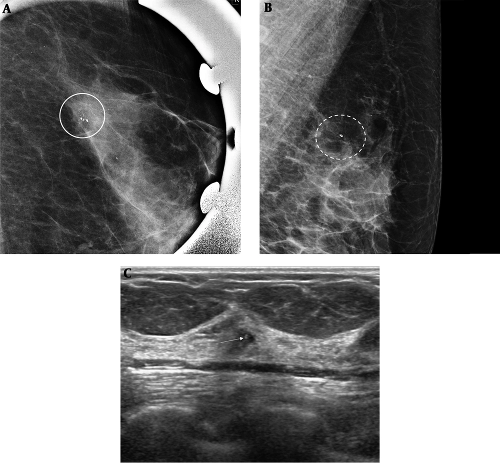
A 63-year-old woman with suspicious microcalcifications in her left breast. A, Magnification mammography of the left breast shows multiple areas of grouped microcalcifications in the left breast, one of which was biopsied (circle), and diagnosed as fibrocystic changes with microcalcifications. B, Follow-up mammography performed 6 months after stereotactic biopsy revealed no remnant microcalcifications at the biopsy site (clip, dotted circle). C, Breast ultrasonography (US) shows a 5-mm sized hypoechoic mass with internal echogenic calcifications (arrow) in the left upper outer quadrant near the biopsy site. This mass was proven as ductal carcinoma in situ (DCIS) on US-guided core biopsy and surgery.
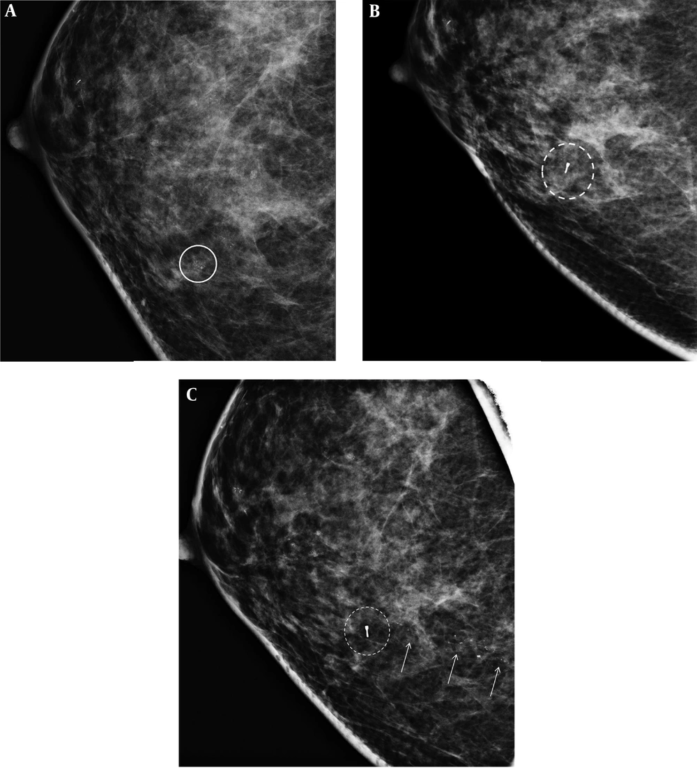
A 50-year-old woman with suspicious microcalcifications in her right breast. A, Magnification mammography of the right breast shows multiple areas of grouped amorphous microcalcifications in the right breast, one of which was biopsied (circle), diagnosed as columnar cell hyperplasia with microcalcifications. B, No significant findings were seen at the biopsy site on the mammogram performed at 6 months after biopsy. C, Follow-up mammography performed 31 months after stereotactic biopsy revealed newly developed fine pleomorphic/fine linear branching microcalcifications (arrows) located posteriorly to the prior biopsy site (dotted circle), which was proven as ductal carcinoma in situ (DCIS) on both stereotactic biopsy and surgery.




