


1. Background
2. Objectives
3. Patients and Methods
3.1. Study Population
3.2. Image Interpretation
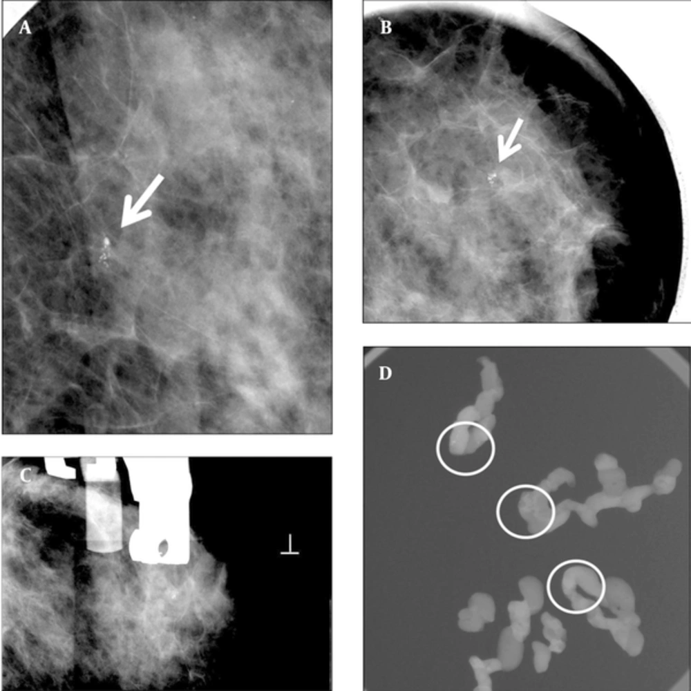
A 55-year-old woman with abnormal left mammogram categorized as 4a. A and B, Left craniocaudal magnification and mediolateral oblique magnification view showed suspicious clustered microcalcifications in the outer breast (arrow). C, Stereotactic biopsy was done. D, Specimen radiography of some samples from an 8 gauge biopsy showed multiple microcalcifications (circled). Pathology of biopsy revealed ductal carcinoma in situ. The imaging and histologic finding was considered concordant. Subsequent surgery was done. Pathologist confirmed no residual malignancy.
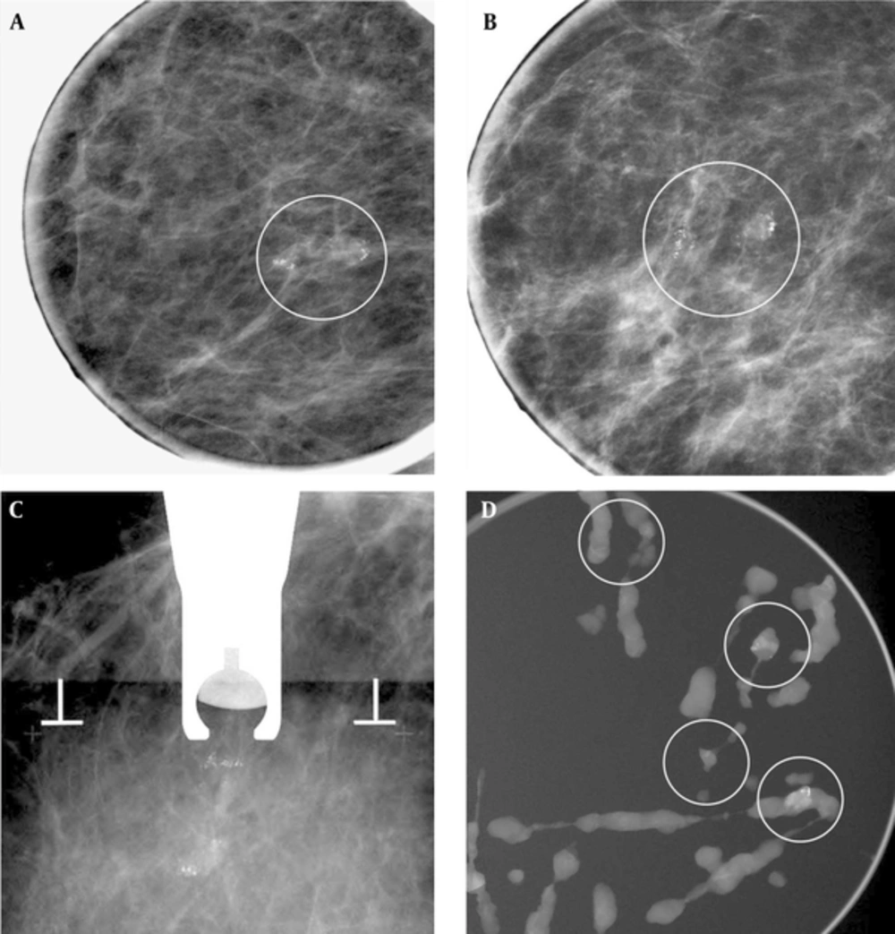
A 62-year-old woman with right mammogram categorized as 4b. A and B, Right craniocaudal magnificantion and mediolateral oblique magnificantion view showed suspicious clustered microcalcifiecation (circled). C, Stereotatic biopsy was done. D, Specimen radiography of some samples from an 8 gauge biopsy showed enough sampling of microcalfication (circled). Pathology of biopsy revealed fibrocystic disease with microcalcifications. Imaging-histologic correlation was considered discordant. Subsequent surgery was planned, but follow-up mammography was performed due to patient’s refuse. There was no interval change occurred on follow up mammography and spot magnification view at least 2 years and regarded as benign.
3.3. Biopsy Technique
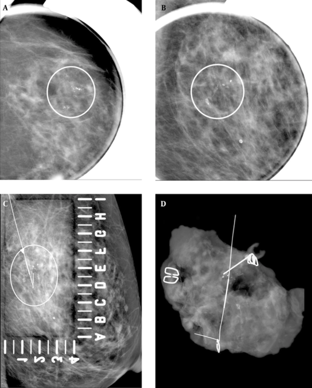
A 69-year-old woman with abnormal left mammogram categorized as 4c. A and B, Craniocaudal magnification and mediolateral oblique magnification view showed suspicious segmental fine linear and branching microcalcifications (circled). C, Mammography-guided needle localization was performed (circled). D, Specimen radiography of surgical specimen revealed adequate sampling of microcalcifications. Pathology of biopsy revealed DCIS. The imaging and histologic finding was considered concordant.
4. Results
| ST-VAB (n = 22) | L-EB (n = 34) | ||
|---|---|---|---|
| Age, year | 46.70 ± 8.78 (Range 28 - 63) | 50.51 ± 10.49 (Range 30 - 69) | |
| Indication | S | 8 | 10 |
| D | 14 | 24 | |
| Lesion size, cm | 8.49 ± 5.39 (Range 1.5 -22) | 5.72 ± 6.71 (Range 1.4 - 21) | |
| MMG-pattern | 1 | 1 (4.54) | 0 |
| 2 | 2 (9.09) | 2 (5.88) | |
| 3 | 15 (68.22) | 17 (50.00) | |
| 4 | 4 (18.18) | 15 (44.12) | |
| BI-RADS | 3 | 3 (13.64) | 1 (2.94) |
| 4a | 17 (77.27) | 32 (94.12) | |
| 4b | 2 (9.09) | 0 | |
| 4c | 0 | 1 (2.94) |
Abbreviations: D, diagnostic exam; MMG, mammography; S, screening exam; ST-VAB, stereotactic vacuum assisted biopsy; L-EB, localization and excisional biopsy; BI-RADS, breast imaging reporting and data system.
aTable numbers are No.(%) except for mean age and mean size.
| Biopsy | ST-VAB | L-EB | ||
|---|---|---|---|---|
| Number of Cases | Histologic Results | Number of Cases | Histologic Results | |
| Malignant lesions | 3 | 3 DCIS | 6 | 5 DCIS; 1 IDC |
| High risk lesion | 1 | 1 sclerosing adenosis | 6 | 1 LCIS 1 sclerosing adenosis; 1 Atypical ductal hyperplasia; 3 Intraductal papilloma |
| Benign lesion | 18 | 18 fibrocystic change | 22 | 21 Fibrocystic change; 1 Pseudocyst and fibrosis |
| Total | 22 | 34 | ||
Abbreviations: ST-VAB, stereotactic vacuum assisted biopsy; L-EB, localization and excisional biopsy; BI-RADS, breast imaging reporting and data system; DCIS, ductal carcinoma in situ; LCIS, lobular carcinoma in situ; IDC, invasive ductal carcinoma.
