The radiologic-pathologic discordance rate has been reported between 2.2% and 6% in the literature (
6,
7). However, open biopsy was performed in 119/667 (17.8%) of clinically or mammographically suspicious lesions in the study by Davidson et al. (
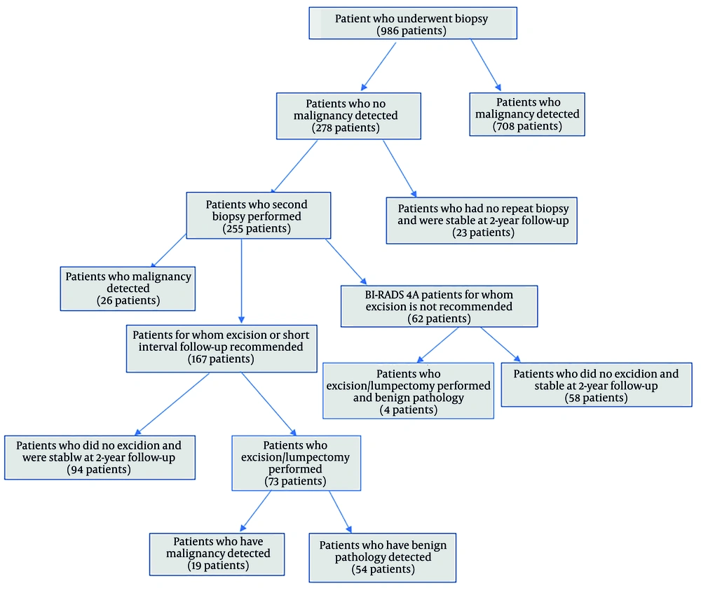
8). In our study, the discordance rate after excision and 2-year follow-up was 1.9% (19/986). Our study showed that there was histopathologic discordance between CNB and excision in 26% (19/73) of the lesions. Liberman et al. (
9) reported the overall malignancy rate as 24.4% in a study on imaging-histological discordance and concluded that discordance is an indication for immediate referral to surgery. In a study by Wan et al. in 2024, the discordance rate was found to be 30.9% (
3). Davidson et al. showed that excision biopsies after CNB detected malignancy in 30% of cases (
8). These rates are similar to our study. Subsequent EB revealed malignancy in up to 64% of discordant benign lesions obtained by image-guided CNB (
10). In our study, older mean age was found to be significantly associated with malignant excision results, similar to Wan et al. (
3). It is generally accepted that breast cancer is more common in older people. Age was not found to be significant in the study of Jung et al. only in BIRADS 3 category lesions. We think that the reason for this is that the majority of the lesions were benign (
11). Positive family history was not different between the groups (P = 0.334). Family history was present in 47.4% (9/19) of patients who were found malignant by excision.
A higher BIRADS category was statistically significantly associated with malignancy at excision. When the lesions were classified according to BIRADS category, it was observed that the rate of malignancy detection increased from BIRADS 4A to BIRADS 5, in accordance with the literature. In our study, after excision, no malignancy was detected in BIRADS 4A lesions; 26.3% of 4B lesions, 50% of 4C lesions, and 50% of BIRADS 5 lesions were found malignant. Soyder et al. reported a carcinoma upgrade rate of 20% in the BIRADS 4 category and 53.8% in the BIRADS 5 category (
6). Suspicious findings in terms of malignancy detected by ultrasound and MRI used in BIRADS categorization were not significantly different between the groups histopathologically. On the other hand, the presence of microcalcifications was significantly higher in the group with malignant excision results. In the study by Wan et al., the relationship between the presence of suspicious microcalcifications and discordance was investigated but not found to be significant (
3). The reason for this is that there is no possibility of stereotactic biopsy in mammography in our hospital; the projection of microcalcifications is marked on the skin with a pencil on mammography, and an ultrasound-guided biopsy is performed from this area. This increases the possibility of failure and leads to discordant benign lesions. There were no significant differences in tumor size and breast composition between the groups (P = 0.646, P = 0.542, respectively). Contrary to expectations, tumor size was slightly higher in the benign group than in the malignant group. This may be due to the fact that the benign group has larger lesions with cystic components instead of pure solid lesions.
In addition to its high diagnostic efficiency, CNB has some disadvantages. The most important one is the difficulty of interpretation due to the small size of the sample, fragmentation, and distortion. Additionally, some benign proliferative, borderline, and in situ lesions, as well as papillary lesions, are challenging in terms of diagnosis (
12,
13). Atypical ductal proliferation with high or intermediate nuclear grade in an intraductal papilloma qualifies as ductal carcinoma in situ (DCIS) regardless of size (
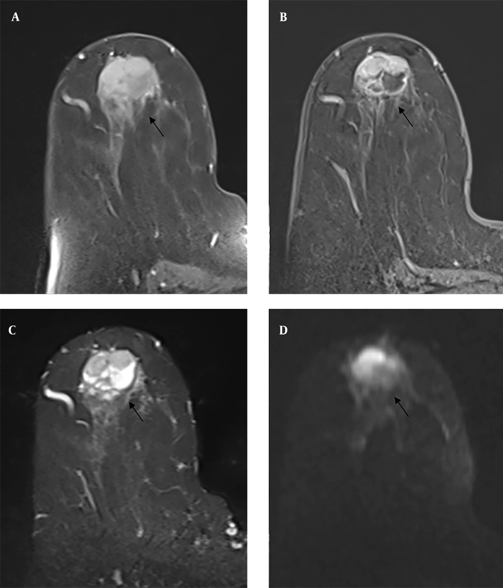
14). In our study, a total of 14 papillary lesions were found after excision, of which 2 (14%) were encapsulated papillary carcinoma (EPC), 3 (21%) were ductal papillomatosis including DCIS and 9 (65%) patients had intraductal papilloma. One of the two lesions defined in the BIRADS 5 category was diagnosed as EPC after excision, while one of the two lesions was interpreted as a papillary lesion by CNB (
Figure 2). In the literature, there are studies suggesting that papillary lesions detected by CNB can be safely followed up (
15,
16), but we think that such lesions should be referred for excision because of the uncertain malignancy potential.
On contrast-enhanced magnetic resonance imaging (MRI), T1-weighted pre-contrast images (A) demonstrate an iso- to hypointense lesion located in the retroareolar region (black arrow). Post-contrast T1-weighted images (B) reveal heterogeneous peripheral enhancement within the solid component of the lesion (black arrow). T2-weighted images (C) show an iso- to hyperintense lesion containing both solid and cystic areas (black arrow). Diffusion-weighted imaging (D) indicates diffusion restriction within the solid portion (black arrow). A core needle biopsy (CNB) classified the lesion as a papillary neoplasm with a Breast Imaging Reporting and Data System (BI-RADS) category 5 rating. Subsequent surgical excision confirmed the diagnosis of encapsulated papillary carcinoma (EPC).
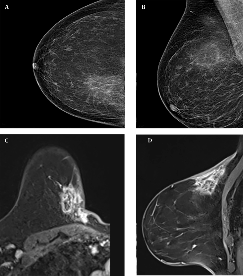
In the other BIRADS 5 lesions in our study, both CNB and excision resulted in fat necrosis. The lesion with fat necrosis is shown in
Figure 3. Fat necrosis consists of solid hypoechoic lesions causing tissue distortion and shadowing posteriorly on ultrasound, which may be radiologically confused with malignancy. On mammography, coarse calcifications, amorphous, linear, fine branching, and pleomorphic microcalcifications, as well as asymmetry, may be observed (
17). In a radiological and clinical study by Bilgen et al. on fat necrosis, increased echogenicity of subcutaneous tissue was observed in 26.9% of lesions, anechoic masses with posterior acoustic shadowing in 16.6%, solid masses in 14.2%, and cysts with internal echoes in 11.1%, regardless of the presence of small cysts (
18). It has a wide range of findings on MRI and may mimic malignancy. The most common finding is round or oval hypointense masses on T1W fat-suppressed images (
19). After intravenous administration of contrast media, it may show highly variable contrast enhancement patterns and kinetic analysis (
20).
Mammography shows irregularly circumscribed asymmetric opacity increase (A, B) in the upper inner quadrant. On contrast-enhanced magnetic resonance imaging (MRI), the irregularly circumscribed heterogeneously contrasted lesion (C, D) with a type 1 time-signal intensity curve on post-contrast T1-weighted (T1W) images was evaluated in Breast Imaging Reporting and Data System (BIRADS) 5 categories with mammography and MRI findings. Both core needle biopsy (CNB) pathology and excision pathology results were reported as fat necrosis.
In a patient with a borderline phylloid tumor as a result of excision, the CNB result was interpreted as fibroadenoma. There are no reliable radiologic features that can distinguish a phylloid tumor from a fibroadenoma. On ultrasound, it is frequently seen as well-circumscribed solid masses with a heterogeneous internal structure. However, a phylloid tumor may be suspected in rapidly growing lesions larger than 4 cm (
21). On MRI, it is usually T1 hypointense and T2 homogeneous hypointense/hyperintense. In post-contrast series, the kinetic curve pattern may be type 1 or type 2 (
22).
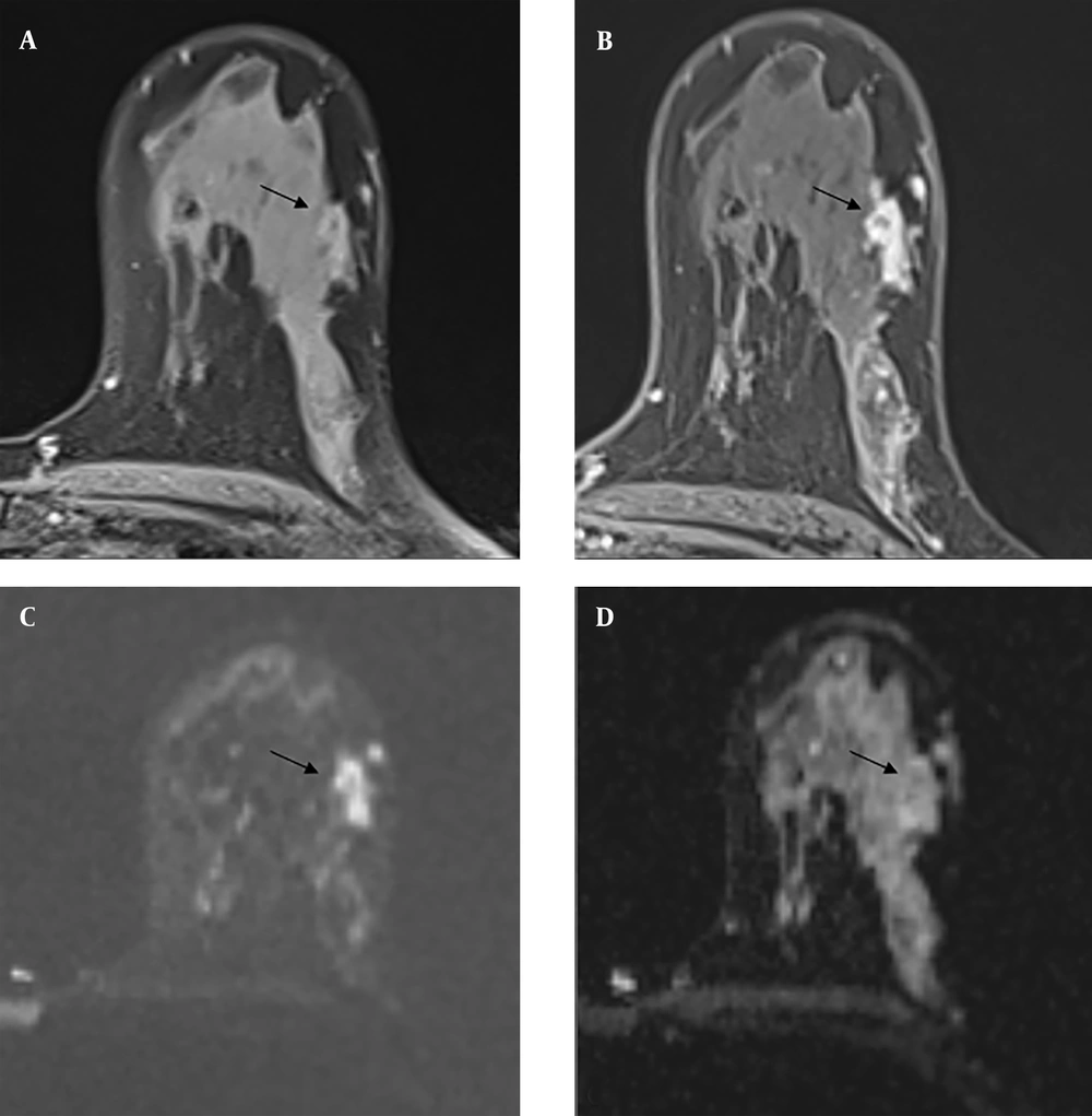
The three lesions with radial scars detected by biopsy were radiologically defined as BIRADS category 4B at the time of diagnosis, and 1 of them (33%) was found to have DCIS after excision (
Figure 4). Radial scars are benign breast lesions of unknown clinical significance that are detected with increasing frequency in women undergoing breast cancer screening (
23). Invasive carcinoma was found in 11% of patients with radial scars in a study by Davidson et al. (
8).
Contrast-enhanced magnetic resonance imaging (MRI) of a Breast Imaging Reporting and Data System (BIRADS) 4B category lesion with a radial scar on core needle biopsy (CNB) pathology and ductal carcinoma in situ on excision pathology. A mild hyperintense lesion is seen on pre-contrast T1-weighted (T1W) images (A), and a homogeneously contrasted irregularly circumscribed lesion is seen on post-contrast T1W images (B) (black arrows). It is shown to be hyperintense on diffusion-weighted imaging (C) and hypointense on apparent diffusion coefficient images (D) due to diffusion restriction. (black arrows)
Some limitations of our study should be mentioned. The retrospective design of the study and the small number of patients can be considered the most important ones. Therefore, the generalizability of the results is limited. Additionally, confounding factors such as the menstrual cycle and hormone use may affect imaging results, especially MRI. Since vacuum biopsy is not covered by social insurance in our country, we do not have the possibility of performing vacuum biopsy. This situation reduces the possibility of making a correct diagnosis before excision. Furthermore, the ultrasound-guided biopsy technique is operator-dependent and causes individual differences. The diagnostic sensitivity of fragmented specimens is reduced. On the other hand, although histopathological evaluation by a single pathologist provides standardization, it may cause diagnostic errors in some lesions that are open to interpretative differences.
In conclusion, the BIRADS system is reliable in determining the malignancy risk of breast masses radiologically. In breast masses where malignancy is suspected based on clinical and imaging findings, CNB is a reliable method with high diagnostic accuracy. However, additional surgery is required when there is clinical and radiologic discordance with the CNB findings. Excision of papillary lesions is necessary to confirm CNB findings and exclude possible malignancy. Multicenter prospective studies in larger populations are necessary to confirm the findings.