
1. Background
Anemia during pregnancy remains a critical global health challenge, affecting approximately 40% of pregnant individuals worldwide; iron deficiency anemia accounts for 60% - 70% of cases, and immunological etiologies account for 10% - 15%. Although iron deficiency dominates clinical discussions, emerging evidence highlights the underrecognized role of immune-mediated mechanisms in maternal anemia. These mechanisms range from classical Rh/ABO incompatibility to complex autoimmune dysregulation, in which maternal antibodies inadvertently target fetal or self red blood cells (RBCs). For instance, in Rh-negative mothers, fetal RBC antigens can trigger IgG antibody production, leading to hemolytic disease of the fetus and newborn (HDFN) in subsequent pregnancies (1). Similarly, autoimmune conditions such as systemic lupus erythematosus (SLE) or autoimmune hemolytic anemia (AIHA) disrupt RBC survival through complement activation and phagocytosis. A less explored but pivotal pathway involves cytokine-mediated iron dysregulation in inflammatory states, such as urinary tract infections, which elevate hepcidin, a master iron regulator that blocks intestinal iron absorption and macrophage iron recycling (2). This intersection of immunology and hematology underscores the need for integrated diagnostic and therapeutic frameworks in prenatal care. Based on recent data up to 2023, the prevalence of anemia among pregnant women in Iran ranges from 25% to 40%, with significant regional disparities. In socioeconomically disadvantaged provinces, such as Sistan and Baluchestan, rates reach up to 45%.
Anemia during pregnancy can cause premature birth, low birth weight, and increased postpartum bleeding. Studies show that iron deficiency anemia in the first trimester is associated with decreased fetal development and negatively affects the development and function of the fetal nervous system, whereas iron deficiency anemia in the second and third trimesters has fewer adverse effects on fetal development (3). According to World Health Organization (WHO) guidelines, anemia in pregnant women is defined as a hemoglobin concentration below 11 g/dL in the first and third trimesters and below 10.5 g/dL in the second trimester (4).
2. Objectives
Because no comprehensive study had been conducted to determine the status of anemia and its associated factors among pregnant women in Dezful, and because findings from other provinces cannot be generalized to this city owing to socioeconomic differences, the present study was designed to determine the prevalence of anemia among pregnant women attending health centers in Dezful. The findings may provide a basis for improved evaluation of anemia during pregnancy.
3. Methods
3.1. Study Design and Setting
This retrospective cross-sectional study was conducted from January 2021 to December 2021 among pregnant women referred to health centers in Dezful, southwest Iran.
3.2. Participants, Sample Size, and Sampling
According to a primary study, the prevalence of anemia was 13.4%, and in another study, the prevalence was reported to be 17.5%. Therefore, to determine the sample size, the average prevalence from 2 similar articles, with an anemia ratio of 15%, was used. With 95% confidence and a maximum estimation error of 3%, the required sample size was estimated at 549 individuals. To reduce the impact of potential sample attrition, 50 additional samples were added, and the final sample size was estimated at 599 individuals. Convenience sampling was used among pregnant women who met the inclusion criteria.
3.3. Inclusion and Exclusion Criteria
Pregnant women with health records in these centers during the study year were included. Women with a registered underlying disease associated with anemia, chronic kidney disease, liver function disorder, or blood donation during the previous 3 months were excluded.
3.4. Data Collection
After obtaining the necessary permits and ethics code, the researcher visited the health centers covered by the university. After presenting a letter of introduction, the researcher explained the study objectives to the officials at the centers and assured them of the confidentiality of the information. Then, from the information recorded in databases in southwest Iran in 2021, 6 of the 13 health centers were randomly selected using an even-number table. The files of 599 individuals were selected, and information on the study variables was collected by the researcher and entered into Excel software using a checklist. The independent variables were the number of pregnancies, education level, number of times care was received before and during pregnancy, employment status, and type of occupation. Hemoglobin and anemia were the dependent variables. Participants were then classified according to hemoglobin level for anemia. Based on WHO guidelines, anemia was classified as severe (< 7 g/dL), moderate (7 - 10 g/dL), or mild (10 - 11 g/dL).
At all stages of the research, trust and scientific integrity were maintained, and participants were assured that the study results would be available to them upon request. The work was reported in accordance with the STROCSS criteria (5).
3.5. Statistical Analysis
The results were statistically described using SPSS software version 24, with the significance level set at P < 0.05. Frequency, percentage, mean, and standard deviation were used to describe the study variables. Kruskal-Wallis, Wilcoxon, and Spearman correlation coefficient tests were used because the data were nonparametric.
4. Results
4.1. Characteristics of the Participants
The results of the present study showed that the mean age of the pregnant women was 24.2 ± 4.1 years, with a minimum age of 15 years and a maximum age of 36 years. Most participants had a bachelor's degree (46.9%), and only 0.7% had less than a diploma. Most participants were housewives (76.5%). The mean number of pregnancies was 1.3, with a minimum of 1 and a maximum of 6 (Table 1).
Table 1.
Background Information of Pregnant Women (N = 599)
| Variables | Values a | Valid Percent | Cumulative Percent |
|---|---|---|---|
| Education | |||
| Less than diploma | 4 (0.7) | 0.7 | 0.7 |
| Diploma | 146 (24.3) | 24.4 | 25.0 |
| Associate degree | 79 (13.2) | 13.2 | 38.2 |
| BSc | 281 (46.8) | 46.9 | 85.1 |
| MSc | 73 (12.2) | 12.2 | 97.3 |
| PhD degree | 16 (2.7) | 2.7 | 100.0 |
| Occupation | |||
| Housewife | 458 (76.5) | 76.5 | 76.5 |
| Self-employed | 39 (6.5) | 6.5 | 83.0 |
| Employee | 102 (17.0) | 17.0 | 100.0 |
| Age, y | 24.00 (24.298 ± 4.30864) | 15.00 - 36.00 b | |
| Number of pregnancies | 1 (1.36 ± 0.655) | 1 - 6 b | |
a Values are expressed as No. (%) or median (mean ± SD).
b Values are expressed as minimum-maximum
Regarding care before and during pregnancy, participants received 0 to 6 types of care. The results showed that the mean number of care types before pregnancy was lower than the mean number during pregnancy (1.2 ± 0.7 vs 2.5 ± 1.2). Overall, 11.8% of pregnant women had not received any care before pregnancy, whereas this rate was 0.5% during pregnancy. In addition, 62.8% of women received only 1 type of care before pregnancy, whereas during pregnancy, 75.3% of women received at least 3 types of care (Table 2 and Figure 1).
Table 2.
Comparison of the Number of Care Types Received Before and During Pregnancy a
| Variables and Number of Care Types | No (%) | Valid Percent | Cumulative Percent |
|---|---|---|---|
| Care before pregnancy | |||
| 0.0 | 71 (11.8) | 11.9 | 11.9 |
| 1.0 | 376 (62.7) | 62.8 | 74.6 |
| 2.0 | 116 (19.3) | 19.4 | 94.0 |
| 3.0 | 29 (4.8) | 4.8 | 98.8 |
| 4.0 | 6 (1.0) | 1.0 | 99.8 |
| 5.0 | 1 (0.2) | 0.2 | 100.0 |
| Care during pregnancy | |||
| 0.0 | 3 (0.5) | 0.5 | 0.5 |
| 1.0 | 150 (25.0) | 25.0 | 25.5 |
| 2.0 | 174 (29.0) | 29.0 | 54.6 |
| 3.0 | 125 (20.8) | 20.9 | 75.5 |
| 4.0 | 97 (16.2) | 16.2 | 91.7 |
| 5.0 | 45 (7.5) | 7.5 | 99.2 |
| 6.0 | 5 (0.8) | 0.8 | 100.0 |
a Wilcoxon Test: Z = -17.413; Asymp. Sig. (2-tailed) = 0.000.
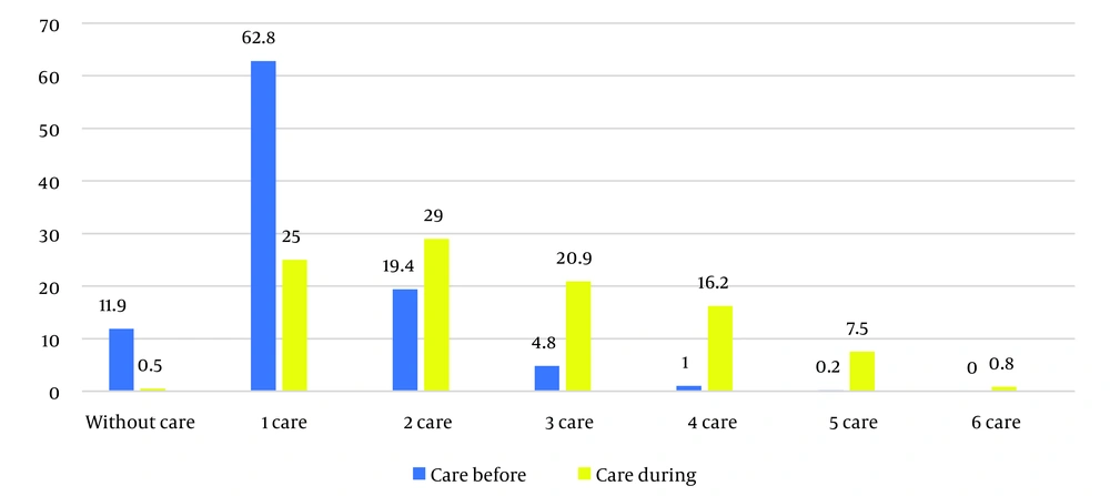
Figure 1.
Frequency and percentage of the number of care types before and during pregnancy
The mean hemoglobin level was 11.7 ± 2.6 and 10.2 ± 1.3 g/dL in the first and third trimester respectiveley. The results showed a significant difference in prevalance of anemia in the first and third trimesters (P < 0.05). Regarding anemia severity in the first trimester, 14 women (2.3%) had moderate anemia (hemoglobin 7 - 10 g/dL), 71 women (11.9%) had mild anemia (hemoglobin 10 - 11 g/dL), and 514 women (85.8%) did not have anemia (hemoglobin above 11 g/dL). None of the women had severe anemia (hemoglobin less than 7 g/dL). The frequency distribution of anemia severity in the third trimester showed that 43 women (7.2%) had moderate anemia (hemoglobin 7 - 10 g/dL), 246 women (41.1%) had mild anemia (hemoglobin 10 - 11 g/dL), and 310 women (51.8%) did not have anemia (hemoglobin above 11 g/dL).
Wilcoxon's nonparametric test was used to assess the significance of the difference in hemoglobin levels between the first and third trimesters of pregnancy. The results showed a significant difference in anemia between the first and third trimesters based on the WHO categorization of hemoglobin level (P < 0.05). Overall, 40 participants (6.6%) had anemia in both the first and third trimesters; mild anemia was more prevalent than moderate anemia in both trimesters (P < 0.05), and none of the cases had severe anemia (Table 3).
Table 3.
Comparison of Anemia Severity in the First and Third Trimesters of Pregnancy a
| Variables | No (%) | Valid Percent | Cumulative Percent |
|---|---|---|---|
| The severity of anemia in the first trimester | |||
| Moderate anemia | 14 (2.3) | 2.3 | 2.3 |
| Mild anemia | 71 (11.8) | 11.9 | 14.2 |
| Normal | 514 (85.7) | 85.8 | 100.0 |
| The severity of anemia in the third trimester | |||
| Moderate anemia | 43 (7.2) | 7.2 | 7.2 |
| Mild anemia | 246 (41.0) | 41.1 | 48.2 |
| Normal | 310 (51.7) | 51.8 | 100.0 |
a Wilcoxon Test: Z = -18.850; Asymp. Sig. (2-tailed) = 0.000.
The Kruskal-Wallis test showed no significant difference between hemoglobin level and age. Spearman's test also showed no significant relationship between hemoglobin level and age among pregnant women (Table 4).
Table 4.
Correlation Between Age Groups and Hemoglobin Levels in the First and Third Trimesters
| Analysis and Variable | Hb Level, First Trimester | Age Group | Kruskal-Wallis Measure | Kruskal-Wallis Value |
|---|---|---|---|---|
| Spearman's rho | ||||
| Hb level, first trimester | ||||
| Correlation coefficient | 1.000 | 0.032 | Chi-square | 1.371 |
| Sig. (2-tailed) | - | 0.435 | df | 3 |
| N | 599 | 599 | Asymp. Sig. | 0.712 |
| Age group, first trimester | Asymp. Sig. | 0.712 | ||
| Correlation coefficient | 0.032 | 1.000 | ||
| Sig. (2-tailed) | 0.435 | - | ||
| N | 599 | 599 | ||
| Age group, third trimester | ||||
| Correlation coefficient | 1.000 | -0.006 | Chi-square | 0.436 |
| Sig. (2-tailed) | - | 0.887 | df | 3 |
| N | 599 | 599 | Asymp. Sig. | 0.933 |
| Hb level, third trimester | Asymp. Sig. | 0.933 | ||
| Correlation coefficient | -0.006 | 1.000 | ||
| Sig. (2-tailed) | 0.887 | - | ||
| N | 599 | 599 |
The Kruskal-Wallis test also showed no relationship between care received before and during pregnancy and anemia (Table 5).
Table 5.
Correlation Between the Number of Care Types Received Before and During Pregnancy and Anemia Level in the First and Third Trimesters
| Grouping Variable and Statistic | Hb Level, First Trimester | Hb Level, Third Trimester |
|---|---|---|
| Care before and during pregnancy | ||
| Chi-square | 5.000 | 2.820 |
| df | 4 | 4 |
| Asymp. Sig. | 0.287 | 0.588 |
| Care during pregnancy | ||
| Chi-square | 8.520 | 3.207 |
| df | 5 | 5 |
| Asymp. Sig. | 0.130 | 0.668 |
5. Discussion
According to the results of the current study of 599 pregnant women in Dezful, the overall prevalence of anemia was 48.3%, with a significant increase from the first trimester (34%) to the third trimester (48.3%). This pattern is consistent with previous studies; however, the higher rate observed in our study may reflect the combined influence of environmental and economic factors and potential immunological mechanisms. Although iron deficiency anemia is considered the primary cause of anemia in pregnancy, global evidence suggests that chronic inflammation due to conditions such as parasitic infections or autoimmune diseases may inhibit iron absorption by increasing hepcidin levels, thereby exacerbating nutritional anemia. Iron deficiency in the context of chronic inflammatory conditions has been discussed in international expert opinion on definition, diagnosis, and management (6, 7).
In addition, the high prevalence of minor thalassemia in Khuzestan Province may have influenced our findings. This factor could partly explain the discrepancy with studies such as Dehghani et al. (8), who reported a 15.71% prevalence of iron deficiency anemia in Iranian pregnant women. Another notable finding was the lack of an association between reproductive age and anemia severity in this study, in contrast to previous reports describing an inverse relationship.
In Khuzestan Province, the frequency of β-thalassemia minor is also high and reaches 10%; alternatively, immune system activation due to frequent exposure to infectious agents might overshadow age-related effects (9). Furthermore, 62.8% of women received only 1 pre-pregnancy care visit, and delayed diagnosis of immunological anemia, such as autoimmune hemolysis, may have affected our findings. In such cases, inappropriate iron supplementation without investigation of underlying causes could exacerbate hemolysis (6).
Other reports have described anemia rates of 8.2%, 28.1%, and 63.7% in the first, second, and third trimesters, respectively. Potential ethnic-genetic differences in immune responses, subsequent decreases in iron absorption, and differences in the prevalence of anemia during pregnancy may contribute to these discrepancies. Pregnancy is associated with inflammation and the activation and proliferation of immune cells. C-reactive protein (CRP), an inflammatory protein, has been reported to be elevated in pregnant women compared with nonpregnant women. This elevation occurs early in the first trimester and increases in the third trimester (10). According to previous studies, genetic polymorphisms related to interleukin-6, for example, influence hepcidin levels and iron stores (11). This hypothesis is reinforced by the lower third-trimester hemoglobin level of 11.2 g/dL compared with 12.2 g/dL in the first trimester, because increased fetal iron demands in late pregnancy strain compensatory mechanisms under chronic inflammation.
Clinically, these findings indicate the need for revised screening protocols. Combined CBC, ferritin, and transferrin saturation testing during pregnancy, along with measurement of hepcidin as a downregulator of iron absorption and iron release from macrophages of the reticuloendothelial system, could help differentiate nutritional from immunological iron deficiency anemia (12). Preventive interventions should include education on iron supplement use and anti-inflammatory dietary adjustments. This integrated approach could reduce both the anemia burden and adverse pregnancy outcomes (13).
5.1. Limitations and Suggestions
This study had several limitations, including the relatively small sample size and the lack of anemia-type classification. Assessment of causes of anemia, such as iron, vitamin B12, vitamin B6, and vitamin B9 deficiency, or incorporation of biomarkers such as hepcidin and cytokine profiling, including interleukin-6, into standard prenatal panels, may help differentiate anemia subtypes. In addition, we did not assess neurodevelopmental outcomes in infants born to mothers with anemia, despite the link between prenatal hypoxia and cognitive deficits.
5.2. Conclusions
This study reveals a critical 48.3% prevalence of anemia among pregnant women in Dezful, Iran. The sharp rise from 14.2% in the first trimester to 48.3% in the third trimester underscores the compounding effects of physiological iron demands and region-specific risk factors. These findings align with global reports of anemia burden in low-resource settings but highlight unique immunological-nutritional interactions with chronic inflammation that exacerbate iron sequestration through hepcidin upregulation. The observed hemoglobin decline from 12.2 g/dL to 11.2 g/dL across trimesters cannot be attributed solely to dietary insufficiency. Emerging evidence suggests that inflammatory mediators, such as interleukin-6, in populations may dysregulate iron metabolism through hepcidin-mediated ferroportin degradation, thereby blocking enteric iron absorption and promoting macrophage iron retention, limiting erythropoiesis, and suppressing erythropoietin sensitivity in proinflammatory microenvironments. This creates a "nutritional trap" in which standard iron supplementation fails to correct anemia unless paired with anti-inflammatory strategies (12, 13).