
1. Context
Dry eye disease (DED) is a multifaceted ocular disorder that substantially impairs patients’ quality of life and affects millions worldwide. Despite extensive research, its diagnosis and management remain challenging because of complex pathophysiology, heterogeneous clinical presentations, and evolving diagnostic modalities (1).
Although conventional assessments, including symptom questionnaires and tear film evaluation, remain central to clinical practice, there is increasing recognition of the need for novel biomarkers and advanced imaging techniques to improve diagnostic accuracy and guide personalized therapy (2). Recent studies have identified matrix metalloproteinase-9 (MMP-9) and other molecular mediators as potential targets for both diagnosis and treatment; however, their integration into routine practice remains limited (3, 4).
This review aims to provide an updated overview of DED, focusing on pathogenesis, classification, diagnostic strategies, and emerging molecular biomarkers and highlighting recent advances that may support precision medicine approaches in clinical management. By synthesizing current evidence, we aim to clarify the clinical relevance of both established and innovative diagnostic modalities, thereby facilitating improved patient outcomes.
2. Evidence Acquisition
A comprehensive literature search was conducted using the PubMed and Scopus databases. Search terms included “dry eye disease,” “pathogenesis,” “classification,” “diagnosis,” “biomarkers,” and “ocular surface”. Eligibility criteria were defined a priori based on study design, population characteristics, diagnostic focus, and outcome measures relevant to dry eye disease. Study selection was performed using a multistage screening process involving title and abstract review followed by full-text assessment for eligibility.
Both intrinsic factors (e.g., age, autoimmune conditions) and extrinsic influences (e.g., environmental exposures, medications) were evaluated. Established diagnostic tools, including tear film breakup time, tear osmolarity, and symptom questionnaires (OSDI, SPEED), were reviewed alongside emerging imaging techniques, lipid layer interferometry, and noninvasive tear breakup analysis. Molecular biomarkers, with particular emphasis on metabolomic studies and MMP-9, were evaluated for their potential diagnostic and therapeutic implications.
A total of 656 records were identified; 234 were screened after duplicate removal, and 62 studies were ultimately included in the final analysis.
Studies were excluded for reasons including irrelevance to the research question, insufficient diagnostic data, lack of full-text availability, nonoriginal study design, or failure to meet predefined inclusion criteria.
3. Results
3.1. Dry Eye Pathogenesis and Classification
Dry eye disease is a common ocular disorder characterized by deficient tear film production or excessive evaporation. Common symptoms include burning sensations or ocular irritation. Other symptoms may include itching or redness due to inflammation caused by an insufficient quantity or quality of tears (2).
Dry eye has a significant impact on patients' quality of life. The chronic nature of the condition often interferes with daily activities, such as reading or driving. Patients may experience difficulty focusing because of blurred vision caused by inadequate tear film lubrication. If left untreated, dry eye may lead to complications such as corneal abrasions and infections (5).
The prevalence of dry eye disease has increased in recent years, with studies estimating that it affects up to 30% of adults worldwide (6). This highlights the importance of understanding its classification and implementing appropriate diagnostic strategies.
The classification of dry eye disease plays a vital role in guiding diagnosis and treatment decisions.
The three main classifications are 1. aqueous-deficient dry eye (ATD) due to decreased tear production, 2. evaporative tear deficiency (ETD) due to increased tear film instability, and 3. mixed aqueous-evaporative tear deficiency (MAED) due to both decreased tear production and increased tear film instability. Each subcategory has distinct underlying causes and contributing factors.
3.1.1. Aqueous-Deficient Dry Eye
Aqueous-deficient dry eye occurs when tear production is reduced, resulting in insufficient lubrication and ocular discomfort. Understanding this condition is crucial for both healthcare providers and patients, as it can significantly affect quality of life.
Several causes and risk factors are associated with aqueous-deficient dry eye. Reduced tear production can be influenced by various underlying conditions. Autoimmune diseases, such as Sjögren's syndrome, can lead to inflammation and damage to the lacrimal glands responsible for tear production. Hormonal changes during menopause or pregnancy may also contribute to decreased tear production. Additionally, certain medications, such as antihistamines and antidepressants, have been linked to dry eye symptoms.
Symptoms of aqueous-deficient dry eye vary but commonly include a gritty or sandy sensation in the eyes, redness, itching, and blurred vision. Diagnosis typically involves assessing patient-reported symptoms along with clinical evaluations, such as measuring tear volume or evaluating tear quality.
Treatment options for aqueous-deficient dry eye aim to alleviate symptoms and improve ocular comfort. Artificial tears provide temporary relief by supplying additional moisture to the ocular surface. Prescription medications, such as anti-inflammatory drugs, may be used for more severe cases or when autoimmune conditions contribute to reduced tear production (7). In some instances, punctal plugs may be inserted into the drainage ducts to slow tear drainage and enhance lubrication (8). Lifestyle modifications, such as avoiding environmental triggers (e.g., smoke) or using humidifiers at home, can also help manage symptoms.
3.1.1.1. Sjögren Syndrome Dry Eye
Sjögren syndrome is a chronic autoimmune disease characterized by lymphocytic infiltration of the exocrine glands, leading to dryness of mucosal surfaces, including the eyes and mouth. Dry eye is one of the most common ocular manifestations of Sjögren syndrome, affecting approximately 50 - 80% of individuals with this condition. The underlying causes of dry eye in Sjögren syndrome are multifactorial but primarily involve immune dysfunction and inflammation. Autoantibodies directed against salivary and lacrimal gland tissues are produced, resulting in gland destruction (9). Impaired innervation due to gland damage leads to decreased tear production, while inflammatory cytokines released further exacerbate local tissue damage and disrupt normal tear film dynamics.
Clinical manifestations commonly observed in individuals with Sjögren-related dry eye include ocular discomfort, redness, blurred vision, foreign body sensation, and photophobia (10). Diagnostic methods such as Schirmer's test can objectively measure tear production; however, ocular surface staining using vital dyes such as fluorescein or rose Bengal may reveal corneal or conjunctival epithelial defects indicative of severe dryness (9).
Management strategies for dry eye in individuals with Sjögren syndrome aim to alleviate symptoms by restoring tear film stability. Artificial tears provide lubrication and moisture to compensate for decreased tear production (10). Punctal plugs can be inserted to prevent tear drainage, thereby prolonging tear presence.
3.1.1.2. Non-Sjögren Syndrome Dry Eye
3.1.1.2.1. Primary Lacrimal Gland Deficiencies
Primary lacrimal gland deficiency (PLGD) occurs due to intrinsic dysfunction or structural abnormalities of the lacrimal glands.
The causes and risk factors for PLGD are multifactorial and can include genetic predisposition as well as environmental factors, such as aging and hormonal changes. CircRNA expression profiles in AQP5–/– mice with PLGD have been shown to differ from those without this condition. (11). This suggests that circRNAs may play a role in regulating tear secretion.
Symptoms of PLGD can range from mild irritation to severe pain and vision impairment. Clinical manifestations may include decreased tear production and increased tear evaporation due to reduced lipid layer thickness or altered mucin composition (12). Diagnosis typically involves evaluating tear volume using techniques such as Schirmer's test or assessing tear film stability through tear breakup time measurements.
3.1.1.2.2. Secondary Lacrimal Gland Deficiencies
Secondary lacrimal gland deficiency (SLGD) is caused by external factors that affect tear production or composition. SLGD can occur due to various systemic conditions, such as Sjögren's syndrome, or certain medications that reduce tear production. Symptoms of SLGD are similar to those of PLGD but may also be accompanied by extraocular manifestations related to the underlying systemic disease. Diagnosis and evaluation of SLGD involve identifying the underlying cause through medical history assessment, blood tests, and imaging techniques.
3.1.1.2.3. Obstruction of the Lacrimal Gland Ducts
The lacrimal gland ducts play a crucial role in tear production, as they secrete tears onto the ocular surface to maintain lubrication and protection.
Obstruction of the lacrimal gland ducts can contribute significantly to dry eye. Various factors can lead to obstruction, including inflammation, infections, or structural abnormalities (13). Inflammation-induced obstruction may result from conditions such as Sjögren's syndrome or chronic conjunctivitis. Infectious obstruction can occur due to bacterial or viral infections affecting the lacrimal gland ducts. Structural abnormalities may involve strictures or blockages within the ductal system.
The consequences of obstructed lacrimal gland ducts on tear production and ocular surface health are substantial. Obstruction hinders proper tear delivery to the ocular surface, leading to an inadequate tear supply. This deficiency results in symptoms commonly associated with dry eye, such as burning sensations, grittiness, foreign body sensation, and blurry vision (14).
To diagnose obstructed lacrimal gland ducts accurately, various methods can be used. Imaging techniques such as dacryoscintigraphy provide valuable information about the functionality of anatomical structures within the nasolacrimal system (13). Tear function tests measure parameters related to tear volume and stability, including Schirmer's test and tear film breakup time tests.
Management options for obstructed lacrimal gland ducts focus primarily on addressing underlying causes and improving tear production. Medications such as anti-inflammatory agents or antibiotics may be prescribed, depending on whether inflammation or infection is present (15).
3.1.1.2.4. Reflex Hyposecretion
Reflex hyposecretion refers to decreased tear production due to altered neural signaling pathways. Several factors can contribute to reflex hyposecretion-induced dry eye. Aging leads to changes in tear film composition and decreased tear production. Hormonal changes during menopause can also result in reduced tear secretion. Medications such as antihistamines or diuretics may further exacerbate dry eye symptoms by inhibiting tear production. Environmental factors such as low humidity or prolonged exposure to air-conditioning can cause tear evaporation and induce reflex hyposecretion.
Clinical manifestations associated with reflex hyposecretion-induced dry eye include ocular discomfort, redness, blurred vision, and light sensitivity. These symptoms are often bothersome and can significantly affect quality of life. Clinicians diagnose reflex hyposecretion through a comprehensive evaluation that includes patient history, physical examination findings, tear film evaluation tests (e.g., Schirmer test), and other diagnostic tools.
Effective management strategies for reflex hyposecretion-induced dry eye involve both nonpharmacologic approaches and pharmacologic interventions. Nonpharmacologic options include lifestyle modifications, such as avoiding triggers that exacerbate dryness, and environmental interventions such as humidification devices or protective eyewear. Pharmacologic interventions aim to provide relief by addressing underlying mechanisms of reflex hyposecretion-induced dry eye. Artificial tears or lubricants help alleviate symptoms by providing moisture, while anti-inflammatory medications such as corticosteroids reduce inflammation associated with dryness. Immunomodulators such as cyclosporine have been found effective in managing chronic cases of dry eye in which inflammation plays a significant role. Secretagogues, which stimulate tear production, may also be prescribed.
3.1.1.2.5. Reflex Sensory Block
Reflex sensory block refers to impaired corneal sensation observed in individuals with dry eye. This block occurs due to dysfunction of corneal nerve fibers responsible for transmitting sensory information to the brain. Several factors contribute to reflex sensory block, including inflammation, neurotrophic factor imbalance, and corneal nerve damage. Patients with dry eye often experience symptoms such as a burning sensation, foreign body sensation, and photophobia due to impaired sensory function (16).
3.1.1.2.6. Reflex Motor Block
Reflex motor block refers to inhibition or reduction of the blink reflex in patients with dry eye. Blinking is essential for tear distribution across the ocular surface and for maintaining ocular surface integrity. Dysfunction of the orbicularis oculi muscle involved in blinking can lead to reduced blink frequency and incomplete eyelid closure. Factors contributing to reflex motor block include meibomian gland dysfunction, decreased lacrimal secretion, and altered neural signaling between lacrimal glands and blink-related muscles (17). The consequences of reflex motor block include decreased tear flow rate and increased evaporation on the ocular surface.
3.1.2. Evaporative Dry Eye
Evaporative dry eye is a chronic condition characterized by an inadequate tear film. The tear film is composed of three layers: lipid, aqueous, and mucin. The lipid layer, the outermost layer of the tear film, is produced by the meibomian glands. The aqueous layer is produced by the lacrimal gland, and the mucin layer is produced by the conjunctival goblet cells. When any tear film layer is deficient, the tear film can become unstable, resulting in evaporative dry eye.
3.1.2.1. Intrinsic Causes of Evaporative Dry Eye
Several intrinsic causes of evaporative dry eye include meibomian gland dysfunction (MGD), disorders of lid aperture and lid/globe congruity or dynamics, and a low blink rate. Meibomian gland dysfunction is an obstruction of the meibomian glands that results in reduced production of the lipid layer of the tear film. This disruption can lead to tear film instability and evaporative dry eye. Disorders of lid aperture and lid/globe congruity or dynamics can also contribute to evaporative dry eye. This refers to eyelid misalignment during blinking, which can disrupt the tear film and accelerate evaporation. Finally, a low blink rate can contribute to evaporative dry eye, as blinking helps replenish the tear film (18).
3.1.2.2. Extrinsic Causes of Evaporative Dry Eye
In addition to intrinsic causes, several extrinsic causes of evaporative dry eye include contact lens wear, ocular surface disorders, allergic conjunctivitis, and environmental factors. Contact lens wear can cause evaporative dry eye due to the reduced blink rate associated with lens use and disruption of the tear film by the contact lens. Ocular surface disorders, such as blepharitis and conjunctivochalasis, can also lead to evaporative dry eye due to tear film disruption. Allergic conjunctivitis can contribute to evaporative dry eye due to ocular surface inflammation. Finally, environmental factors such as dry air, wind, and smoke can irritate the ocular surface and contribute to evaporative dry eye (18).
3.1.3. Mixed Aqueous-Evaporative Tear Deficiency
Mixed aqueous-evaporative tear deficiency combines elements of both ATD and ETD, in which patients exhibit characteristics of both insufficient tear production and increased evaporation. This classification is significant because it reflects the complexity of DED and necessitates a tailored treatment approach that addresses both underlying causes. Treatment may include a combination of artificial tears, punctal occlusion, and therapies targeting meibomian gland function (19).
3.2. Diagnosis of Dry Eye Disease
3.2.1. Dry Eye Symptom Assessment Questionnaires and Their Limitations
For initial diagnosis, patient questionnaires are commonly used to evaluate and record the consistency of changes in clinical symptoms. Symptom assessment is typically conducted according to the criteria set forth by the Japan Dry Eye Society (JDES) and the Asia Dry Eye Society (ADES).
Several questionnaires are commonly used for assessing dry eye symptoms, including the Ocular Surface Disease Index (OSDI), a 12-question survey that assesses symptom severity, ocular symptoms, and their impact on visual function; the Standard Patient Evaluation of Eye Dryness (SPEED), a 4-question questionnaire; the McMonnies Dry Eye Index, a 12-question survey; the NEI-VFQ25, a 25-question vision-targeted health-related quality of life questionnaire; the Dry Eye Questionnaire, a 21-question survey; and the IDEEL Questionnaire, a 57-question instrument (20). Functional Visual Acuity (FVA) is an emerging test that utilizes questionnaires to assess visual function. It has been observed that FVA decreases with increasing severity of dry eye disease and is also associated with aging (21). Kazuo Tsubota has explained this phenomenon through the use of the DryeyeKT mobile application, a smartphone tool for evaluating dry eye symptoms and visual acuity (22).
Although patient questionnaires provide valuable information about symptoms, they have several limitations, including lack of standardization and repeatability issues. Current questionnaires are not fully standardized across countries and populations, making it difficult to compare results and establish universal diagnostic criteria. In addition, dry eye symptom questionnaires are not repeatable diagnostic tests, meaning that results may vary between administrations even in the absence of significant changes in the patient's condition (23, 24).
Ongoing efforts by the JDES, ADES, and other international societies aim to develop more robust and standardized symptom assessment tools for diagnosing and monitoring DED. Future research should focus on improving the reliability and validity of these questionnaires to enhance the accuracy of DED diagnosis and management.
3.2.2. Signs and Objective Evaluation
Unfortunately, some tests are invasive, with low accuracy and non-reproducibility (25). Clinical signs are assessed using diagnostic tools that correlate with tear film layers. Tear film osmolarity was among the first proposed objective measures (26).
Kojima et al. proposed a comprehensive approach to diagnosing dry eye disease, encompassing several key assessments. These include evaluating tear film stability through tests such as tear film breakup time (TBUT), measuring visual disturbances with functional visual acuity (FVA) tests, and analyzing tear volume using tear meniscus height measurements. Additionally, the approach involves cellular and tissue evaluations through methods such as impression cytology, assessing tear breakup patterns with a focus on short tear film breakup time-type linked to meibomian gland dysfunction (MGD), and diagnosing MGD itself. Tear osmolarity measurement is also included as an indicator of tear film instability. This multifaceted strategy combines subjective and objective diagnostic tests, facilitating accurate diagnosis and targeted treatment of dry eye disease (23).
Accurate diagnosis relies on a variety of diagnostic approaches tailored to specific subcategories or severity grades. Hyperosmolarity is considered the primary mechanism underlying ocular surface disorder and dry eye disease (2, 26). Two commonly used diagnostic tests are the Schirmer test and the tear film breakup time (TFBUT) (26). Schirmer's test measures tear production, whereas tear breakup time evaluates tear stability (27). These diagnostic methods help clinicians classify dry eye disease and identify the most suitable treatment options.
The diagnostic criteria established by the Japan Dry Eye Society (JDES) and the Asia Dry Eye Society (ADES) are based on the tear-film oriented diagnosis (TFOD) approach. This method evaluates various tear film-related parameters, including tear secretion volume, tear stability, lipid layer thickness, and vital staining of the ocular surface (23). Although the TFOD approach provides valuable insights into tear film dynamics, the signs and symptoms of dry eye disease often show poor correlation (26). This discrepancy highlights the need for a comprehensive assessment that incorporates both objective findings and subjective patient experiences.
3.2.2.1. Tear Film Osmolarity in Dry Eye Diagnosis
Tear film osmolarity is considered one of the most important tests for assessing tear film composition (28). Hyperosmolarity is a fundamental mechanism in the pathogenesis of dry eye disease (DED), and measurement of tear osmolarity can help detect mild severity and early stages of the condition. Although tear film osmolarity can provide an estimate of DED severity, it has limitations. The test requires consumable materials and has low repeatability, making it less practical than some other diagnostic methods (24).
Both types of dry eye disease—aqueous-deficient dry eye (ADDE) and evaporative dry eye (EDE)—are characterized by increased osmolarity. Freezing point depression (FPD) is a suitable method for evaluating tear osmolarity in a clinical setting (24). FPD measurements can be confirmed through techniques such as impedance, electrical conductivity, and vapor pressure analysis (29). In this context, osmolarity values greater than 308 mOsm/L indicate mild DED, whereas values exceeding 312 mOsm/L suggest moderate to severe DED, with reported sensitivity of 73% and specificity of 92%. However, the cost associated with these evaluations can be a significant burden for patients (24).
3.2.2.2. Tear Volume Test
The Schirmer test is a traditional method used to measure tear volume and evaluate tear secretion. Although this test can provide valuable information, it is invasive and has low reproducibility (23). Tear meniscus studies involve various parameters, such as tear meniscus area (TMA), tear meniscus curvature (TMR), tear meniscus height (TMH), and tear meniscus depth (30). Strip meniscometry, based on fluorophotometry, is another technique used in this context. However, it is important to note that the results indicate that the SMTube method is invasive for diagnosing aqueous-deficient dry eye (ADDE) and typically requires no anesthesia (31).
Emerging alternatives for assessing tear volume include tear meniscus measurements using anterior optical coherence tomography (OCT) and fluorophotometry (20).
3.2.2.3. Tear Film Stability
Tear film breakup time (TFBUT) is an invasive method used to evaluate tear film abnormalities. It is important to note that abnormalities in one tear film layer, such as the aqueous layer, can lead to disruptions in other layers, including the lipid and mucin components (23). The TFBUT test is influenced by various factors, such as temperature, air circulation, the presence of fluorescein, and humidity levels (11).
The use of TFBUT is recommended by several organizations, including the Tear Film and Ocular Surface Society Dry Eye Workshop II (TFOS DEWS II), the Japan Dry Eye Society (JDES), the Asia Dry Eye Society (ADES), and the new definition of tear film oriented diagnosis (TFOD) in Asian countries. These recommendations highlight the importance of TFBUT in the diagnosis and management of dry eye disease (32).
It is crucial to understand the limitations and potential confounding factors associated with TFBUT, as they can affect the accuracy and reliability of test results. Factors such as the volume of fluorescein instilled, practitioner skill, patient cooperation, and the properties of the fluorescein solution (preservatives, pH, and concentration) can influence TFBUT measurements. Despite these limitations, TFBUT remains a widely used and valuable tool for assessing tear film stability and diagnosing dry eye disease.
To assess tear film stability, the Tear Stability Analysis System (TSAS) and the grid xeroscope are recommended as suitable diagnostic tools (33). These methods provide valuable insights into tear film integrity and help differentiate among the various forms of DED.
3.2.2.4. Noninvasive Tear Film Breakup Time and Lipid Layer Interferometry
Two promising emerging tests for evaluating dry eye disease are non-invasive tear film breakup time (NIBUT) and lipid layer interferometry (20). Non-invasive tear breakup time (NIBUT) is assessed using various techniques, including corneal topography, aberrometry, interferometry, confocal microscopy, and functional visual acuity assessment. The test involves recording the first discontinuity of the reflected image on the corneal surface after a blink. NIBUT has been shown to be superior to the standard invasive tear breakup time (TBUT) test in detecting dry eye, with higher sensitivity and specificity (24). Lipid layer interferometry is performed by visualizing the lipid layer using vital stains such as fluorescein and lissamine green. The observed colored fringes represent interference between light reflected from the lipid layer and the interface between the lipid and aqueous layers. The TFOS DEWS II guidelines recommend performing NIBUT after tear osmolarity testing. However, it is important to note that NIBUT alone may not be sufficient for evaluating the mucin layer on the ocular surface (23).
3.2.2.5. Tear Turnover Rate and Tear Clearance Assessment
Tear turnover rate and tear clearance are important aspects of dry eye disease assessment. Emerging tests for evaluating these parameters include fluorescein clearance, tear film instability (TFI), and fluorophotometry (20). In fluorophotometry, external radiation is used to excite the transparent layer over the lacrimal gland, enabling measurement of tear clearance and turnover rates (33). This technique provides valuable insights into tear production and drainage dynamics, contributing to a better understanding of tear film stability and ocular surface health.
3.2.2.6. Corneal Evaluation
The traditional method for corneal evaluation is fluorescein staining, whereas emerging techniques include measuring epithelial thickness, confocal microscopy, and thermography (20). Inflammation plays a significant role in the development of dry eye disease (DED) by promoting the maturation and migration of dendritic cells. These cells can activate T-cells, which contribute to epithelial damage through the secretion of cytokines and matrix metalloproteinases.
In vivo confocal microscopy (IVCM) is particularly useful for detecting dendritic cells in the corneal epithelium. IVCM encompasses two main types of technology: laser scanning confocal microscopy (LSCM) and slit scanning confocal microscopy (SSCM) (34). These advanced imaging techniques provide valuable insights into the cellular and structural changes associated with DED, enhancing diagnostic accuracy and facilitating improved management of the condition.
3.2.2.7. Conjunctival Evaluation
Traditional tests for assessing the conjunctiva in dry eye disease include lissamine green and rose Bengal staining. However, more advanced techniques, such as biopsy, impression cytology, and confocal microscopy, are emerging as valuable tools for conjunctival evaluation. Biopsy allows direct examination of conjunctival tissue, providing detailed information about cellular and structural changes. Impression cytology involves collecting cells from the conjunctival surface using a specialized filter paper or membrane, which can then be analyzed for signs of inflammation, goblet cell density, and other markers of dry eye disease. In vivo confocal microscopy (IVCM) is also increasingly used to evaluate the conjunctiva. This non-invasive imaging technique enables visualization of conjunctival epithelial cells, goblet cells, and inflammatory cells, providing a more comprehensive assessment of conjunctival health than traditional staining methods (20). These emerging techniques, particularly impression cytology and IVCM, have the potential to enhance understanding of dry eye disease pathophysiology and guide targeted treatment strategies. However, further research is needed to standardize these methods and establish their clinical utility in the routine diagnosis and management of dry eye disease.
3.2.2.8. Lid Evaluation
Traditional methods for evaluating the eyelids include slit lamp examination, meibomian gland expression, meiboscopy, and blink rate assessment. These techniques provide valuable insights into eyelid structure and function and meibomian gland status.
Emerging tests, such as meibography and evaluation of lid wiper epitheliopathy, provide additional tools for assessing eyelid health. Meibography uses advanced imaging techniques to visualize the meibomian glands, allowing detection of gland dropout or dysfunction. Assessment of lid wiper epitheliopathy focuses on identifying changes in epithelial cells of the lid wiper region, which can indicate mechanical irritation or inflammation associated with dry eye disease (20). Together, these traditional and emerging methods support a comprehensive evaluation of dry eye disease.
3.3. Metabolite Assessment
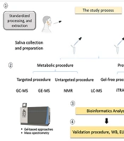
Metabolites play a crucial role in the pathogenesis of dry eye disease (DED). Androgens are one significant group of metabolites associated with DED; serum androgens have been linked to the disease, suggesting their potential involvement in its development (22). Distinct metabolites are also being studied through metabolomics profiling in DED. This approach aims to identify specific metabolites, selected ratios, or multicomponent patterns that can provide insights into disease progression, diagnosis, prognosis, and treatment options (35). Analysis of tear metabolite profiles can help physicians identify DED at earlier stages. However, studies focusing on these metabolites remain limited. Current single-omics technologies do not adequately capture the interactions among RNAs, proteins, and phenotypes required for diagnosing DED. Metabolomics involves comprehensive research on small molecules, particularly those soluble in water, which are important intermediates in metabolic pathways (36).
As tissues undergo stress, tear metabolite composition changes, particularly with age (11). Integrating genomics, epigenomics, transcriptomics, and proteomics with metabolomics can enhance the diagnosis, prognosis, treatment selection, and monitoring of dry eye disease (36). Approximately 98% of tears consist of water, whereas about 2% contains around 500 soluble proteins (e.g., lysozyme, matrix metalloproteinases, secretory immunoglobulin A, lactoferrin, and lipocalin), various metabolites and electrolytes (e.g., Ca²⁺, Na⁺, K⁺, and Cl⁻), carbohydrates (such as mucins and N-acetyl neuraminic acid), and lipids (including cholesterol, triglycerides, and monounsaturated fatty acids). It is critical to halt metabolic pathways immediately after tear collection to preserve sample integrity (36). A study comparing 48 different metabolites between DED patients and a control group found that the most prevalent metabolites were glucose, amino acids, and lipids. Patients with DED exhibited elevated metabolic activity and energy levels, with initial increases in glucose followed by lactate and creatine. Conversely, significant decreases in glutathione, N-acetylglucosamine, and cholesterol were observed. Additionally, certain biochemical factors, such as lipocalin, lipophilin AC-1, and lactoferrin, were found to be elevated, along with kynurenine (11), which is implicated in the inflammatory response associated with Sjögren’s syndrome DED (37). Aging is a risk factor for primary DED and is linked to decreased tear secretion. According to the TFOS DEWS II Epidemiology Report, specific metabolites related to dry eye disease have been identified in individuals aged 50 - 60 years (1).
In recent studies, various countries have implemented different diagnostic tests for dry eye disease (DED), evaluating a range of metabolites and biomarkers to determine disease severity. Table 1 provides a comprehensive overview of the diagnostic landscape for DED, showcasing the diverse methodologies and findings, which can inform clinical practice and research in the field (Table 1).
Table 1.
Overview of Diagnostic Tests, Evaluated Metabolites, Biomarkers, and Severity of Dry Eye Disease Across Various Countries
| Countries Commonly Reporting the Diagnostic Test | The Diagnostic Test | The Evaluated Metabolites | The Evaluated Biomarkers | Severity DED | The Emerging Test |
|---|---|---|---|---|---|
| Australia, Canada, Italy, Japan, Switzerland, United Kingdom, United States (28%) (26) Korea (37.1%) (26) | History DED /questionnaire/symptoms | − CH3 lipids, cholesterol/lipids, N-acetylglucosamine, glutamate, amino-n-butyrate, choline, glucose, phenylalanine, and formate (38) | ↑ Diadenosine polyphosphates, Ap4A and Ap5 (36) Collagen IL1B MAPK (39) | Moderate, Severe | FVA (20) |
| United states (18%), (40) Australia (< 10%) (41) | Tear osmolarity | MMP-9 Glucose Lactate Creatinine (11) | Collagen IL1B MAPK (39) | Moderate, Severe (42) | FPD (20) |
| Philippines (95%) (43) United states (49%) (44) Ghana (62%) (45) Australia and the United Kingdom (56.2%) (46) | Fluorescein break up time (FBUT) | MMP-9 (39) Glucose Lactate Creatine and 42 numbered factors (11) | Collagen IL1B MAPK (39) | Moderate, Severe (47) | Non-invasive TBUT, Lipid layer interferometry (20) |
| Korea (13.2%) (26) Philippines (70%) (43) | Schirmer test (Tear volume test) | Cortisol, corticosterone, 11-deoxycortisol, 4-androstene-3,17-dione, testosterone, 17α-hydroxyprogesterone, and progesterone (48) MMP-9 (22) | ↑Diadenosine polyphosph, Ap4A and Ap5 (36) Collagen IL1B MAPK (39) | Moderate, Severe (49) | Anterior OCT and fluorophotometry (20) |
| Australia and the United Kingdom (59.6%) (46) Philippines (84%) (43) | Meibomian gland evaluation | Glucose Lactate Creatine (11) Unrelated to MMP-9 (49) | Collagen IL1B MAPK (39) | Moderate, Severe (49) | Meibography and lid wiper epitheliopath (20) |
| Philippines (91%) (43) | Fluorescein corneal stain | HbA1c Glucose Lactate Creatine and 26 numbered factors (11) MMP-9 (39) | Collagen IL1B MAPK (39) | Moderate, Severe (44) | Epithelial thickness, confocal microscopy and thermography (20) |
| Various countries and populations (43) | OSDI (the most current used questionnaire) | MMP-9 (50) Glucose Lactate Creatine (11) 42 numbered factors (11) −CH3 lipids, cholesterol/lipids, N-acetylglucosamine, glutamate, amino-n-butyrate, choline, phenylalanine, and formate (3, 38) | Collagen IL1B MAPK (39) | Moderate, Severe (44) | Digital OSDI-based symptom assessment and electronic patient-reported outcome tools |
| Korea (49) | MMP-9 POCT (49) | Glucose Lactate Creatine (11) cholesterol/lipids, N-acetylglucosamine, glutamate, phenylalanine (3, 38) | Collagen IL1B MAPK (39) | Early stage Mild, Moderate, Severe (49) | _ |
3.3.1. Role of MMP-9 in Dry Eye Disease
MMP-9 is a proteolytic enzyme that plays a significant role in the pathophysiology and diagnosis of DED. Increased levels and activity of MMP-9 have been detected in tears, ocular surface epithelial cells, and inflammatory cells in patients with dry eye.
3.3.1.1. MMP-9 in Dry Eye Disease Pathogenesis
Inflammatory factors play a crucial role in DED. Desiccation of the ocular surface epithelium can activate the nuclear factor (NF)-κB and mitogen-activated protein kinase (MAPK) pathways (39). This activation leads to the release of inflammatory mediators such as interleukin (IL)-1β, IL-6, IL-8, tumor necrosis factor (TNF)-α, and MMP-9. MMP-9, recognized as an ideal biomarker for early inflammation, is activated by stimulation from IL-1β, TNF-α, IL-6, IL-8, and IL-17 (39). MMP-9 is a particularly important biomarker because it is the most rapidly acting enzyme in transforming inactive interleukin-1 (IL-1) into its active form, thereby inducing inflammation on the ocular surface. Pathologically, MMP-9 is the primary protease responsible for matrix degradation and destruction. Inactive MMP-9 (pro-MMP-9) is produced in the corneal epithelium and is activated in the tear film. The main activator of MMP-9 in the tear film is MMP-2, also known as gelatinase A (50). Increased MMP-9 expression and activity promote lysis of tight junction proteins, accelerating detachment of apical corneal epithelial cells. Elevated MMP-9 levels are associated with increased ocular surface inflammation, including blepharitis, allergic conjunctivitis, corneal ulceration, and conjunctivochalasis, as evidenced by correlations with other inflammatory markers such as MMP-8 and myeloperoxidase (51). Oxidative stress can elevate inflammatory mediators such as MMP-9, which is a functional metalloenzyme regulated alongside MMP-2 (22).
3.3.1.2. MMP-9 as a Biomarker for Dry Eye Disease
Tear MMP-9 levels have been found to correlate with the severity of dry eye symptoms, making it a potential biomarker for disease monitoring and assessment of treatment response (51).
In DED, tear MMP-9 levels are typically ≥ 40 ng/mL (39). MMP-9 positivity is correlated with dry eye disease (DED) severity across various stages, including early, mild, moderate, and severe, making it a valuable biomarker compared with other diagnostic tests (48). It has been demonstrated that MMP-9 point-of-care testing (POCT) can detect elevated MMP-9 levels in secondary DED associated with conditions such as Sjögren’s syndrome, diabetes, lupus erythematosus, and certain cancers (22, 42).
Tear MMP-9 concentration is directly correlated with tear osmolarity and inversely correlated with Schirmer test scores, a measure of tear production (51).
Commercially available assays such as InflammaDry (RPS, Sarasota, FL), which detect MMP-9, show high sensitivity and specificity for diagnosing dry eye disease (51, 52), compared with clinical assessments such as the Ocular Surface Disease Index (OSDI), Schirmer test, tear breakup time (TBUT), and keratoconjunctival staining (3). A novel silicon nanowire-based biosensor achieved 86.96% sensitivity and 90% specificity in detecting tear MMP-9 in dry eye patients, demonstrating its potential as an alternative diagnostic tool (53).
The use of anti-inflammatory treatments, including corticosteroids, doxycycline, azithromycin (in animal studies), and tetracycline, can reduce MMP-9 levels on the ocular surface, potentially leading to false-negative results in immunoassay tests (3).
Antibodies produced against the epitope of MMP-9 can react with the entire MMP-9 protein with high affinity. The diagnostic performance of this assay has shown 96.7% sensitivity, 80% specificity, 91% accuracy, 91.66% precision, and an area under the curve (AUC) of 0.827 (P < 0.001), with an 83% positive predictive value and a 96% negative predictive value.
3.3.1.3. Therapeutic Implications of MMP-9 in Dry Eye Disease
Elevated MMP-9 levels can disrupt tight junctions and corneal barrier function, resulting in epithelial cell migration and desquamation. Oxidative stress can further exacerbate this inflammatory response. Decreased MMP-9 expression is associated with improvements in the ocular surface epithelium. The expression and level of MMP-9 have been found to decrease with anti-inflammatory treatments such as cyclosporine (51, 52), suggesting that targeting MMP-9 may be beneficial in managing dry eye. Deletion or inhibition of the MMP-9 gene in mouse models has been shown to protect against dry eye-induced corneal changes, whereas exogenous administration of MMP-9 can recapitulate acute corneal barrier disruption (54).
The molecular structure of MMP-9 consists of three main domains: the N-terminal propeptide domain, the catalytic domain, and the C-terminal hemopexin-like domain. The catalytic domain of MMP-9, which lacks fibronectin repeats, shares a common structure with other MMPs, featuring a five-stranded beta-sheet and three alpha-helices. The molecular structure of MMP-9 is essential for understanding its function and for designing specific inhibitors for therapeutic interventions (55).
3.3.2. Metabolomic Profiles Associated With Dry Eye Disease
Jiang et al. identified significant metabolites linked to DED (11). They are listed below, and the most notable metabolites are italicized.
3.3.2.1. Metabolites Associated With Fluorescein Breakup Time
Gamma-Aminobutyric acid, L-Tyrosine, Oxidized glutathione, Cholesterol, Glutamate, fumarate, Butyrylcarnitine, Lactate, Formate, acetylcholine, L-Isoleucine, Oleamide, 1-Stearoyl-2-Arachidonoyl PC, Inosine, Uracil, Uridine, Guanine, L-Valine, Dibutyl phthalate, L-Methionine, Cytosine, Uridine, Squalene, L-Kynurenine, Malate, Spermine, Citric acid, L-Serine, Glucose, Pyroglutamic acid, Pyruvic acid, Adenine, Glycocholic acid, L-Phenylalanine, Alpha-dimorphecolic acid, Arginine, Creatine, Glycine, Choline, Niacinamide, 4-Hydroxycitrulline, PS (20:2(11Z,14Z)/16:0), S-Glutathionyl-L-cysteine, PS (22:4(7Z,10Z,13Z,16Z)/17:0), Oleoyl Oxazolopyridine, Triisobutyl phosphate, 2′-Hydroxy-a-naphthoflavone, 1-Oleoyl-2-acetyl-sn-glycerol, (1,2-Dierucoyl-SN-Glycero-3-Phosphoethanolamine), S-Acetyldihydrolipoamide-E, Tetrahydropteroyltri-L-glutamate, Streptidine 6-phosphate, PE (16:0/0:0), MG (8:2(9Z,12Z)/0:0/0:0, Succinoadenosine and Tributyl phosphate.
3.3.2.2. Metabolites Associated With Fluorescein Staining
Betaine, L-Tyrosine, Oxidized glutathione, N-Acetylglucosamine, Lithocholic acid, Lactate, Pyrrolidonecarboxylic acid, L-Methionine, L-Tryptophan, Urocanic acid, Uracil, (8)-Shogaol, L-Proline, Purine, Glucose, Pyruvic acid, Adenine, Alpha-dimorphecolic acid, Phenylalanyl-Arginine, Dideoxymycobactin, Glycerol tripropanoate, Phenylpyruvic acid, Creatine, 3-Formylsalicylic acid, 4-oxo-docosanoic acid, 3-Pyrimidin-2-yl-Propionic Acid, Palmitoyl glucuronide, N-Acetylvanilalanine, Myristoylcarnitine, PG(P-18:0/0:0), 3′-Keto-3′-deoxy-AMP, 2-glyceryl-PGF2α, Palmitoyl Ethanolamide, Niacinamide, N-palmitoyl leucine, 1-Octen-3-yl glucoside, Inosine 2′-phosphate, Prosopinine, Tetrandrine, N-arachidonoyl tyrosine, 3,7-Dimethyloctanal, 3-Amino-L-Tyrosine, PS (12:0/18:3(6Z,9Z,12Z)), N-ethyl N-(2-hydroxy-ethyl) arachidonoyl amine, Oleoyl Ethanolamide, 2-Methyl-1,3-oxathiane, 2,8-Dihydroxyadenine, 3S-Aminodecanoic acid, DL-Glycerol 1-phosphate, Maprotiline glucuronide, 4-Methylene-L-glutamate, N,N-Dimethylaniline N-oxide, Cystamine, L-Homocysteic acid, PI(P-18:0/15:1(9Z)), Propylthiouracil glucuronide, Butyl butyryllactate.
3.3.2.3. Metabolites Contributing to Dry Eye Disease
Glutamate*, N-Acetylglucosamine*, Urocanic acid*, Dibutyl phthalate*, Cholesterol*, Fumarate*, Lactate*, L-Tyrosine*, Betaine*, L-Valine*, Amino-n-butyrate*, Gamma-Aminobutyric acid*, Oxidized glutathione*, Thiodiacetic acid*, L-Methionine*, Pyrrolidonecarboxylic acid*, Octadecanamide*, L-Proline*, Inosine*, L-Tryptophan*, Hypoxanthine*, L-Phenylalanine*, Purine*, Uracil*, Glucose*, Uridine*, Cytosine*, Oleamide*, Adenine*, L-Isoleucine*, Squalene*, (8)-Shogaol*, Malate*, Creatine*, Niacinamide*, Acetylcholine*, pyruvic acid*, L-Serine*, Lithocholic acid*, Citric acid*, Glycine*, Spermine*, Guanine*, Alpha-Dimorphecolic acid*, Formate*, Butyrylcarnitine*, L-Kynurenine*, Pyroglutamic acid*, Arginine, Choline, Glycocholic acid.
Among them, 31 metabolites were decreased (11):
Glutamate, N-Acetylglucosamine, Urocanic acid, Cholesterol, L-Tyrosine, Betaine, L-Valine, Amino-n-butyrate, Gamma-Aminobutyric acid, Oxidized glutathione, Thiodiacetic acid, L-Methionine, Pyrrolidonecarboxylic acid, Octadecanamide, Inosine, L-Tryptophan, Hypoxanthine, Purine, Uracil, Uridine, Adenine, L-Isoleucine, Squalene, (8)-Shogaol, L-Serine, Lithocholic acid, Glycine, Spermine, Guanine, Butyrylcarnitine, Pyroglutamic acid.
And 17 metabolites were increased (11):
Dibutyl phthalate, Fumarate, Lactate, L-Proline, L-Phenylalanine, Glucose, Cytosine, Oleamide, Malate, Creatine, Niacinamide, pyruvic acid, Acetylcholine, Citric acid, Alpha-Dimorphecolic acid, Formate, L-Kynurenine.
3.3.2.4. Implications
Increased levels of glucose, lactate, and creatine suggest a link to high energy metabolism in DED (11). Additionally, elevated kynurenine levels may indicate an inflammatory response in conditions such as Sjögren’s syndrome (56).
Low levels of lactoferrin, lipocalin, and lipophilin AC-1 can increase susceptibility to infections in DED by reducing antimicrobial proteins and increasing oxidative stress. Studies indicate that decreased levels of adenine, uridine, pyrimidine, and uracil may contribute to diminished corneal sensitivity and reduced tear fluid secretion. Conversely, elevated acetylcholine levels, through activation of muscarinic receptors, can stimulate tear secretion (11). Overall, insights from tear metabolomics may enhance prognosis, diagnosis, and therapeutic strategies for DED (11).
3.3.3. Protein Concentration in Tears
The concentration of proteins in tears increases to 8 - 10 μg/μL, with S100 A8 and S100 A9 being related to ocular surface inflammation (57).
3.3.4. Role of Lactoferrin
Lactoferrin, an iron-binding protein and antimicrobial factor, is associated with aqueous-deficient dry eye. Various peptides derived from proline-rich proteins and other sources have been identified in tears, indicating their potential role in dry eye pathology (57); additionally, 234 peptides derived from 25 proteins in basal tear were reported (58).
Significantly, the decreased proteins in DED include lactotransferrin (LTF), lysozyme (LYZ), zinc-alpha-2-glycoprotein 1 (AZGP1), lipocalin 1 (LCN1), secretoglobin, member 1(SCGB2A1), family 2A, lacritin (LACRT), deleted in malignant brain tumors 1 (DMBT1), and proline rich receptor 4 (PRR4) (40). By contrast, keratin 1 (KRT1), transferrin (TF), complement component 3 (C3), polymeric immunoglobulin receptor (PIGR), S100A8, S100A9, orosomucoid 1 (ORM1), immunoglobulin J polypeptide (IGJ), Annexin A1 (ANXAI), and heat shock 27kDa protein 1 (HSPB1) were increased in DED (40).
4. Discussion
The pathogenesis of DED is influenced by a combination of intrinsic and extrinsic factors. Aging, autoimmune diseases such as Sjögren's syndrome, and environmental conditions are prominent contributors to tear film instability and ocular surface inflammation. Inflammatory mediators, particularly MMP-9, play a pivotal role in the progression of DED (59).
Moreover, the impact of medications on tear production cannot be overlooked. Commonly prescribed drugs, including antihistamines and certain antidepressants, have been shown to exacerbate dry eye symptoms by affecting lacrimal gland function. This highlights the need for healthcare providers to consider the ocular side effects of medications when treating patients, particularly those with pre-existing dry eye conditions (60).
The classification systems for DED provide a framework for diagnosing and managing the condition. The distinction between aqueous-deficient and evaporative dry eye is essential, as it informs treatment strategies. For instance, patients with aqueous-deficient dry eye may benefit from artificial tears and punctal occlusion, whereas those with evaporative dry eye may require interventions targeting meibomian gland dysfunction (59).
Accurate assessment of dry eye severity is crucial for tailoring treatment plans. Integrating patient-reported outcomes with clinical evaluations can enhance understanding of how DED affects daily activities and overall well-being. The limitations of conventional diagnostic tools highlight the need for emerging approaches such as tear biomarkers, advanced imaging, and AI-based analysis.
Metabolomic profiles in tears reveal significant metabolites linked to DED, such as elevated lactate and glucose levels, indicating high energy metabolism. These findings enhance understanding of DED pathophysiology and may improve diagnostic and therapeutic strategies (11).
The concentration of MMP-9 in tears serves as a potential biomarker for assessing the severity of DED. Notably, point-of-care testing for MMP-9 has shown high sensitivity and specificity in diagnosing DED, outperforming traditional clinical assessments such as the Schirmer test and tear breakup time. The correlation between MMP-9 levels and tear osmolarity further emphasizes its utility in monitoring disease progression and treatment response (60).
DED is increasingly recognized as an immune-mediated inflammatory disorder, making anti-inflammatory and immunomodulatory therapies central to its management. Therapeutic agents such as corticosteroids provide rapid suppression of acute inflammation, whereas long-term immunomodulatory treatments, including cyclosporine A and lifitegrast, address chronic T-cell–mediated inflammatory responses and improve lacrimal gland function. Emerging biologics and cytokine-targeted therapies offer additional promise by selectively modulating specific inflammatory mediators involved in disease progression. Targeting the underlying inflammatory cascade not only alleviates symptoms but also helps restore tear film homeostasis and protect ocular surface integrity. Elevated MMP-9 levels correlate with increased ocular surface inflammation, suggesting that targeting this pathway may offer therapeutic potential for managing DED symptoms and preventing complications such as corneal abrasions and infections.
Additionally, lifestyle modifications and environmental adjustments play a critical role in managing DED symptoms. Patients should be educated on the importance of maintaining a humid environment, taking regular breaks from screens, and avoiding irritants that can exacerbate their condition.
Notably, this study is limited by heterogeneity in diagnostic criteria, biomarker assays, patient populations, and clinical settings across included studies. Variations in diagnostic thresholds, laboratory methods, and population characteristics may affect the consistency and comparability of findings. These limitations emphasize the need for standardized diagnostic criteria and biomarker assessment methods in future dry eye disease research.
4.1. Conclusions
A comprehensive understanding of the multifactorial etiology, clinical heterogeneity, and emerging biomarkers of dry eye disease is essential for effective patient management. Integrating traditional assessment tools with advanced imaging and molecular diagnostics may facilitate precision medicine approaches, optimize outcomes, and guide future research.